Comparative Study
. 2020 Oct;184(2):1042-1055.
doi: 10.1104/pp.19.01248. Epub 2020 Aug 5.

The OXA2a Insertase of Arabidopsis Is Required for Cytochrome c Maturation

Affiliations
Comparative Study

The OXA2a Insertase of Arabidopsis Is Required for Cytochrome c Maturation

Renuka Kolli et al. Plant Physiol. 2020 Oct.

Abstract

In yeast (Saccharomyces cerevisiae) and human (Homo sapiens) mitochondria, Oxidase assembly protein1 (Oxa1) is the general insertase for protein insertion from the matrix side into the inner membrane while Cytochrome c oxidase assembly protein18 (Cox18/Oxa2) is specifically involved in the topogenesis of the complex IV subunit, Cox2. Arabidopsis (Arabidopsis thaliana) mitochondria contain four OXA homologs: OXA1a, OXA1b, OXA2a, and OXA2b. OXA2a and OXA2b are unique members of the Oxa1 superfamily, in that they possess a tetratricopeptide repeat (TPR) domain at their C termini. Here, we determined the role of OXA2a by studying viable mutant plants generated by partial complementation of homozygous lethal OXA2a transfer-DNA insertional mutants using the developmentally regulated ABSCISIC ACID INSENSITIVE3 (ABI3) promoter. The ABI3p:OXA2a plants displayed growth retardation due to a reduction in the steady-state abundances of both c-type cytochromes, cytochrome c 1 and cytochrome c The observed reduction in the steady-state abundance of complex III could be attributed to cytochrome c 1 being one of its subunits. Expression of a soluble heme lyase from an organism with cytochrome c maturation system III could functionally complement the lack of OXA2a. This implies that OXA2a is required for the system I cytochrome c maturation of Arabidopsis. Due to the interaction of OXA2a with Cytochrome c maturation protein CcmF C-terminal-like protein (CCMFC) in a yeast split-ubiquitin based interaction assay, we propose that OXA2a aids in the membrane insertion of CCMFC, which is presumed to form the heme lyase component of the cytochrome c maturation pathway. In contrast with the crucial role played by the TPR domain of OXA2b, the TPR domain of OXA2a is not essential for its functionality.


Figures

Figure 1.
Figure 1.
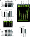
Phenotypes of oxa2a partial complementation plants. A, Plate-based growth progression analysis. Arrows indicate the time taken by wild-type plants to reach the developmental stages: 0.1 = imbibition; 0.5 = radicle emergence; 0.7 = hypocotyl and cotyledon emergence; 1.0 = cotyledons fully open; 1.02 = two rosette leaves > 1 mm in length; and 1.04 = four rosette leaves > 1 mm in length. Boxes represent time between the growth stages. Data are given as averages for 60 plants. Asterisks indicate statistical significance based on Student's t test with reference to Col-0 (**P < 0.00005). B, Primary root lengths of plants grown vertically for 14 d. Data are given as averages ± se. n = 30, 36, 26, 34, and 33 for Col-0, oxa2a+ABI3p:OXA2a-1, oxa2a+ABI3p:OXA2a-2, oxa2a+35Sp:OXA2a, and oxa2a+35Sp:OXA2a∆245, respectively. Asterisks indicate statistical significance based on Student's t test and a specified P-value. C, Soil-based growth progression analysis. Developmental stages: 1.10: 10 rosette leaves > 1 mm in length; 5.10: First flower buds visible; 6.00: First flower open; 6.90: Flowering complete. Data are given as averages for 24 plants. Asterisks indicate statistical significance based on Student's t test with reference to Col-0 (*P < 0.0005 and **P < 0.00005). D, Representative pictures of plants grown for the soil-based phenotyping. Pictures were taken after the indicated days of growth. E, Plant height measured at stage 6.90. Data are given as averages ± se for 24 plants. Asterisks indicate statistical significance based on Student's t test and a specified P-value.
Figure 2.
Figure 2.
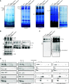
Analysis of mitochondrial complexes in oxa2a partial complementation plants. A, BN-PAGE analysis of mitochondrial complexes. On the left, Coomassie-stained complexes of mitochondria isolated from 4-week-old plants are represented on a polyvinylidene difluoride membrane. Adjacently, gels containing the 4-week-old samples were stained for NADH dehydrogenase (Complex I) activity, cytochrome c oxidase (complex IV) activity, and ubiquinol-cytochrome c oxidoreductase (complex III) activity, which was subsequently destained for better visibility. Complexes and supercomplexes are indicated where appropriate. B, Immunoblot analysis of mitochondrial complexes of 4-week-old plants after BN-PAGE using the antibodies against carbonic anhydrase 2 (CA2, complex I), three complex III subunits (MPPα, CYC1, and RISP), COX1, COX2 and COX3 (complex IV), F1 (complex V), and TOM20-3 (TOM complex). C, Immunoblot analysis of complex III-containing supercomplexes separated by BN-PAGE using the antibody against COB. Upon longer exposure of the blot, a putative assembly intermediate of complex III (intermediate II) was detected exclusively in the complementation plants. D, Immunoblot analysis of the specified complex III subunits after 2D-BN/SDS-PAGE of mitochondria from 4-week-old plants. The identified proteins are indicated between the blots of Col-0 and oxa2a+ABI3p:OXA2a-2 while the respective complexes are indicated on the top. I, Complex I; V, complex V; III2, dimeric complex III; I+III2, supercomplex composed of complex I and dimeric complex III;, I2+III4, supercomplex composed of two complex I monomers and two copies of dimeric complex III; Intermediate II, an assembly intermediate of complex III.
Figure 3.
Figure 3.
Analysis of mitochondrial proteins in oxa2a partial complementation plants. Immunoblot analysis of the indicated proteins involved in respiration, CCM, protein import, and other functions using mitochondria isolated form 4-week-old plants. In all the sections, 30 μg mitochondrial sample was loaded, the antibody used is mentioned on the left side and the molecular weight in kilodaltons is located on the right. The correct band is indicated with an arrowhead next to the protein name. In some cases, more than one isoform was detected. Bands not indicated are nonspecific reactions of the antibody. Where protein abundance is significantly different as shown in Supplemental Figure S8, it is indicated on the right side by either an up (up-regulated) or a down (down-regulated) arrow. For a full list of antibodies, see Supplemental Table S2.
Figure 4.
Figure 4.
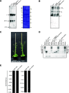
Analyses of the CCM process in ABI3p:OXA2a plants. A, Mitochondrial proteins of 4-week-old ABI3p:OXA2a plants separated by SDS-PAGE were subjected to heme staining. The stained proteins are indicated. The asterisk indicates a nonspecific band. Adjacently, the Coomassie-stained polyvinylidene difluoride membrane is shown along with molecular weight markers in kilodaltons. B, Mitochondrial complexes of 4-week-old ABI3p:OXA2a plants separated by BN-PAGE were subjected to heme staining. The stained complexes are indicated. III2, Dimeric complex III; I+III2, supercomplex composed of complex I and dimeric complex III; ?, an unknown complex. C, Representative plants of the specified genotype at 43 d of growth. D, Genotyping PCR analysis of oxa2a complementation line. The plants were genotyped for the T-DNA insert and for the presence of the inserted complementation construct. The sizes of the PCR products are as follows: LP2-RP2 = 1082 bp, LB2-RP2 = ∼800 bp, and F1-R1 = 1035 bp. Asterisk indicates a nonspecific PCR product, which was also found in Supplemental Figure S1C. E, Mating-based split-ubiquitin system was used to determine the interaction partner of OXA2a. Diploid yeast expressing the indicated fusion proteins was dropped at OD600 = 0.1 on vector-selective (CSM-Leu-, Trp-, Ura-) and interaction-selective (CSM-Leu-, Trp-, Ura-, Met-, Ade-, His-, +500 µm Met) media. NubWt was used as a positive control and NubG as a negative control. The Oligosaccharyltransferase4 (OST4) transmembrane domain was used to anchor the Cub fusion to the endoplasmic reticulum membrane.

Similar articles

Cited by

References

    1. Allen JW, Jackson AP, Rigden DJ, Willis AC, Ferguson SJ, Ginger ML(2008) Order within a mosaic distribution of mitochondrial c-type cytochrome biogenesis systems? FEBS J 275: 2385–2402 - PubMed
    1. Anghel SA, McGilvray PT, Hegde RS, Keenan RJ(2017) Identification of Oxa1 homologs operating in the eukaryotic endoplasmic reticulum. Cell Rep 21: 3708–3716 - PMC - PubMed
    1. Asseck LY, Grefen C(2018) Detecting interactions of membrane proteins: The split-ubiquitin system. Methods Mol Biol 1794: 49–60 - PubMed
    1. Babbitt SE, Sutherland MC, San Francisco B, Mendez DL, Kranz RG(2015) Mitochondrial cytochrome c biogenesis: No longer an enigma. Trends Biochem Sci 40: 446–455 - PMC - PubMed
    1. Baerenfaller K, Hirsch-Hoffmann M, Svozil J, Hull R, Russenberger D, Bischof S, Lu Q, Gruissem W, Baginsky S(2011) pep2pro: A new tool for comprehensive proteome data analysis to reveal information about organ-specific proteomes in Arabidopsis thaliana. Integr Biol 3: 225–237 - PubMed

Publication types