Review
. 2020 Jun 1;4(2):NS20200008.
doi: 10.1042/NS20200008. eCollection 2020 Jun.

Mechanisms and roles of mitochondrial localisation and dynamics in neuronal function

Affiliations
Review

Mechanisms and roles of mitochondrial localisation and dynamics in neuronal function

Richard Seager et al. Neuronal Signal. .

Abstract

Neurons are highly polarised, complex and incredibly energy intensive cells, and their demand for ATP during neuronal transmission is primarily met by oxidative phosphorylation by mitochondria. Thus, maintaining the health and efficient function of mitochondria is vital for neuronal integrity, viability and synaptic activity. Mitochondria do not exist in isolation, but constantly undergo cycles of fusion and fission, and are actively transported around the neuron to sites of high energy demand. Intriguingly, axonal and dendritic mitochondria exhibit different morphologies. In axons mitochondria are small and sparse whereas in dendrites they are larger and more densely packed. The transport mechanisms and mitochondrial dynamics that underlie these differences, and their functional implications, have been the focus of concerted investigation. Moreover, it is now clear that deficiencies in mitochondrial dynamics can be a primary factor in many neurodegenerative diseases. Here, we review the role that mitochondrial dynamics play in neuronal function, how these processes support synaptic transmission and how mitochondrial dysfunction is implicated in neurodegenerative disease.

Keywords: DRP1; fission; fusion; mitochondria; mitochondrial dynamics; post translational modification.


Conflict of interest statement

The authors declare that there are no competing interests associated with the manuscript.

Figures

Figure 1
Figure 1. Neuronal mitochondria exhibit compartment-specific morphologies
(A) Primary rat hippocampal neuron expressing a mitochondrially targeted fluorescent protein (MitoDS-Red). Ankyrin-G staining (magenta) shows the axonal initial segment, used to identify the axon. Highlighted within the boxes are the axonal (i) and dendritic (ii) compartments (enlarged beneath); scale bar: 20 µm, 10 µm in enlargements. (B) Schematic showing the compartments of a neuron, depicting the long axon and multiple dendrites from the cell body (soma), containing the nucleus. Axonal mitochondria are small and sparse, whereas dendritic mitochondria are larger and occupy a greater volume of the process. Mitochondria are densely packed within the soma.
Figure 2
Figure 2. Roles of mitochondrial dynamics and calcium in support of neuronal activity and plasticity
(A) Axonal mitochondria are very dynamic, exhibiting both anterograde and retrograde movement. At the presynapse, glycolysis and OXPHOS support basal activity of the synapse. (B) Dendritic mitochondria are larger than axonal mitochondria and exhibit more stabilised behaviour, with a small number of spines containing mitochondria. (C) During an action potential, an influx of Ca2+ rapidly increases cytoplasmic [Ca2+], also increasing mitochondria [Ca2+], which stimulates OXPHOS activity and increased ATP generation. The enhanced ATP generation powers energy demanding processes such as vesicle recycling, endocytosis and mobilisation of the reserve pool. Local mitochondria stop trafficking and are stabilised by the action of syntaphilin (anchoring protein) and rearrangements of the Miro-TRAK complex. Axonal mitochondria also buffer Ca2+ to prevent sustained elevations of [Ca2+]i, thus regulating continuous rounds of firing, while also preventing asynchronous transmission. (D) Ca2+ influx at the postsynapse causes dendritic mitochondria to protrude into the spine, and also causes mitochondrial fission, which regulates Ca2+ transients in the mitochondria during activity. Dendritic mitochondria provide ATP for actin-dynamics, supporting growth of the postsynaptic density, surface expression of neurotransmitter receptors and supports local translation. (E) Schematic of mitochondrial compartments and Ca2+ influx. Mitochondria consist of two membranes: the mitochondrial outer membrane (MOM) and the mitochondrial inner membrane (MIM). The region formed between the MOM and MIM is called the intermembrane space (IMS). The MIM has extensive folds, which form the cristae, increasing the surface area of the membrane which houses the electron transport chain (ETC) components. Substrates from the tricarboxylic acid (TCA) cycle act as electron donors to the ETC, which couples electron transfer with the shuttling of protons across the MIM into the IMS, forming a higher concentration of protons in the IMS compared to the matrix. This generates a concentration gradient and an electrical potential (due to charge separation), termed the mitochondrial membrane potential (∆ψm). The ∆ψm is the driving force for ATP synthesis, as protons pass down their concentration and electrical gradient, passing through the enzyme ATP Synthase, forming ATP. ∆ψm is also a driving force for Ca2+ sequestration in the matrix. Ca2+ must traverse both the MOM and MIM to enter the matrix. The voltage-dependent anion-selective channel (VDAC) and the mitochondrial Ca2+ uniporter (MCU) make the MOM and MIM permeable to Ca2+, respectively. Matrix localised Ca2+ stimulates the TCA cycle and enhances mitochondrial OXPHOS function.
Figure 3
Figure 3. Mitochondrial transport in axons and dendrites
(A) Mitochondria are transported in an anterograde and retrograde fashion along microtubules via kinesin and dynein/dynactin motor proteins. Microtubules are arranged from minus to plus-ends away from the cell body, whereas microtubules in the dendrite are arranged non-uniformly. The adaptor protein TRAK1 preferentially localises within axons, while TRAK2 preferentially localises to dendrites. (B) Upon an action potential or reaching a site of high energy demand characterised by an increase in [Ca2+]i, a reversible rearrangement of the motor and adaptor complex occurs, causing axonal mitochondria to pause. There are a number of models of how this occurs (i) Miro binds to Ca2+ and induces a conformational change in kinesin, releasing it from the microtubule tract and (ii) Miro binding to Ca2+ causes it to dissociate from kinesin. Syntaphilin also helps to immobilise mitochondria to the microtubule. How/if dendritic mitochondria are stabilised in a similar manner remains to be established. (C) Disruption of the neuronal cytoskeleton (actin and microtubules) stabilises axonal mitochondria, whereas dendritic mitochondria become shorter and more dynamic.
Figure 4
Figure 4. Regulation of mitochondrial dynamics proteins
(A) DRP1 constantly cycles from the cytosol to the MOM. Phosphorylation at DRP1S616, mediated by PKCδ and CaMKII, promotes mitochondrial association and fission. Phosphorylation at DRP1S637 is mediated by PKA, which promotes mitochondrial elongation by inhibiting DRP1 GTPase activity and reducing MOM association, which is antagonised by the phosphatases calcineurin and PP2A. CDK5 has been demonstrated to phosphorylate DRP1S616, with reportedly contrasting effects on neuronal mitochondrial morphology, while CaMKIα-mediated phosphorylation at DRP1S637 has been shown to promote fission. Further research is required to confirm the roles of CDK5 and CaMKIα in DRP1 phosphorylation and function. S-Nitrosylation enhances pDRP1S616 levels, whereas O-GlcNAcylation reduces pDRP1S637 levels. Modification by SUMO1 promotes fission, whereas SUMO2/3 modified DRP1 sequesters DRP1 in the cytosol, preventing fission. The deSUMOylating enzymes SENP5 and SENP3 remove SUMO1 and SUMO2/3 from DRP1, respectively. (B) Following bioenergetic stress, leading to a reduction in the ATP/ADP ratio, AMPK is activated. AMPK phosphorylates MFF at S155 and S172, leading to fragmentation of the mitochondria. (C) Parkin activation in response to damaged mitochondria (i.e. dissipation of the ∆ψm) ubiquitinates many substrates on the MOM, leading to recruitment of the mitophagy machinery and degradation of the mitochondria. The two mitofusin proteins and MFF have been identified as parkin targets. Parkin-mediated ubiquitination of Mfn1/2 results in degradation, thus prevents fusion. Parkin-mediated ubiquitination of MFF recruits the mitophagy adaptor p62, leading to elimination of damaged mitochondria. (D) Under mitochondrial stress, MARCH5 ubiquitinates Mfn1, promoted by Mfn1 acetylation, which leads to Mfn1 degradation and fission. ERK-mediated phosphorylation of Mfn1 promotes mitochondrial fragmentation and apoptosis. Fis1 can bind to Mfn1 and inhibit its GTPase activity, thus promoting fission. (E) MiD proteins can impair the GTPase activity of DRP1, while MFF promotes DRP1 activity. DRP1-MiD-MFF exist as a trimeric complex, whereby MiD proteins facilitate binding between DRP1 and MFF; however, a mechanism of how this is regulated remains to be determined.
Figure 5
Figure 5. Effects of manipulating the fusion/fission proteins on neuronal mitochondria
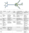
Summary of the effects on axonal and dendritic mitochondria upon manipulation (knockdown/knockout (loss) or overexpression (OE)) of the fusion and fission machinery. See text for details.

Similar articles

Cited by

References

    1. Liesa M., Palacín M. and Zorzano A. (2009) Mitochondrial Dynamics in Mammalian Health and Disease. Physiol. Rev. 89, 799–845 10.1152/physrev.00030.2008 - DOI - PubMed
    1. Eisner V., Picard M. and Hajnóczky G. (2018) Mitochondrial dynamics in adaptive and maladaptive cellular stress responses. Nat. Cell Biol. 20, 755–765 10.1038/s41556-018-0133-0 - DOI - PMC - PubMed
    1. Tilokani L., Nagashima S., Paupe V. and Prudent J. (2018) Mitochondrial dynamics: overview of molecular mechanisms. Essays Biochem. 62, 341–360 10.1042/EBC20170104 - DOI - PMC - PubMed
    1. Spinelli J.B. and Haigis M.C. (2018) The multifaceted contributions of mitochondria to cellular metabolism. Nat. Cell Biol. 20, 745–754 10.1038/s41556-018-0124-1 - DOI - PMC - PubMed
    1. Nunnari J. and Suomalainen A. (2012) Mitochondria: In sickness and in health. Cell 148, 1145–1159 10.1016/j.cell.2012.02.035 - DOI - PMC - PubMed