[Preprint]. 2020 Jun 16:3628297.
doi: 10.2139/ssrn.3628297.

Mouse model of SARS-CoV-2 reveals inflammatory role of type I interferon signaling

Affiliations

Mouse model of SARS-CoV-2 reveals inflammatory role of type I interferon signaling

Benjamin Israelow et al. SSRN. .

Update in

Abstract

Severe Acute Respiratory Syndrome- Coronavirus 2 (SARS-Cov-2) has caused over 5,000,000 cases of Coronavirus disease (COVID-19) with significant fatality rate.1-3 Due to the urgency of this global pandemic, numerous therapeutic and vaccine trials have begun without customary safety and efficacy studies.4 Laboratory mice have been the stalwart of these types of studies; however, they do not support infection by SARS-CoV-2 due to the inability of its spike (S) protein to engage the mouse ortholog of its human entry receptor angiotensin-converting enzyme 2 (hACE2). While hACE2 transgenic mice support infection and pathogenesis,5 these mice are currently limited in availability and are restricted to a single genetic background. Here we report the development of a mouse model of SARS-CoV-2 based on adeno associated virus (AAV)-mediated expression of hACE2. These mice support viral replication and antibody production and exhibit pathologic findings found in COVID-19 patients as well as non-human primate models. Moreover, we show that type I interferons are unable to control SARS-CoV2 replication and drive pathologic responses. Thus, the hACE2-AAV mouse model enables rapid deployment for in-depth analysis following robust SARS-CoV-2 infection with authentic patient-derived virus in mice of diverse genetic backgrounds. This represents a much-needed platform for rapidly testing prophylactic and therapeutic strategies to combat COVID-19.


Conflict of interest statement

Competing Interests None of the authors declare interests related to the manuscript.

Figures

Extended Data Fig 1.
Extended Data Fig 1.. AAV-hACE2 infection makes WT mice susceptible to SARS-CoV-2.
a, Immunofluorescence staining of ACE2 in mice infected with AAV-hACE2, 20 days post infection. b, Flow cytometry gating strategy for figures 1g, 3g–i, extended data fig 1c, and extended data fig 3a–c. c, Representative flow cytometry plots for figure 1g T cells and NK cells. d, Representative flow cytometry plots for figure 1g myeloid cells. e, Different macrophage populations in the lungs of SARS-CoV-2 infected mice two days post infection by flow cytometry. f, Serum of mice were collected 7 days after infection with SARS-CoV-2 and limiting dilutions were made to measure reactivity against S1 protein of SARS-CoV-2 using ELISA. g, Serum of mice were collected 14 days after infection with SARS-CoV-2 and limiting dilutions were made to measure reactivity against S1 protein of SARS-CoV-2 using ELISA.
Extended Data Fig 2.
Extended Data Fig 2.. Myeloid cell infiltration in SARS-CoV-2 infected mice.
a, Flow cytometry gating strategy for figures 1g, 3d–f, and extended data fig 1b, 1e and extended data fig. 2b-d. b, Relative percentage of different cell populations in different knockout mice infected with SARS-CoV-2 two days post infection. c, Representative flow cytometry plots for figure 3d and e. d, Relative percentage of myeloid cell populations in wildtype mice infected with AAV-hACE2 at days 2 and 4 post SARS-CoV-2 infection.
Extended Data Fig 3.
Extended Data Fig 3.. T cell and NK cell infiltration in SARS-CoV-2 infected mice.
a, Representative flow cytometry plots for figure 3g–i. b, Relative percentage of different lymphoid cell populations in different knockout mice infected with SARS-CoV-2 two days post infection. c, Relative percentage of lymphoid cell populations in wildtype mice infected with AAV-hACE2 at days 2 and 4 post SARS-CoV-2 infection.
Figure 1
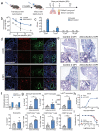
Figure 1. AAV-hACE2 transduction allows for productive SARS-CoV-2 infection in vivo.
a, Schematic of experimental plans. C57BL/6J mice were transduced intratracheally with an adeno-associated vector coding for hACE2 (AAV-hACE2) or control (AAV-GFP or PBS) and infected with SARS-CoV-2 two weeks after. Lung and blood samples were collected at days 2, 4, 7, and 14 days for analysis. b, Viral RNA from lung homogenates were measured using qPCR against SARS-CoV-2 N (CDC N1 primers). c, Viral titer from lung homogenates were performed by plaque assay on VeroE6 cells. d, Frozen lung tissue was stained for SARS-CoV-2 N protein (red) and epithelial cells (EpCAM, green). e, Fixed lung tissue was paraffin embedded and stained with H&E. f, Images from e were scored by a pulmonary pathologist for perivenular score. g, At two days post infection, single cell suspensions of lung were analyzed by flow cytometry. Data are shown as frequency of CD45+ cells (monocyte-derived macrophages, Ly6Chi monocytes, and neutrophils), frequency of parent cells (CD44+CD69+ CD4+ T cells, CD44+CD69+ CD8+ T cells, and CD69+ NK cells), or mean fluorescence intensity of CD64 (Ly6Chi monocytes). h, Serum antibodies were measured against spike protein using an ELISA. i, Day 7 and 14 sera from h was used to perform a plaque reduction neutralization assay on VeroE6 cells incubated with SARS-CoV-2.
Figure 2
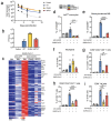
Figure 2. AAV-hACE2 mice infected with SARS-CoV-2 show similar interferon signatures as COVID19 patients.
a, Volcano plot showing differential expression of genes from whole lungs of mice infected with SARS-CoV-2 with and without AAV-hACE2 at day two post infection. Gray indicates significantly differentially upregulated genes, and blue indicates subsets of genes that are known ISGs. b, Significantly upregulated genes were put into Interferome (www.interferome.org) to identify how many genes are stimulated by type I, type II, or type III interferons. Subset of differentially expressed genes in a that were significantly upregulated in lungs of COVID19 patients (from Blanco-Melo et al 2020)., d, Upregulated gene list from human samples (Blanco-melo et. al. 2020) was graphed (left panel) and used to perform hierarchical clustering for differentially expressed genes from lungs of AAV-hACE2 SARS-CoV-2 infected mice (blue, mouse ISGs; gray, other genes)., e, Go Enrichment Analysis was performed on significantly upregulated genes to identify enriched cellular processes.
Figure 3
Figure 3. AAV-hACE2 mice infected with SARS-CoV-2 exhibit Type I Interferon dependent immune cell infiltration.
C57BL/6J (WT), IFNAR knockout and IRF3/7 double knockout mice were transduced intratracheally with an adeno-associated vector coding for hACE2 (AAV-hACE2) and infected with SARS-CoV-2 two weeks later. a, Viral titers in the lung of mice were measured using qPCR against SARS-CoV-2 N (Control and WT were from the same experiment as Figure 1b). b, Lung homogenates tittered on VeroE6 cells. c, Heat map of top 100 upregulated genes in SARS-CoV-2 infected C57BL/6J mice transduced with AAV-hACE2 versus SARS-CoV-2 infected AAV-GFP transduced mice. d-i At day two post infection, lungs of mice were made into single cell suspensions for flow cytometry.

Similar articles

References

    1. Zhu N. et al. A Novel Coronavirus from Patients with Pneumonia in China, 2019. N Engl J Med 382, 727–733, doi:10.1056/NEJMoa2001017 (2020). - DOI - PMC - PubMed
    1. Wang D. et al. Clinical Characteristics of 138 Hospitalized Patients With 2019 Novel Coronavirus-Infected Pneumonia in Wuhan, China. JAMA, doi:10.1001/jama.2020.1585 (2020). - DOI - PMC - PubMed
    1. Dong E., Du H. & Gardner L. An interactive web-based dashboard to track COVID-19 in real time. Lancet Infect Dis, doi:10.1016/S1473-3099(20)30120-1 (2020). - DOI - PMC - PubMed
    1. Callaway E. Coronavirus vaccines: five key questions as trials begin. Nature 579, 481, doi:10.1038/d41586-020-00798-8 (2020). - DOI - PubMed
    1. Bao L. et al. The pathogenicity of SARS-CoV-2 in hACE2 transgenic mice. Nature, doi:10.1038/s41586-020-2312-y (2020). - DOI - PubMed

Publication types

LinkOut - more resources