

Loss of progesterone receptor is associated with distinct tyrosine kinase profiles in breast cancer
- PMID: 32710281
- PMCID: PMC7497693
- DOI: 10.1007/s10549-020-05763-7
Loss of progesterone receptor is associated with distinct tyrosine kinase profiles in breast cancer
Abstract
Purpose: The aim of this study was to assess protein tyrosine kinase profiles in primary breast cancer samples in correlation with the distinct hormone and growth receptor profiles ER, PR, and HER2.
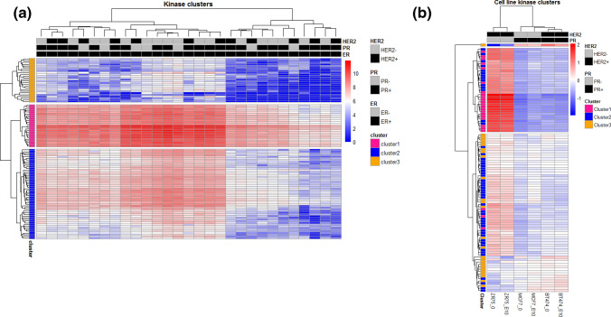
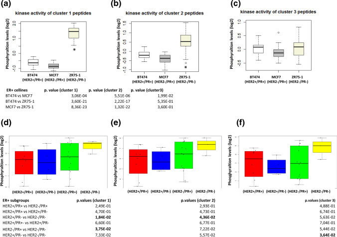
Experimental design: Pamchip® microarrays were used to measure the phosphorylation of 144 tyrosine kinase substrates in 29 ER+ breast cancer samples and cell lines MCF7, BT474 and ZR75-1. mRNA expression data from the METABRIC cohort and publicly available PR chip-sequencing data were used for validation purposes, together with RT-PCR.
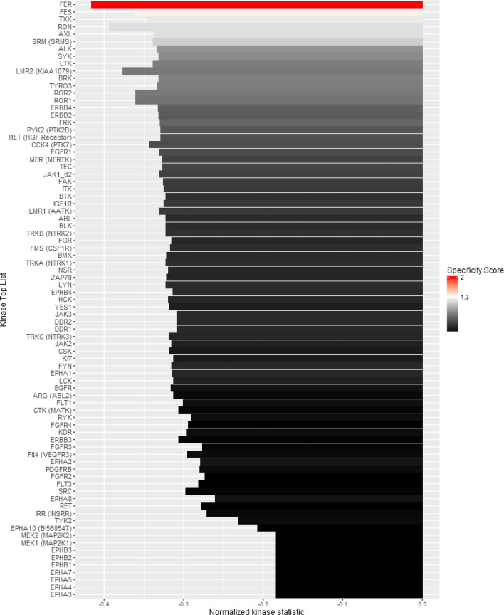
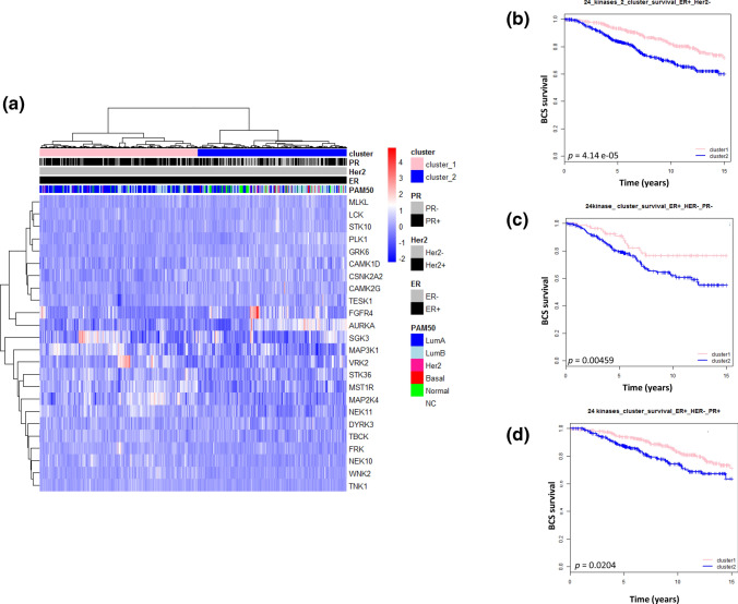
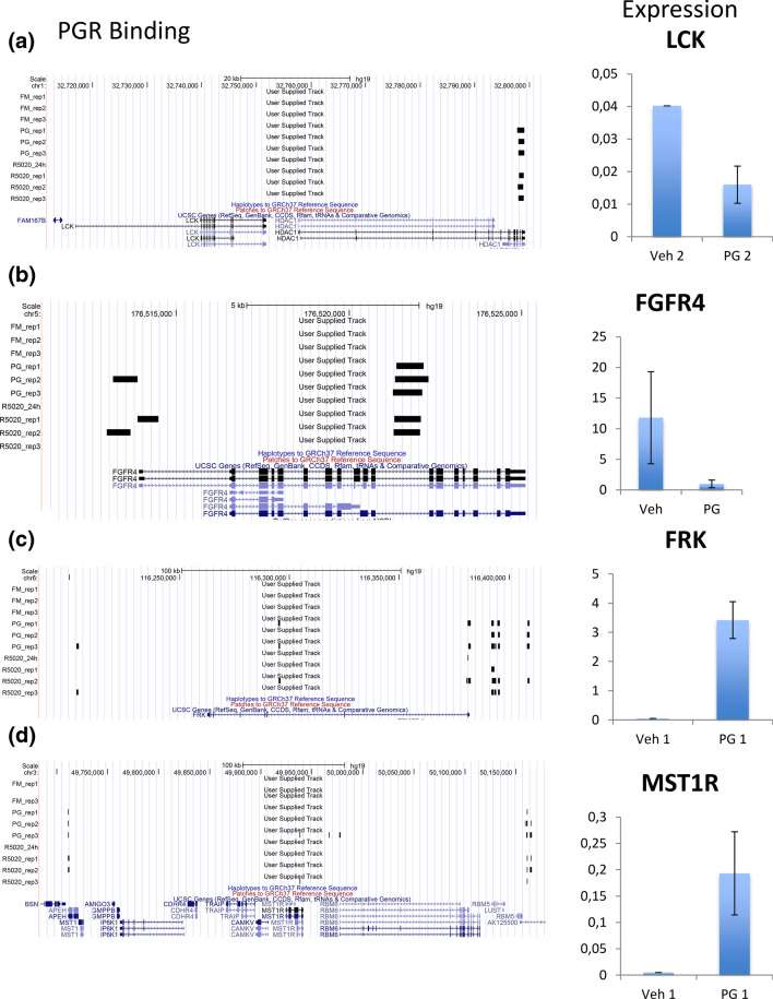
Results: In ER+ breast tumors and cell lines, we observed that the loss of PR expression correlated to higher kinase activity in samples and cell lines that were HER2-. A number of kinases, representing mostly proteins within the PI3K/AKT pathway, were identified as responsible for the differential phosphorylation between PR- and PR+ in ER+/HER2- tumors. We used the METABRIC cohort to analyze mRNA expression from 977 ER+/HER2- breast cancers. Twenty four kinase-encoding genes were identified as differentially expressed between PR+ and PR-, dividing ER+/HER2- samples in two distinct clusters with significant differences in survival (p < 0.05). Four kinase genes, LCK, FRK, FGFR4, and MST1R, were identified as potential direct targets of PR.
Conclusions: Our results suggest that the PR status has a profound effect on tyrosine kinases, especially for FGFR4 and LCK genes, in ER+/HER2- breast cancer patients. The influence of these genes on the PI3K/AKT signaling pathway may potentially lead to novel drug targets for ER+/PR- breast cancer patients.
Keywords: Breast cancer; ER; HER2; PI3K; PR; Tyrosine kinase.
Conflict of interest statement
Rik DeWijn works for Pamgene. No potential conflicts of interest were disclosed by the authors.
Figures
Similar articles
-
Genomic Landscape and Endocrine-Resistant Subgroup in Estrogen Receptor-Positive, Progesterone Receptor-Negative, and HER2-Negative Breast Cancer.Theranostics. 2018 Dec 8;8(22):6386-6399. doi: 10.7150/thno.29164. eCollection 2018. Theranostics. 2018. PMID: 30613307 Free PMC article.
-
Relapse-free survival of statistically standardized continuous RT-PCR estrogen receptor (ER), progesterone receptor (PR), and human epidermal growth factor receptor 2 (HER2): NCIC CTG MA.14.Breast Cancer Res Treat. 2016 May;157(1):101-8. doi: 10.1007/s10549-016-3806-z. Epub 2016 Apr 26. Breast Cancer Res Treat. 2016. PMID: 27116182 Clinical Trial.
-
Coexistence of the loss of heterozygosity at the PTEN locus and HER2 overexpression enhances the Akt activity thus leading to a negative progesterone receptor expression in breast carcinoma.Breast Cancer Res Treat. 2007 Mar;101(3):249-57. doi: 10.1007/s10549-006-9295-8. Epub 2006 Sep 28. Breast Cancer Res Treat. 2007. PMID: 17006756
-
Mutations in the phosphatidylinositol 3-kinase pathway: role in tumor progression and therapeutic implications in breast cancer.Breast Cancer Res. 2011;13(6):224. doi: 10.1186/bcr3039. Epub 2011 Nov 1. Breast Cancer Res. 2011. PMID: 22114931 Free PMC article. Review.
-
The multigene classifiers 95GC/42GC/155GC for precision medicine in ER-positive HER2-negative early breast cancer.Cancer Sci. 2021 Apr;112(4):1369-1375. doi: 10.1111/cas.14838. Epub 2021 Feb 26. Cancer Sci. 2021. PMID: 33544932 Free PMC article. Review.
Cited by
-
Association Between GATA3 and Histopathological and Immunohistochemical Parameters in Early-Infiltrating Breast Carcinomas.Eur J Breast Health. 2022 Jul 1;18(3):229-234. doi: 10.4274/ejbh.galenos.2022.2022-3-9. eCollection 2022 Jul. Eur J Breast Health. 2022. PMID: 35855199 Free PMC article.
-
The active kinome: The modern view of how active protein kinase networks fit in biological research.Curr Opin Pharmacol. 2022 Feb;62:117-129. doi: 10.1016/j.coph.2021.11.007. Epub 2021 Dec 27. Curr Opin Pharmacol. 2022. PMID: 34968947 Free PMC article. Review.
-
Kinase activity profiling in renal cell carcinoma, benign renal tissue and in response to four different tyrosine kinase inhibitors.Oncotarget. 2022 Aug 4;13:970-981. doi: 10.18632/oncotarget.28257. eCollection 2022. Oncotarget. 2022. PMID: 36093296 Free PMC article.
-
Discovering biomarkers for hormone-dependent tumors: in silico study on signaling pathways implicated in cell cycle and cytoskeleton regulation.Mol Genet Genomics. 2022 Jul;297(4):947-963. doi: 10.1007/s00438-022-01900-7. Epub 2022 May 9. Mol Genet Genomics. 2022. PMID: 35532795
-
Molecular characterization of low-grade serous ovarian carcinoma identifies genomic aberrations according to hormone receptor expression.NPJ Precis Oncol. 2022 Jun 29;6(1):47. doi: 10.1038/s41698-022-00288-2. NPJ Precis Oncol. 2022. PMID: 35768582 Free PMC article.
References
-
- Perou CM, Sorlie T, Eisen MB, van de Rijn M, Jeffrey SS, Rees CA, Pollack JR, Ross DT, Johnsen H, Akslen LA, Fluge O, Pergamenschikov A, Williams C, Zhu SX, Lonning PE, Borresen-Dale AL, Brown PO, Botstein D. Molecular portraits of human breast tumours. Nature. 2000;406(6797):747–752. doi: 10.1038/35021093. - DOI - PubMed
-
- Sorlie T, Perou CM, Tibshirani R, Aas T, Geisler S, Johnsen H, Hastie T, Eisen MB, van de Rijn M, Jeffrey SS, Thorsen T, Quist H, Matese JC, Brown PO, Botstein D, Lonning PE, Borresen-Dale AL. Gene expression patterns of breast carcinomas distinguish tumor subclasses with clinical implications. Proc Natl Acad Sci USA. 2001;98(19):10869–10874. doi: 10.1073/pnas.191367098. - DOI - PMC - PubMed
-
- Blows FM, Driver KE, Schmidt MK, Broeks A, van Leeuwen FE, Wesseling J, Cheang MC, Gelmon K, Nielsen TO, Blomqvist C, Heikkila P, Heikkinen T, Nevanlinna H, Akslen LA, Begin LR, Foulkes WD, Couch FJ, Wang X, Cafourek V, Olson JE, Baglietto L, Giles GG, Severi G, McLean CA, Southey MC, Rakha E, Green AR, Ellis IO, Sherman ME, Lissowska J, Anderson WF, Cox A, Cross SS, Reed MW, Provenzano E, Dawson SJ, Dunning AM, Humphreys M, Easton DF, Garcia-Closas M, Caldas C, Pharoah PD, Huntsman D. Subtyping of breast cancer by immunohistochemistry to investigate a relationship between subtype and short and long term survival: a collaborative analysis of data for 10,159 cases from 12 studies. PLoS Med. 2010;7(5):e1000279. doi: 10.1371/journal.pmed.1000279. - DOI - PMC - PubMed
MeSH terms
Substances
LinkOut - more resources
Full Text Sources
Medical
Research Materials
Miscellaneous
