. 2020 Aug 5;12(555):eabc9396.
doi: 10.1126/scitranslmed.abc9396. Epub 2020 Jul 20.

An Alphavirus-derived replicon RNA vaccine induces SARS-CoV-2 neutralizing antibody and T cell responses in mice and nonhuman primates

Affiliations

An Alphavirus-derived replicon RNA vaccine induces SARS-CoV-2 neutralizing antibody and T cell responses in mice and nonhuman primates

Jesse H Erasmus et al. Sci Transl Med. .

Abstract

The coronavirus disease 2019 (COVID-19) pandemic, caused by infection with the severe acute respiratory syndrome coronavirus-2 (SARS-CoV-2), is having a deleterious impact on health services and the global economy, highlighting the urgent need for an effective vaccine. Such a vaccine would need to rapidly confer protection after one or two doses and would need to be manufactured using components suitable for scale up. Here, we developed an Alphavirus-derived replicon RNA vaccine candidate, repRNA-CoV2S, encoding the SARS-CoV-2 spike (S) protein. The RNA replicons were formulated with lipid inorganic nanoparticles (LIONs) that were designed to enhance vaccine stability, delivery, and immunogenicity. We show that a single intramuscular injection of the LION/repRNA-CoV2S vaccine in mice elicited robust production of anti-SARS-CoV-2 S protein IgG antibody isotypes indicative of a type 1 T helper cell response. A prime/boost regimen induced potent T cell responses in mice including antigen-specific responses in the lung and spleen. Prime-only immunization of aged (17 months old) mice induced smaller immune responses compared to young mice, but this difference was abrogated by booster immunization. In nonhuman primates, prime-only immunization in one intramuscular injection site or prime/boost immunizations in five intramuscular injection sites elicited modest T cell responses and robust antibody responses. The antibody responses persisted for at least 70 days and neutralized SARS-CoV-2 at titers comparable to those in human serum samples collected from individuals convalescing from COVID-19. These data support further development of LION/repRNA-CoV2S as a vaccine candidate for prophylactic protection against SARS-CoV-2 infection.


Figures

Fig. 1
Fig. 1. repRNA-CoV2S vaccine design and formulation.
(A) Shown is the codon-optimized full length spike (S) protein open reading frame, including the S1, S2, transmembrane (TM), and cytoplasmic (CD) domains, corresponding to positions 21,536 to 25,384 in the S protein of SARS-CoV-2 isolate Wuhan-Hu-1 (GenBank: MN908947.3). This construct was fused to a C-terminal v5 epitope tag and then was cloned into an alphavirus replicon encoding the 4 nonstructural protein (nsP1-4) genes of Venezuelan equine encephalitis virus, strain TC-83. Following RNA transcription and capping, repRNA-CoV2S was transfected into BHK cells. 24 hours later, the transfected BHK cells were analyzed by (B) anti-v5 immunofluorescence and (C) Western blot using either convalescent human serum or anti-v5 serum for immunodetection. Recombinant SARS-CoV2 spike protein (rCoV2-Spike) and repRNA-GFP were used as positive and negative controls, respectively. (D) Shown is a graphical representation of LION and formation of the vaccine complex after mixing with repRNA. (E) Shown is the evolution of LION particle size over 15 weeks measured by dynamic light scattering during storage at 4°C, 25°C or 42°C. (F) After mixing LION particles and repRNA, complex formation was confirmed by a shift in size distribution. (G) Gel electrophoresis analysis of triplicate preparations of repRNA extracted from LION particles after a concentrated RNase challenge showed substantial protection relative to a triplicate preparation of a dose-matched naked RNA following RNAse challenge. The formulated vaccine was stable for at least one week after mixing and storage at 4°C or 25°C as determined by (H) gel electrophoresis of repRNA extracted by phenol-chloroform treatment and (I) particle size of the complex. Data in B and C are representative of 2 independent experiments. Data in E, H, and I are from a single experiment, whereas data in F and G are representative of 3 independent experiments. Data in E, G, and I are shown as mean ± s.d. of 3 technical replicates. Scale bar in B is 100 μm.
Fig. 2
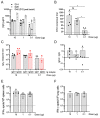
Fig. 2. The LION/repRNA-CoV2S vaccine induces Th1-biased and neutralizing antibodies in C57BL/6 mice.
Six to eight-week old C57BL/6 mice (n=5/group) received 10, 1, or 0.1 μg LION/repRNA-CoV2S via the intramuscular route on days 0 and 28. (A) Anti-S IgG antibody concentrations were determined by enzyme linked immunosorbent assay (ELISA) on days 14, 28, and 40. For day 14 samples, (B) 50% inhibitory concentrations (IC50) were determined by pseudovirus (SARS-CoV-2 Wuhan-Hu-1 pseudotype) neutralization assays. For day 14 samples, (C) anti-S IgG1 and IgG2c antibody endpoint titers and (D) ratios were determined by ELISA. On day 40, 12 days after a booster immunization, (E) spleens and (F) lungs were harvested and IFN-γ responses were measured by enzyme-linked immune absorbent spot (ELISpot) assay following an 18-hour stimulation with 10 peptide pools encompassing the S protein and consisting of 15 mers overlapping by 11 amino acids (see Fig. S1). Data in A, C, and D are representative of 3 independent experiments; data in B, E, and F are from a single experiment. Dotted lines in A, B, E, and F represent the lower limit of detection. All data are represented as individual values as well as mean ± s.d. *p<0.05 as determined by one-way ANOVA with Tukey’s multiple comparison test.
Fig. 3
Fig. 3. LION/repRNA-CoV2S induces Th1-biased antibodies in aged BALB/c mice.
Two-, eight-, or seventeen-month old BALB/c mice (n=5/group) received 10 or 1 μg LION/repRNA-CoV2S via the intramuscular route on days 0 and 28. On day 14 after prime and day 12 after boost (A) anti-S IgG was measured by ELISA. On day 40, 12 days after the boost, spleens were harvested and (B) IFN-γ responses were measured by enzyme-linked immune absorbent spot (ELISpot) assay following an 18 hour stimulation with 10 peptide pools encompassing the S protein and consisting of 15 mers overlapping by 11 amino acids (see Fig. S1). (C) Anti-S IgG1 and IgG2a antibody endpoint titers and (D) ratios were determined by enzyme-linked immunosorbent assay (ELISA) 14 days after the prime immunization. Data in 17-, 8-, and 2-month old BALB/c mice are from a single experiment; data for the 2-month old BALB/c mice were replicated in a second experiment. All data are represented as individual values as well as mean ± s.d. *p<0.05 as determined by one-way ANOVA with Tukey’s multiple comparison test between the 17-month old animals and either the 8- or 2-month old animals.
Fig. 4
Fig. 4. LION/repRNA-CoV2S induces neutralizing antibody responses in pigtailed macaques.
(A) Pigtail macaques were vaccinated with 250 μg (n=3) or with 50 μg (n=2) LION/repRNA-CoV2S via the intramuscular route and serum was collected on days 10, 14, 28, 42, 56, and 70. The 50 μg dose group received a boost vaccination on day 28 and blood was collected 14, 28, and 42 days later. (B) Using pre-immunization blood draws to establish a baseline, serum anti-S IgG enzyme linked immunosorbent assays (ELISAs) were performed on the post-immunization serum samples. (C) Pseudovirus (SARS-CoV-2 Wuhan-Hu-1 pseudotype) neutralization assays were performed on serum samples collected on days 14, 28, and 42 to determine mean 50% inhibitory concentrations (IC50) of each sample. Additionally, (D) 80% plaque-reduction neutralizing antibody titers (PRNT80) against the SARS-CoV2/WA/2020 isolate were measured at days 28 and 42 alongside 7 human convalescent serum samples collected from confirmed COVID-19 patients (see Table S1). The experiment was performed once. Each line in B and C represents each individual animal. Data in D are reported as individual values as well as mean ± s.d. *p<0.05 as determined by students t test comparing 250 μg dose groups at days 14 and 28. There was no significant difference (ns) between mean PRNT80 titers in all 5 animals at day 42 and titers in sera from 7 convalescent humans, as measured by Mann-Whitney U test.
Fig. 5
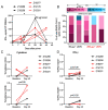
Fig. 5. LION/repRNA-CoV2S induces T-cell responses in pigtail macaques.
Pigtail macaques were vaccinated with 250 μg (n=3) or with 50 μg (n=2) LION/repRNA-CoV2S via the intramuscular route. PBMCs were isolated from blood at baseline and on days 10, 14, 28, and 42 after prime immunization for T-cell analysis. Shown are (A) magnitude and (B) breadth of IFN-γ responses measured in PBMCs by ELISpot assay following 24 hour stimulation with 11 peptide pools encompassing the spike (S) protein including the full-length S protein, and S1, S2 and RBD domains. (B) Data are presented as percent of total full-length S protein response. (C, D). The frequency of S-specific CD4+ or CD8+ T cells producing any cytokine (IFNγ, IL-2, IL-17A, TNFα, and/or MIP-1β, Granzyme B/CD107a) or IFN-γ alone was determined using cryopreserved PBMCs stimulated overnight with S protein peptides. Shown are the frequencies of S-specific CD4+ or CD8+ T cells after subtraction of background (DMSO vehicle). Data are from a single experiment. In A, C, D, each symbol/line is an individual animal. Data in B are representative of each individual animal. (A) Friedman test with multiple comparisons, *p<0.05; (C, D) Wilcoxon matched-pairs signed rank test; p-values are shown.

Similar articles

  • Single-dose replicating RNA vaccine induces neutralizing antibodies against SARS-CoV-2 in nonhuman primates.
    Erasmus JH, Khandhar AP, Walls AC, Hemann EA, O'Connor MA, Murapa P, Archer J, Leventhal S, Fuller J, Lewis T, Draves KE, Randall S, Guerriero KA, Duthie MS, Carter D, Reed SG, Hawman DW, Feldmann H, Gale M, Veesler D, Berglund P, Fuller DH. Erasmus JH, et al. bioRxiv [Preprint]. 2020 May 28:2020.05.28.121640. doi: 10.1101/2020.05.28.121640. bioRxiv. 2020. PMID: 32511417 Free PMC article. Preprint.
  • Evaluation of the mRNA-1273 Vaccine against SARS-CoV-2 in Nonhuman Primates.
    Corbett KS, Flynn B, Foulds KE, Francica JR, Boyoglu-Barnum S, Werner AP, Flach B, O'Connell S, Bock KW, Minai M, Nagata BM, Andersen H, Martinez DR, Noe AT, Douek N, Donaldson MM, Nji NN, Alvarado GS, Edwards DK, Flebbe DR, Lamb E, Doria-Rose NA, Lin BC, Louder MK, O'Dell S, Schmidt SD, Phung E, Chang LA, Yap C, Todd JM, Pessaint L, Van Ry A, Browne S, Greenhouse J, Putman-Taylor T, Strasbaugh A, Campbell TA, Cook A, Dodson A, Steingrebe K, Shi W, Zhang Y, Abiona OM, Wang L, Pegu A, Yang ES, Leung K, Zhou T, Teng IT, Widge A, Gordon I, Novik L, Gillespie RA, Loomis RJ, Moliva JI, Stewart-Jones G, Himansu S, Kong WP, Nason MC, Morabito KM, Ruckwardt TJ, Ledgerwood JE, Gaudinski MR, Kwong PD, Mascola JR, Carfi A, Lewis MG, Baric RS, McDermott A, Moore IN, Sullivan NJ, Roederer M, Seder RA, Graham BS. Corbett KS, et al. N Engl J Med. 2020 Oct 15;383(16):1544-1555. doi: 10.1056/NEJMoa2024671. Epub 2020 Jul 28. N Engl J Med. 2020. PMID: 32722908 Free PMC article.
  • A vaccine targeting the RBD of the S protein of SARS-CoV-2 induces protective immunity.
    Yang J, Wang W, Chen Z, Lu S, Yang F, Bi Z, Bao L, Mo F, Li X, Huang Y, Hong W, Yang Y, Zhao Y, Ye F, Lin S, Deng W, Chen H, Lei H, Zhang Z, Luo M, Gao H, Zheng Y, Gong Y, Jiang X, Xu Y, Lv Q, Li D, Wang M, Li F, Wang S, Wang G, Yu P, Qu Y, Yang L, Deng H, Tong A, Li J, Wang Z, Yang J, Shen G, Zhao Z, Li Y, Luo J, Liu H, Yu W, Yang M, Xu J, Wang J, Li H, Wang H, Kuang D, Lin P, Hu Z, Guo W, Cheng W, He Y, Song X, Chen C, Xue Z, Yao S, Chen L, Ma X, Chen S, Gou M, Huang W, Wang Y, Fan C, Tian Z, Shi M, Wang FS, Dai L, Wu M, Li G, Wang G, Peng Y, Qian Z, Huang C, Lau JY, Yang Z, Wei Y, Cen X, Peng X, Qin C, Zhang K, Lu G, Wei X. Yang J, et al. Nature. 2020 Oct;586(7830):572-577. doi: 10.1038/s41586-020-2599-8. Epub 2020 Jul 29. Nature. 2020. PMID: 32726802
  • Potential adjuvants for the development of a SARS-CoV-2 vaccine based on experimental results from similar coronaviruses.
    Gupta T, Gupta SK. Gupta T, et al. Int Immunopharmacol. 2020 Sep;86:106717. doi: 10.1016/j.intimp.2020.106717. Epub 2020 Jun 18. Int Immunopharmacol. 2020. PMID: 32585611 Free PMC article. Review.
  • SARS-CoV-2: A New Song Recalls an Old Melody.
    Subbarao K. Subbarao K. Cell Host Microbe. 2020 May 13;27(5):692-694. doi: 10.1016/j.chom.2020.04.019. Cell Host Microbe. 2020. PMID: 32407706 Free PMC article. Review.

Cited by

References

    1. Lu H., Stratton C. W., Tang Y.-W., Outbreak of pneumonia of unknown etiology in Wuhan, China: The mystery and the miracle. J. Med. Virol. 92, 401–402 (2020). 10.1002/jmv.25678 - DOI - PMC - PubMed
    1. Wu F., Zhao S., Yu B., Chen Y.-M., Wang W., Song Z.-G., Hu Y., Tao Z.-W., Tian J.-H., Pei Y.-Y., Yuan M.-L., Zhang Y.-L., Dai F.-H., Liu Y., Wang Q.-M., Zheng J.-J., Xu L., Holmes E. C., Zhang Y.-Z., A new coronavirus associated with human respiratory disease in China. Nature 579, 265–269 (2020). 10.1038/s41586-020-2008-3 - DOI - PMC - PubMed
    1. Wang C., Horby P. W., Hayden F. G., Gao G. F., A novel coronavirus outbreak of global health concern. Lancet 395, 470–473 (2020). 10.1016/S0140-6736(20)30185-9 - DOI - PMC - PubMed
    1. Hamming I., Timens W., Bulthuis M. L. C., Lely A. T., Navis G., van Goor H., Tissue distribution of ACE2 protein, the functional receptor for SARS coronavirus. A first step in understanding SARS pathogenesis. J. Pathol. 203, 631–637 (2004). 10.1002/path.1570 - DOI - PMC - PubMed
    1. Letko M., Marzi A., Munster V., Functional assessment of cell entry and receptor usage for SARS-CoV-2 and other lineage B betacoronaviruses. Nat. Microbiol. 5, 562–569 (2020). 10.1038/s41564-020-0688-y - DOI - PMC - PubMed

Publication types

MeSH terms