. 2020 Dec;129(3-4):227-242.
doi: 10.1007/s00412-020-00740-x. Epub 2020 Jul 17.

Pericentromere clustering in Tradescantia section Rhoeo involves self-associations of AT- and GC-rich heterochromatin fractions, is developmentally regulated, and increases during differentiation

Affiliations

Pericentromere clustering in Tradescantia section Rhoeo involves self-associations of AT- and GC-rich heterochromatin fractions, is developmentally regulated, and increases during differentiation

Hieronim Golczyk et al. Chromosoma. 2020 Dec.

Abstract

A spectacular but poorly recognized nuclear repatterning is the association of heterochromatic domains during interphase. Using base-specific fluorescence and extended-depth-of-focus imaging, we show that the association of heterochromatic pericentromeres composed of AT- and GC-rich chromatin occurs on a large scale in cycling meiotic and somatic cells and during development in ring- and bivalent-forming Tradescantia spathacea (section Rhoeo) varieties. The mean number of pericentromere AT-rich domains per root meristem nucleus was ca. half the expected diploid number in both varieties, suggesting chromosome pairing via (peri)centromeric regions. Indeed, regular pairing of AT-rich domains was observed. The AT- and GC-rich associations in differentiated cells contributed to a significant reduction of the mean number of the corresponding foci per nucleus in relation to root meristem. Within the first 10 mm of the root, the pericentromere attraction was in progress, as if it was an active process and involved both AT- and GC-rich associations. Complying with Rabl arrangement, the pericentromeres preferentially located on one nuclear pole, clustered into diverse configurations. Among them, a strikingly regular one with 5-7 ring-arranged pericentromeric AT-rich domains may be potentially engaged in chromosome positioning during mitosis. The fluorescent pattern of pachytene meiocytes and somatic nuclei suggests the existence of a highly prescribed ring/chain type of chromocenter architecture with side-by-side arranged pericentromeric regions. The dynamics of pericentromere associations together with their non-random location within nuclei was compared with nuclear architecture in other organisms, including the widely explored Arabidopsis model.

Keywords: Chromocenters; Interphase; Meiosis; Pericentromere; Rhoeo; Tradescantia spathacea.


Conflict of interest statement

The authors declare that they have no conflict of interest.

Figures
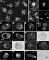
Fig. 1
Fig. 1
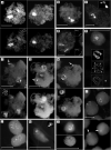
ap AT-rich (DAPI/AMD technique) pericentromeric heterochromatic domains of the ring-forming (ad, fg, i, k, m–n, p) and bivalent-forming variety (e, h, j, l, o) of T. spathacea. bars = 10 μm; nu, nucleolus. ac Pachytene nuclei and compound structure of their chromocenters; dash lined boxes of a are magnified in b and c with right panels obtained by capturing the same objects using low exposure time settings of the camera; arrows in b point to four AT-rich domains; each of the three meiotic chromocenters in c is composed of two AT-rich domains. dm Nuclei of the root meristem (ef, hm), 10-mm root sector (d) and 1-mm root sector (g); arrows in right panels of j and k point to pericentromere pairs. np Nuclei of leaf parenchyma (n), leaf epidermis (o), and root hair (p)
Fig. 2
Fig. 2
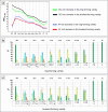
ac Changes in basic parameters: mean number of AT- or GC-rich domains per nucleus (a) and frequency (%) of the nuclei with a given number of AT-rich domains in the ring-forming (b) or bivalent-forming (c) variety. MNDAT, GC, mean number of AT-rich or GC-rich domains per nucleus; MP, meiotic prophase (pachytene); RM, root meristem; 1 mm–5 mm, 10 mm = 1 mm–5 mm, 10 mm root sectors; RH, root hairs; LP, leaf parenchyma; LE, leaf epidermis; 1–2, 3–4, 5–6, etc. = nuclear classes characterized by the presence of 1–2, 3–4, 5–6, etc. domains per nucleus. Vertical bars in b and c are standard deviations; to render the graph lines visible, standard deviations for a are not included. They are given in Table S1
Fig. 3
Fig. 3
al AT-rich (ae) and GC-rich (fl) heterochromatic domains (CMA3/DA/DAPI technique) in cycling and differentiated nuclei of the ring-forming variety (bh, l) and bivalent-forming variety (a, ik) of T. spathacea; bars = 10 μm; nu, nucleolus; additional dash line boxes represent graphical interpretation or structural details viewed under low exposure time and alleviated contrast of the camera. a Pachytene nuclei viewed under normal (top panels) and low exposure (bottom panels). b and c Strongly squashed nuclei representing pachytene (b) and root meristem (c) with thin fibers connecting clustered AT-rich prericentromeric domains. d Root meristem nucleus with two chains consisting of side-by-side positioned pericentromeres. e Leaf parenchyma nucleus with a chain-like arrangement pericentromeres; the chain consists of six (1–6) domains, two of them (domain 1 and 6) consisting clearly of two side-by-side arranged subdomains which most likely represent single pericentromeres (top and middle box); such a chromatin organization can be interpreted as a ring-type collective chromocenter disturbed by squashing (bottom box). fh Pachytene nuclei with their pericentromeric GC-rich foci (top panels) scattered (f) or ring-arranged (g, h); bottom panels show the same nuclei viewed in the DAPI channel; to see clearly the correlation between ring-type arrangement of the DAPI-positive heterochromatin and formation of the peripheral circle by the GC-rich pericentromeric domains, compare f with h; typically however the DAPI-rings could not be satisfactory resolved because of the high fluorescent haze of the UV illumination—see g as an example; arrows point to terminal GC-rich NOR sites typically fused into one spot localized opposite to the centromere pole. i Nuclei from the root 10th mm possessing one collective chromocenter (bottom panel, arrows) with a clear ring of CMA3-foci (top panel) localized peripherally around the AT-rich chromocenter core; such ring-type chromocenters were seen in those somatic nuclei of the root meristem and root sectors which possessed clearly one big AT-rich pericentromeric domain; in nuclei with more AT-rich domains, the CMA3-foci were scattered, as seen in j. j Root meristem nuclei with scattered CMA3-positive fluorescence foci. k and l CMA3-fluorscence of root hair (k), leaf parenchyma (l top), leaf epidermis (l bottom) nuclei; the ring-arrangement of the CMA3-foci as seen in l (bottom panel) was frequently observed in the three types of terminally differentiated nuclei in both varieties; however, it was not possible to state whether the foci are arranged around AT-rich heterochromatic core. Due to small size of these nuclei and a high density of their chromatin, it was not possible to distinguish their pericentromeric chromocenters in the DAPI channel when CMA3/DA/DAPI technique was applied
Fig. 4
Fig. 4
ad Multiple nuclear constraints operate in Tradescantia section Rhoeo. a Pericentromere structure. b A general universal Rabl nuclear organization with heterochromatic pericentromeres clustered on one pole to form collective chromocenter(s) and differently fused terminal NORs forming joint nucleolus or nucleoli. c Planar view of a ring-type pericentromeric collective chromocenter; when GC-rich pericentromeric domains are seen peripherally around the AT-rich chromocenter core, a ring of side-by-side positioned pericentromeres formed within the Rabl organization is the only possibly explanation (top); ideally such an arrangement can be viewed as the AT-rich heterochromatic ring in the DAPI channel (middle); frequently however such rings could not be satisfactorily resolved because of the high fluorescent haze of the UV illumination (bottom); GC-rich domains can be subjected to different degrees of fusion reducing the number of the corresponding fluorescent spots. d Pericentromere elongation during interphase (top panel) increases the fusigenic potential of the GC-rich domains by allowing their multiple contacts (bottom panel)

Similar articles

Cited by

References

    1. Alcobia I, Quina AS, Neves H, Clode N, Parreira L. The spatial organization of centromeric heterochromatin during normal human lymphopoiesis: evidence for ontogenically determined spatial patterns. Exp Cell Res. 2003;290:358–369. doi: 10.1016/s0014-4827(03)00335-5. - DOI - PubMed
    1. Anamthawat-Jónsson K, Heslop-Harrison JS. Centromeres, telomeres and chromatin in the interphase nucleus of cereals. Caryologia. 1990;43:205–213. doi: 10.1080/00087114.1990.10796999. - DOI
    1. Baroux C, Pecinka A, Fuchs J, Kreth G, Schubert I, Grossniklaus U. Non-random chromosome arrangement in triploid endosperm nuclei. Chromosoma. 2017;126:115–124. doi: 10.1007/s00412-016-0578-5. - DOI - PubMed
    1. Barr HJ, Ellison JR. Ectopic pairing of chromosome regions containing chemically similar DNA. Chromosoma. 1972;39:53–61. doi: 10.1007/bf00320590. - DOI - PubMed
    1. Bartholdi M. Nuclear distribution of centromeres during the cell cycle of human diploid fibroblasts. J Cell Sci. 1991;99:255–263. - PubMed

Publication types

Substances

LinkOut - more resources