. 2021 Aug 1;106(8):2147-2160.
doi: 10.3324/haematol.2020.253716.

Endothelial damage and dysfunction in acute graft-versus-host disease

Affiliations

Endothelial damage and dysfunction in acute graft-versus-host disease

Steffen Cordes et al. Haematologica. .

Abstract

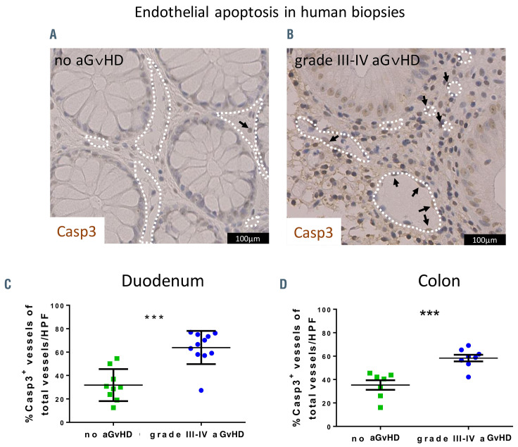
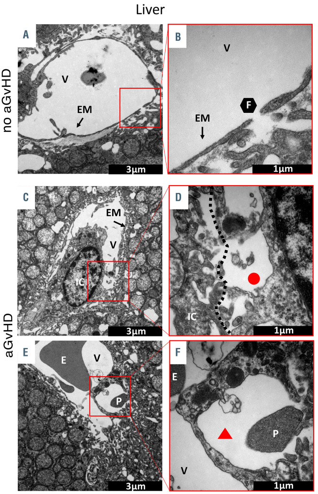
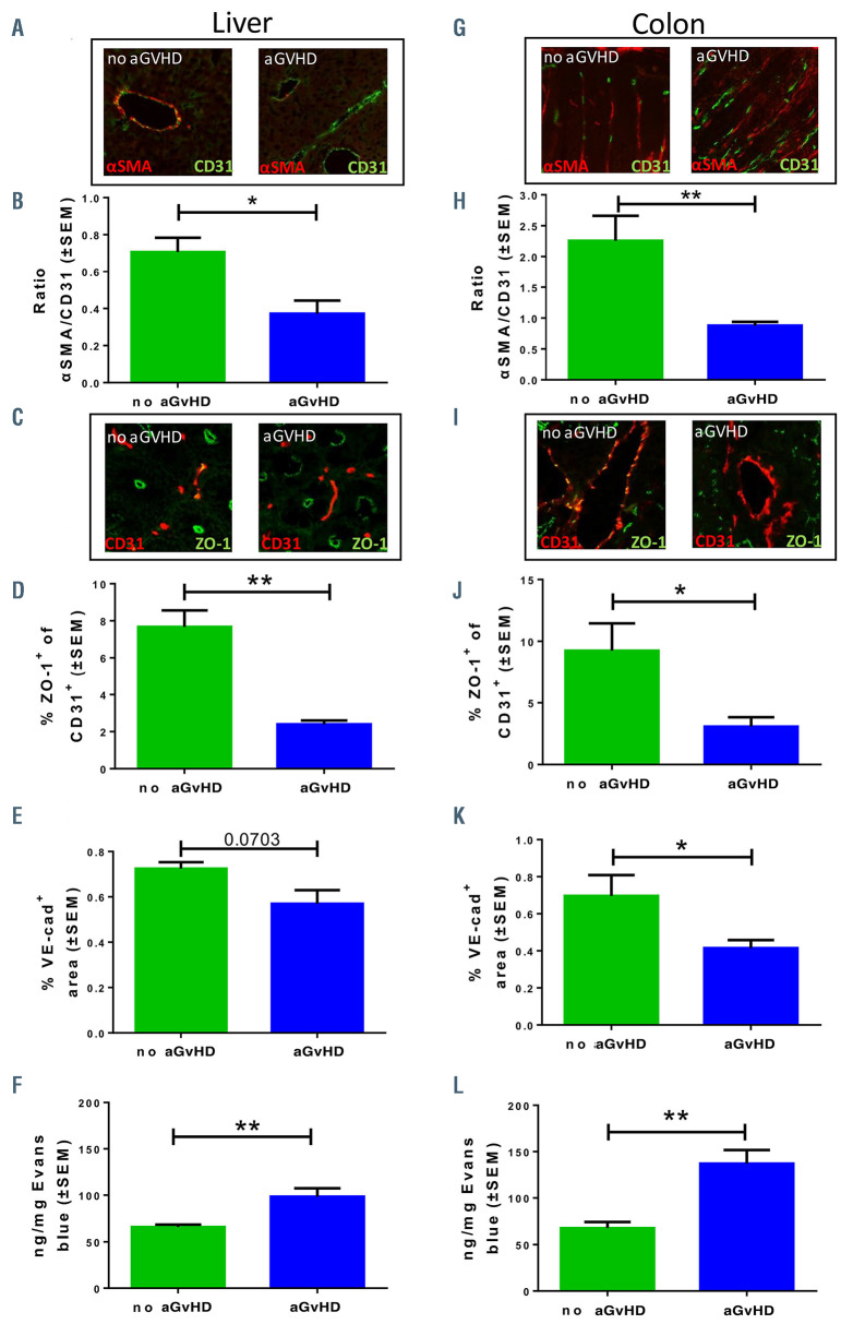
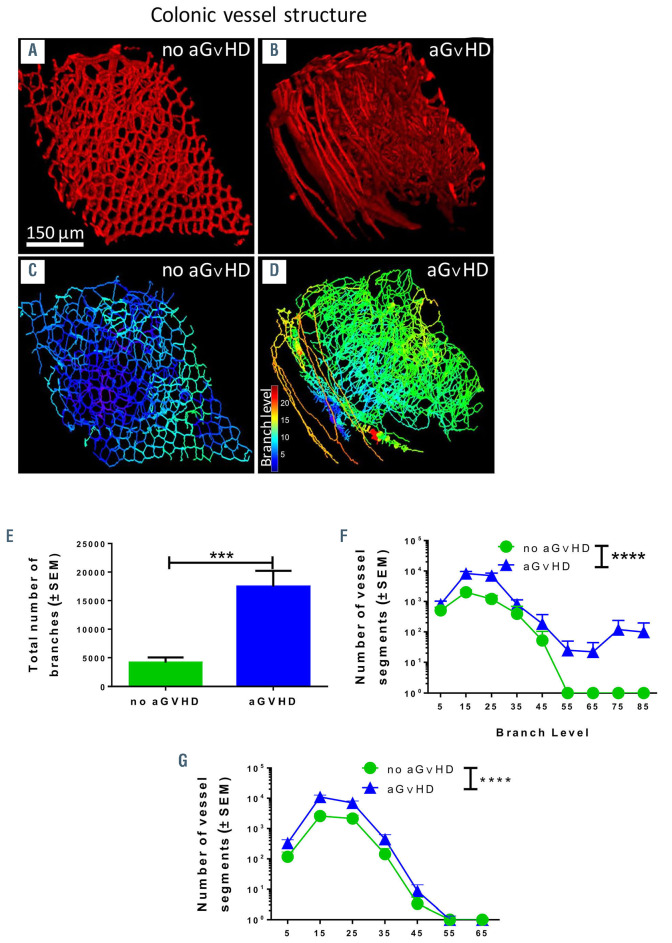
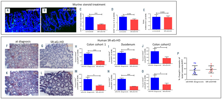
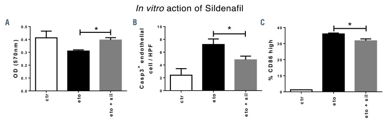
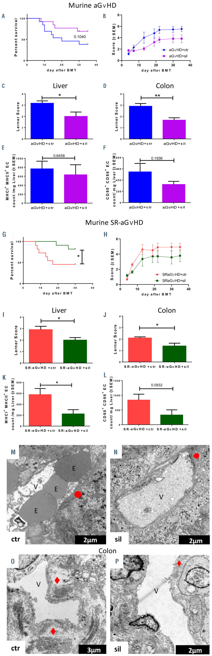
Clinical studies suggested that endothelial dysfunction and damage could be involved in the development and severity of acute graft-versus-host disease (aGVHD). Accordingly, we found increased percentage of apoptotic Casp3+ blood vessels in duodenal and colonic mucosa biopsies of patients with severe aGVHD. In murine experimental aGVHD, we detected severe microstructural endothelial damage and reduced endothelial pericyte coverage accompanied by reduced expression of endothelial tight junction proteins leading to increased endothelial leakage in aGVHD target organs. During intestinal aGVHD, colonic vasculature structurally changed, reflected by increased vessel branching and vessel diameter. Because recent data demonstrated an association of endothelium-related factors and steroid refractory aGVHD (SR-aGVHD), we analyzed human biopsies and murine tissues from SR-aGVHD. We found extensive tissue damage but low levels of alloreactive T cell infiltration in target organs, providing the rationale for T-cell independent SR-aGVHD treatment strategies. Consequently, we tested the endothelium-protective PDE5 inhibitor sildenafil, which reduced apoptosis and improved metabolic activity of endothelial cells in vitro. Accordingly, sildenafil treatment improved survival and reduced target organ damage during experimental SR-aGVHD. Our results demonstrate extensive damage, structural changes, and dysfunction of the vasculature during aGVHD. Therapeutic intervention by endothelium-protecting agents is an attractive approach for SR-aGVHD complementing current anti-inflammatory treatment options.


Figures

Figure 1.
Figure 1.
Endothelial damage in human intestinal biopsies. (A) Exemplary picture of a colon biopsy of a patient after allogeneic hematopoietic stem cell transplantation (allo-HSCT) without histologic evidence of acute graft-versus-host disease (aGvHD) and low level of endothelial apoptosis. The white dotted lines indicate vessel lumen and the arrow indicates one apoptotic caspase 3 positive (Casp3+) endothelial cell. (B) Exemplary picture of a colon biopsy of a patient with grade III-IV intestinal aGvHD and increased endothelial apoptosis. The white dotted lines indicate vessel lumen and the arrows indicate apoptotic Casp3+ endothelial cells. (C) Quantification of Casp3+ events in duodenal endothelium of allo-HSCT recipients given in percent of vessels in high-power fields (HPF). (D) Quantification of Casp3+ events in colonic endothelium of allo-HSCT recipients given in percent of vessels in HPF. Percentage of Casp3+ vessels was tested for significance by Student’s t-test (***P<0.001; n=7-11 patients per group). Error bars indicate mean ± standard error of the mean.
Figure 2.
Figure 2.
Visualization of acute graft-versus-host disease- associated ultrastructural changes in the liver by transmission electron microscopy. Representative pictures of sections from liver taken at day+15 after experimental hematopoietic stem cell transplantation (HSCT) in the chemotherapy based B6→BDF model. (A and B) Liver sinusoidal endothelial monolayer after syngeneic-HSCT (syn-HSCT) without acute graft-versus- host disease (aGvHD). (A) Normal, fenestrated sinusoidal blood vessel completely covered with endothelial monolayer. (B) Higher magnification of a 100 nm large fenestration of the endothelium in the liver. (C to-F) Sinusoidal liver endothelial monolayer after allo-HSCT during aGvHD. (C) Liver sinusoidal vessel with destroyed and unregularly shaped endothelial monolayer in contact with an immune cell. (D) Higher magnification of contact zone between immune cell and endothelial cell. (E) Blistering of the endothelial monolayer with a platelet in the region of injury. (F) Higher magnification of endothelial blistering. The perivascular space is marked by a red triangle. V: vessel lumen; EM: endothelial monolayer; F: fenestrated endothelium; IC: immune cell; E: erythrocyte; P: platelet; red circle: loss of endothelium; red triangle: endothelial blistering. Control groups (no aGvHD) were transplanted with the same bone marrow cell numbers and T-cell numbers from syngeneic donors.
Figure 3.
Figure 3.
Pericyte coverage, tight junctions and endothelial leakiness during acute graft-versus-host disease. Organs were harvested at day+15 after experimental hematopoietic stem cell transplantation (HSCT) in the chemotherapy based LP/J→B6 model. Control groups (no acute graft-versus-host disease [aGvHD]) were transplanted with the same bone marrow cell numbers and T-cell numbers from syngeneic donors. (A, B, and G to H) Quantification of pericyte coverage of vessels. (A and G) Representative pictures of staining for pericyte marker α smooth muscle actin (αSMA) in red and endothelial cell marker CD31 in green. Right organs of animals without aGvHD and left organs of animals suffering from aGvHD. (B and H) Ratio of αSMA positive area and CD31 positive area in (B) liver sinusoidal endothelium and (H) in colonic mucosal vessels in aGvHD versus no aGvHD. (C, D, I and J) Quantification of endothelial tight junction protein expression ZO-1. (C and I) Representative pictures of staining ZO-1 in green and CD31 in red. Right organs of animals without aGvHD and left organs of animals suffering from aGvHD. (D and J) Percentage of ZO- 1+ CD31+ area in (D) liver sinusoidal endothelium and (J) colonic mucosal vessels in aGvHD versus no aGvHD. (E and K) Quantification of endothelial adherence junction protein expression VE-cadherin. Percentage of VE-cadherin positive area in (E) liver sinusoidal endothelium and (K) colonic mucosal vessels in aGvHD versus no aGvHD. (F and L) Measurement of Evans blue extravasation in ng Evans blue per mg at day+15 after experimental allo-HSCT in the chemotherapy based B6→BDF model. (F) Liver and (L) colon of aGvHD versus no aGvHD. Significance was tested with Student’s t-test (*P<0.05; **P<0.01; n=5 animals per group). All experiments were reproduced in a biological independent experiment and shown are representative results of one experiment. Error bars indicate mean ± standard error of the mean.
Figure 4.
Figure 4.
Structural changes of vasculature in target organs during acute graft-versus-host disease by scanning light sheet fluorescence microscopy. We analyzed organs at day+15 after experimental hematopoietic stem cell transplantation (HSCT) in the chemotherapy based B6→BDF model. Control groups (no aGvHD) were transplanted with the same bone marrow (BM) cell numbers and T-cell numbers from syngeneic donors. (A and B) VE-cadherin signal of vasculature in colon of (A) allogeneic- HSCT (allo-HSCT) recipients without aGvHD and (B) allo-HSCT recipients with aGvHD. (C-D) Computed three-dimensional (3D) model of colonic vasculature with number of branches (blue= low branch levels; red= high branch levels) of (C) allo-HSCT recipients without aGvHD and (D) allo-HSCT recipients with aGvHD. (E to G) Analysis of colonic vasculature parameters. Assessment of (E) total number of branches (F) distribution of vessels branching level and (G) vessel diameter distribution in allo-HSCT recipients with aGvHD versus without aGvHD. Significance of total number of branches was tested by Student’s t-test (***P<0.001; n=3 animals per group) and significance of vessel branching level was tested by twoway ANOVA with Tukey’s multiple comparison test (***P<0.001; n=3 animals per group). Error bars indicate mean ± standard error of the mean.
Figure 5.
Figure 5.
Inflammatory infiltration and endothelial damage in human steroid-refractory acute graft-versus-host disease (SR-aGvHD) and in an experimental model of SR-aGvHD. (A to E) Immune cell infiltration in colon samples at day+15 after experimental allogeneic hematopoietic stem cell transplantation (allo-HSCT) in the chemotherapy based B6→BDF model. Exemplary pictures of CD4+ infiltrates (green) in colon mucosa of (A) control phosphate buffered saline (PBS) treated (untreated) aGvHD and (B) Dexamethasone treated aGvHD (SR-aGvHD, 0.5mg/kg/day Dexamethasone starting at day+4). (C) Quantification of CD4+ area in colonic mucosa of untreated aGvHD versus SR-aGvHD. (D) Quantification of CD8+ area in colonic mucosa of untreated aGvHD versus SR-aGvHD. (E) Ratio of pericyte marker α smooth muscle actin (αSMA) positive area and endothelial cell marker CD31 positive area in in colonic mucosa of untreated aGvHD versus SR-aGvHD. Significance was tested by Student’s t-test (***P<0.001; n=5 animals per group). Error bars indicate mean ± standard error of the mean. (F to P) Human intestinal biopsies at aGvHD diagnosis and during the course of SR-aGvHD. (F to J) Intestinal biopsies at aGvHD diagnosis and SR-aGvHD stained against pan leukocyte marker CD45 (F) Colon biopsy stained against CD45 at aGvHD diagnosis with massive infiltrates. (G) Colon biopsy stained against CD45 during SR-aGvHD with low infiltrates. (H to J) Quantification of CD45 infiltrates at aGvHD diagnosis versus SR-aGvHD in (H) colon and (K) duodenum biopsies of cohort 1 and (J) in colon biopsies of cohort 2. (K-O) Intestinal biopsies at aGvHD diagnosis and SR-aGvHD stained against T-cell receptor marker CD3 (K) Colon biopsy stained against CD3 at aGvHD diagnosis with massive infiltrates. (L) Colon biopsy stained against CD3 during SR-aGvHD with low infiltrates. (M to O) Quantification of CD3 infiltrates at aGvHD diagnosis versus SR-aGvHD in (M) colon and (N) duodenum biopsies of cohort 1 and (O) in colon biopsies of cohort 2. (P) Quantification of capspase 3 positive (Casp3+) events in colonic endothelium of allo-HSCT recipients of cohort 2 at aGvHD diagnosis and during SR-aGvHD given in percent of vessels per high power field (HPF). Significance was tested by Student’s ttest (*P<0.05; **P<0.01; ***P<0.001; n= 4-6 patients per group). Error bars indicate mean ± standard error of the mean.
Figure 6.
Figure 6.
Reduction of endothelial apoptosis and endothelial activation by sildenafil in vitro. Mouse cardiac endothelial cells (MCEC) were incubated with either phosphate buffered saline (PBS)/ 0,1% dimethylsulfoxide (DMSO) (control [ctr]), 100 nm etoposide (eto), an inducer of cell death, or with 100 nm etoposide and 34 nm sildenafil (eto+sil) for 24 hours before analysis. (A) MTT assay showed higher optical density of eto+sil group versus eto-only group. (B) Staining for apoptotic cell marker caspase 3 (Casp3) showed reduced Casp3+ cells per high-power field (HPF) in eto+sil group compared to eto-only group. (C) Flow cytometry analysis of CD86, a costimulatory and endothelial activation marker, showed reduced percentage of endothelial CD86high cells in eto+sil group compared to eto-only group. Signif icance was tested by Student’s t-test (*P<0.05; n=2-3 experiments with at least triplicates per condition). Error bars indicate mean ± standard error of the mean.
Figure 7.
Figure 7.
In vivo treatment of acute graft-versus-host disease (aGvHD) and steroid-refractory-aGvHD with sildenafil. (A to G) Treatment of aGvHD with 10 mg/kg/d sildenafil after experimental allogeneic hematopoietic stem cell transplantation (allo-HSCT) at day+15 after experimental allo-HSCT in the radiation based B6→BALB/c model. (A) Survival analysis and (B) clinical aGvHD manifestations of sildenafil (sil) treated allo-HSCT recipients with aGvHD versus control substance phosphate buffered saline/dimethylsulfoxide (PBS/DMSO, control [ctr]) treated allo-HSCT recipients with aGvHD. Histopathological assessment of aGvHD manifestations in (C) liver and (D) colon in sildenafil versus control substance treated allo-HSCT recipients at day+15. Flow cytometry quantification of (E) major histocompatibility complex I (MHCI) and MHCII expression and (F) CD80 and CD86 expression of isolated liver sinusoidal endothelial cells of sildenafil versus control substance treated allo-HSCT recipients at day+15. (G to L) Treatment with 10 mg/kg/d sildenafil in a murine model of SR-aGvHD at day+15 after experimental allo-HSCT in the radiation based B6→BALB/c model. (G) Sur vival analysis and (H) clinical aGvHD manifestations of sildenafil (SR-aGvHD+sil) versus control substance t rea te d (SR -aGvHD +ct r) SR-aGvHD. Histopathological assessment of aGvHD severity in (I) liver and (J) colon in SRaGvHD+ sil and SR-aGvHD+ctr at day+15 after allo-HSCT. Flow cytometr y quantification of (K) MHCI and MHCII expression and (L) CD80 and CD86 expression of isolated liver sinusoidal endothelial cells of SR-a Gv HD+sil and SR-aGvHD+ctr at day+15 after allo-HSCT. Significance was tested by Kaplan-Meier method and compared with the Mantel-Cox log-rank test (*P<0.05; n=8-10 animals per group) and S tudent’ s t -test (*P<0.0 5; ** P <0.01; n=6 animals per group). Error bars indicate mean ± standard error of the mean. Survival data was pooled from two experiments. All experiments were reproduced in a biological independent experiment and shown are representative results of one experiment. (M to P) Visualization of S R-aGvHD a nd silden afil tr eated SRaGvHD a ssoc iated ultr astr u ctur al changes in the liver and the colon by transmission electron microscopy. Shown are typical pictures of sections from liver and colon taken at day+15 after experimental allo-HSCT in the chemotherapy based 129→B6 mo del. (M and N) Sinusoidal liver endothe lial m onolayer during SR-aGvHD and sildeanfil (sil) treated SR-aGvHD. (M) Liver sinusoidal vessel of untreated SR-aGvHD with destroyed and unregularly shaped en dot helia l monolayer, marked by a red circle. (N) Liver sinusoidal vessel of Sildenafil treated SR-aGvHD with small ruptures of the endothelial monolayer, marked by a red circle. (O and P) Colonic mucosa endothelium duri n g SR-aGvH D and sildean fil treated SR- aGvHD. (O) The vessel of untreated SR-aGvHD is surrounded by massive perivascular fibrinogen deposits m arked by a re d rhomb us. (P) Occasionally little perivascular fibrinogen deposits, marked by a red rhombus, could be detected in silden afil treated SRaGvHD. V: vessel lumen; E: erythrocyte; N: nucleus; red rhombus: perivascular fibrinogen de posits; red circl e: loss of endothelium; red trapezoid: endothelial convolution).

Similar articles

Cited by

References

    1. Garnett C, Apperley JF, Pavlu J. Treatment and management of graft-versus-host disease: improving response and survival. Ther Adv Hematol. 2013;4(6):366-378. - PMC - PubMed
    1. Deeg HJ. How I treat refractory acute GVHD. Blood. 2007;109(10):4119-4126. - PMC - PubMed
    1. Westin JR, Saliba RM, De Lima M, et al. . Steroid-refractory acute GVHD: predictors and outcomes. Adv Hematol. 2011;2011: 601953. - PMC - PubMed
    1. Gloude NJ, Khandelwal P, Luebbering N, et al. . Circulating dsDNA, endothelial injury, and complement activation in thrombotic microangiopathy and GVHD. Blood. 2017;130(10):1259-1266. - PMC - PubMed
    1. Fan CQ, Crawford JM. Sinusoidal obstruction syndrome (hepatic veno-occlusive disease). J Clin Exp Hepatol. 2014;4(4):332-346. - PMC - PubMed

Publication types

Grants and funding

Funding: MB was funded by the European Training and Research in Peritoneal Dialysis Program, funded by the European Union within the Marie Curie Scheme (287813). AB received funding from the German Research Foundation (DFG), collaborative research center TRR221 (B11, Z02) and ZM was funded by the DFG collaborative research center TRR225 (B08).