. 2020 Jul 13;39(1):134.
doi: 10.1186/s13046-020-01626-7.

M2 macrophage-derived extracellular vesicles promote gastric cancer progression via a microRNA-130b-3p/MLL3/GRHL2 signaling cascade

Affiliations

M2 macrophage-derived extracellular vesicles promote gastric cancer progression via a microRNA-130b-3p/MLL3/GRHL2 signaling cascade

Yu Zhang et al. J Exp Clin Cancer Res. .

Retraction in

Abstract

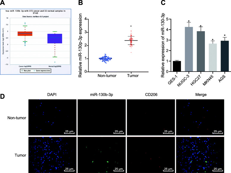
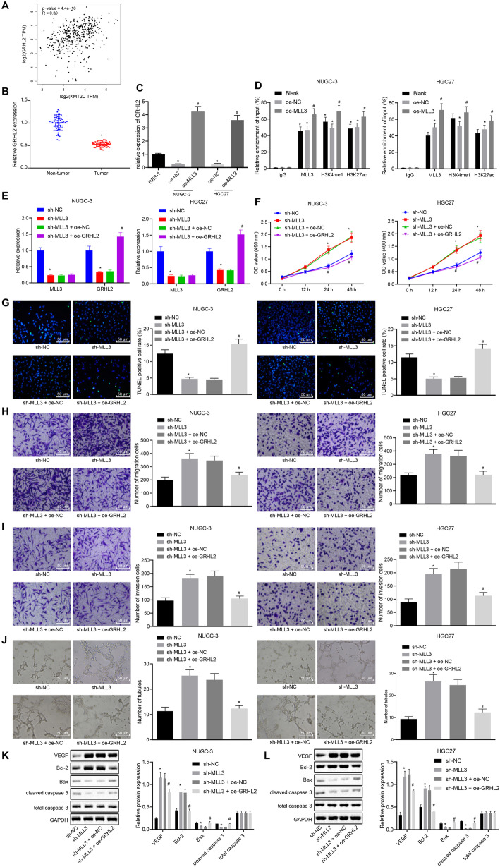
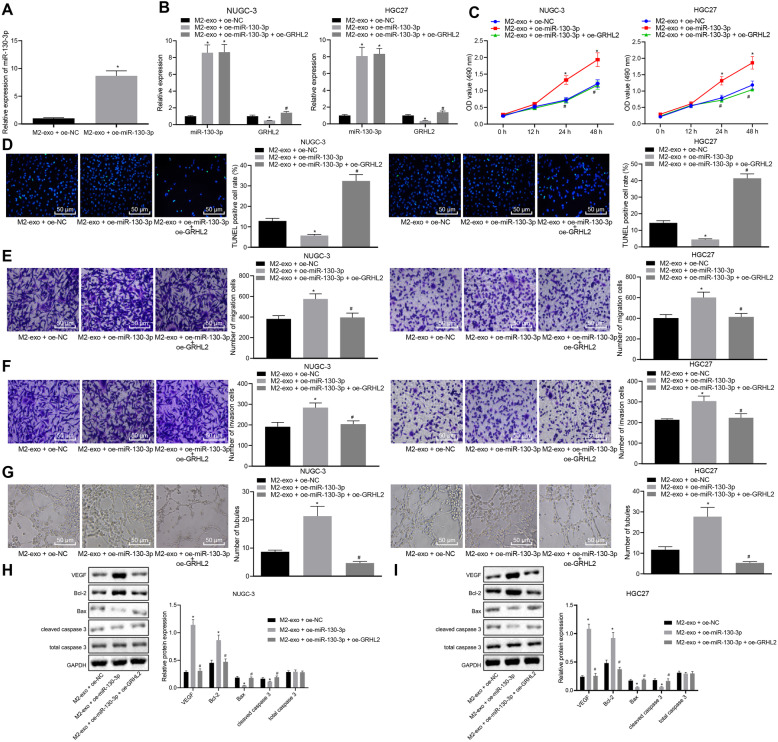
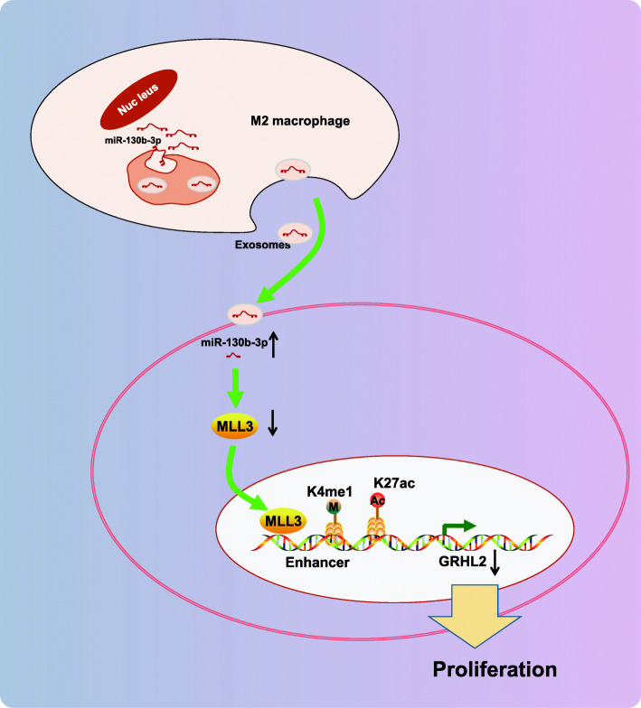
Background: Transfer of noncoding microRNAs (miRNAs) by extracellular vesicles (EVs) promotes the development of chemoresistance in many tumor types. Additionally, restoration or depletion of several miRNAs has been observed in multiple cancer types including gastric cancer (GC). In this present study, we aimed to investigate the mechanism of miR-130b-3p in M2 macrophage-derived EVs in the development of GC through regulation of mixed lineage leukemia 3 (MLL3) and grainyhead-like 2 (GRHL2).

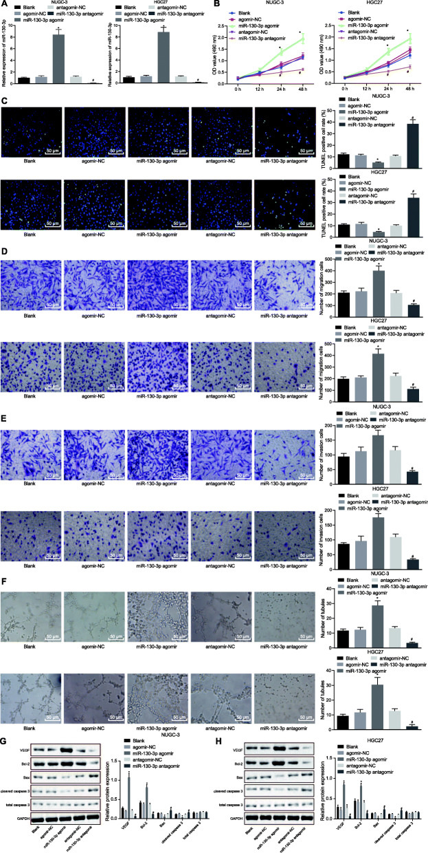
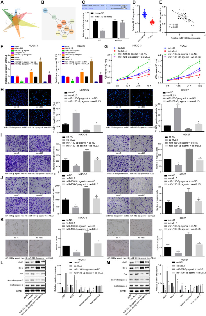
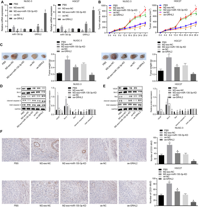
Methods: Expression of miR-130b-3p and GRHL2 was quantified in 63 pairs of cancerous and noncancerous gastric tissues. The predicted binding between miR-130b-3p and MLL3, together with the enrichment of MLL3, H3K4me1, and H3K27ac in gene enhancer region, was verified by luciferase activity assay and chromatin immunoprecipitation. Effects of miR-130b-3p on GC cell proliferation, apoptosis, migration and invasion, as well as tube formation of human umbilical endothelial vein cells (HUEVCs) were further determined by gain- and loss-of function assays in vitro.

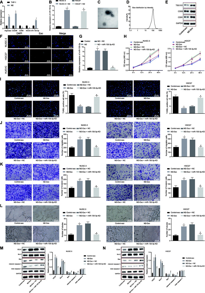
Results: miR-130b-3p was upregulated in GC tissues, and miR-130b-3p promoted survival, metastasis and angiogenesis of GC cells as well as enhanced tumor formation and angiogenesis in GC in vivo. Additionally, miR-130b-3p delivered in M2 macrophage-derived EVs promoted survival, migration, invasion, and angiogenesis of GC cells. Notably, MLL3 inhibited GC cell proliferation, migration, invasion, and vessel-like tube formation of HUEVCs by increasing GRHL2. Furthermore, downregulation of miR-130b-3p in M2 macrophage-derived EVs or upregulation of GRHL2 inhibited tumor formation and angiogenesis in GC.

Conclusion: This study highlights that EVs loaded with the specific miRNA cargo miR-130b-3p mediate communication between M2 macrophages and cancer cells in the tumor microenvironment through the modulation of MLL3 and GRHL2 in GC.

Keywords: Extracellular vesicles; GRHL2; Gastric cancer; M2 macrophages; MLL3; microRNA-130b-3p.


Conflict of interest statement

The authors declare that they have no conflict of interest.

Figures

Fig. 1
Fig. 1
miR-130b-3p is highly expressed in GC. a The starBase database (http://www.sysu.edu.cn) predicts the expression of miR-130b in GC. The red box on the left indicates the expression of GC samples, and the purple box on the right indicates the expression of normal samples. b RT-qPCR was used to detect the expression of miR-130b-3p in GC tissues and adjacent normal tissues. * p < 0.05 vs. adjacent normal tissues. c RT-qPCR was used to detect the expression of miR-130b-3p in GC cells. * p < 0.05 vs. GES-1 cells. d Expression of CD206 and miR-130b-3p in GC tissues and adjacent normal tissues detected by immunofluorescence and FISH assay, respectively. Paired t-test was utilized for the comparison between the GC tissues and the adjacent normal tissues, and unpaired t-test for the comparison between other two groups
Fig. 2
Fig. 2
Downregulation of miR-130b-3p inhibits GC cell survival, metastasis, and tube formation of HUEVCs in vitro.a The expression of miR-130b-3p in cells was detected by RT-qPCR. b CCK-8 assay was adopted to detect the viability of cells. c TUNEL assay was used to detect apoptosis of cells (200 ×). d Transwell assay was utilized to detect the migration of cells (200 ×). e Transwell assay was conducted to detect the invasion of cells (200 ×). f Matrigel-based angiogenic assays was performed to detect numbers of vessel-like tubes formed in vitro (100 ×) g, h: Western blot assay was used to detect the expression of proteins, normalized to GAPDH in each group. * p < 0.05 vs. agomir-NC-transfected cells; # p < 0.05 vs. antagomir-NC-transfected cells. One-way ANOVA was used among multiple groups, and two-way ANOVA was performed at different time points
Fig. 3
Fig. 3
miR-130b-3p induces GC tumor formation and angiogenesis in vivo.a RT-qPCR was used to detect the expression of miR-130b-3p in tumors after injection of NUGC-3 and HGC27 cells. b Tumor volume changes in each group of nude mice. c Tumor weight of each group in the nude mice. d, e Western blot assay was used to detect the expression of each protein in the tumor normalized to GAPDH after injection of NUGC-3 and HGC27 cells. f Immunohistochemistry was used to detect CD31 expression in tumors of nude mice (200 ×). p < 0.05 vs. agomir-NC-injected nude mice; # p < 0.05 vs. antagomir-NC-injected nude mice. One-way ANOVA was used for statistical analysis among multiple groups, and repeated measure ANOVA was performed at different time points. n = 5 for each mouse group
Fig. 4
Fig. 4
M2 macrophage-derived EVs mediate miR-130b-3p to promote survival, migration, invasion and angiogenesis of GC cells. a The expression of M1 and M2 macrophage marker genes was detected by RT-qPCR. b RT-qPCR was used to detect miR-130b-3p expression in M2 macrophages. c The morphology of M2 macrophage-derived EVs was observed by a transmission electron microscope (scale bar = 100 nm). d Nanoparticle tracking analysis of EVs size distribution. e Western blot analysis for the expression of EV marker proteins TSG101, CD63 and CD81 normalized to GAPDH. f Fluorescence staining of PKH-67-labeled M2 macrophage-derived EVs were captured by NUGC-3 and HGC27 cells and HUEVCs (400 ×). g The expression of miR-130b-3p in M2 macrophages was detected by RT-qPCR. h The CCK-8 assay was adopted to detect the viability of cells. i The TUNEL assay was used to detect apoptosis of cells (200 ×). j Transwell assay was utilized to detect the migration of cells (200 ×). k Transwell assay was conducted to detect the invasion of cells (200 ×). l Matrigel-based angiogenic assays were performed to detect numbers of vessel-like tubes formed in vitro (100 ×). m, n Western blot assay was used to detect the expression of proteins normalized to GAPDH in each group. * p < 0.05 vs. untreated EV group; # p < 0.05 vs. M2-EV + NC group. Unpaired t test was used for the comparison between the two groups, and two-way ANOVA was performed at different time points
Fig. 5
Fig. 5
miR-130b-3p targets MLL3 to promote GC cell proliferation, migration, invasion, and vessel-like tube formation of HUEVCs. a Venn plots of miR-130b’s downstream target gene results predicted by TargetScan (http://www.targetscan.org/vert_71/), miRWalk (http://mirwalk.umm.uni-heidelberg.de/), DIANA TOOLS (http://diana.imis.athena-innovation.gr/DianaTools), StarBase, miDIP (http://ophid.utoronto.ca/mirDIP/) and miRDB (http://www.mirdb.org/). b The PPI network diagram obtained in the String. The connection between genes indicates a relationship between genes. The color indicates the core degree. The red means the higher core degree, and the blue means lower core degree. c Dual luciferase reporter gene assay to detect the binding site between miR-130b-3p and MLL3. * p < 0.05 the mimic-NC group. d RT-qPCR was used to detect the expression of miR-130b-3p in GC tissues and adjacent normal tissues. * p < 0.05 vs. adjacent normal tissues. e Correlation analysis between miR-130b-3p and MLL3 by Pearson’s correlation coefficient. f MLL3 expression in NUGC-3 and HGC27 cells was detected by RT-qPCR. * p < 0.05 vs. agomir-NC-transfected cells; # p < 0.05 vs. antagomir-NC-transfected cells; & p < 0.05 vs. oe-NC-transfected cells; @ p < 0.05 vs. miR-130b-3p agomir + oe-NC-transfected cells. g CCK-8 assay was adopted to detect the viability of cells. h TUNEL assay was used to detect apoptosis of cells (200 ×). i Transwell assay was utilized to detect the migration of cells (200 ×). j Transwell assay was conducted to detect the invasion of cells (200 ×). k Matrigel-based angiogenic assays were performed to detect numbers of vessel-like tubes formed in vitro (100 ×). l, m Western blot assay was used to detect the expression of proteins normalized to GAPDH in each group. g-m * p < 0.05 vs. oe-NC-transfected cells; # p < 0.05 vs. miR-130b-3p agomir + oe-NC-transfected cells. Unpaired t test was used for the comparison between the two groups, and two-way ANOVA and Bonferroni test were performed for the comparison among multiple groups at different time points
Fig. 6
Fig. 6
MLL3 elevates GRHL2 to restrict GC cell proliferation, migration, invasion, and vessel-like tube formation of HUEVCs. a Correlation between MLL3 and GRHL2 expression in GC obtained by GEPIA (p = 4.4e-16). b RT-qPCR was used to detect the expression of GRHL2 in GC tissues. * p < 0.05 vs. Adjacent normal tissues. c The effect of MLL3 on GRHL2 expression in NUGC-3 and HGC27 cells was detected by RT-qPCR. * p < 0.05 vs. GES-1 cells; # p < 0.05 vs. oe-NC-transfected cells (NUGC-3); & p < 0.05 vs. oe-NC-transfected cells (HGC27). d ChIP assay to detect MLL3, H3K4me1 and H3K27ac enrichment in GRHL2 gene enhancer region. * p < 0.05 vs. the IgG group; # p < 0.05 vs. the oe-NC group. e RT-qPCR was used to detect the expression of MLL3 and GRHL2 in each group of cells. f CCK-8 assay was adopted to detect the viability of cells. g TUNEL assay was used to detect apoptosis of cells (200 ×). h Transwell assay was utilized to detect the migration of cells (200 ×). i Transwell assay was conducted to detect the invasion of cells (200 ×). j Matrigel-based angiogenic assays were performed to detect numbers of vessel-like tubes formed in vitro (100 ×). k, l Western blot assay was used to detect the expression of proteins normalized to GAPDH in each group. e-l * p < 0.05 vs. sh-NC-transfected cells; # p < 0.05 vs. sh-MLL3 + oe-NC-transfected cells. Unpaired t test was used for the comparison between the two groups, and two-way ANOVA and Bonferroni test were performed at different time points
Fig. 7
Fig. 7
Overexpression of GRHL2 in GC cells alleviates the effects of miR-130b-3p from M2 macrophage-derived EVs. a RT-qPCR detection of miR-130b-3p expression in M2 macrophage-derived EVs. * p < 0.05 vs. cells with M2-EV + oe-NC treatment. b RT-qPCR detection of miR-130b-3p and GRHL2 expression in NUGC-3 and HGC27 cells treated with EVs. c CCK-8 assay was adopted to detect the viability of cells. d TUNEL assay was used to detect apoptosis of cells (200 ×). e Transwell assay was utilized to detect the migration of cells (200 ×). f Transwell assay was conducted to detect the invasion of cells (200 ×). g Matrigel-based angiogenic assays were performed to detect numbers of vessel-like tubes formed in vitro (100 ×). h, i Western blot assay was used to detect the expression of proteins normalized to GAPDH in each group. b-i * p < 0.05 vs. cells with M2-EV + oe-NC treatment; # p < 0.05 vs. cells with M2-EV + oe-miR-130b-3p treatment. One-way ANOVA and Tukey’s test were used for the comparison among multiple groups, and two-way ANOVA and Bonferroni test were performed at different time points
Fig. 8
Fig. 8
Knockdown of miR-130b-3p in M2 macrophage-derived EVs or overexpression of GRHL2 inhibits GC tumor growth and angiogenesis in vivo. a RT-qPCR detection of miR-130b-3p and GRHL2 expression in tumors after injection of NUGC-3 and HGC27 cells. b Tumor volume change in each group. c Tumor weight change in each group. d, e Western blot analysis to detect the expression of each protein in tumor tissues normalized to GAPDH after injection of NUGC-3 and HGC27 cells. f CD31 expression in each group of tumors (200 ×). * p < 0.05 vs. mice without treatment; # p < 0.05 vs. mice injected with M2-EV-NC; & p < 0.05 vs. mice injected with oe-NC. One-way ANOVA and Tukey’s test were used for the comparison among multiple groups, and two-way ANOVA and Bonferroni test were performed at different time points. n = 5 for each mouse group
Fig. 9
Fig. 9
miR-130b-3p from M2 macrophage-derived EVs regulates the expression of GRHL2 by MLL3, thereby participating in GC progression. In GC, miR-130b-3p from M2 macrophage-derived EVs can target MLL3 expression. The downregulation of MLL3 promotes the reduction of H3K4me1 and H3K27ac enrichment in the GRHL2 enhancer, thereby inhibiting the expression of GRHL2, and ultimately promoting the survival of GC cells

Similar articles

Cited by

References

    1. Zhang XY, Zhang PY. Gastric cancer: somatic genetics as a guide to therapy. J Med Genet. 2017;54(5):305–312. - PubMed
    1. Hamashima C. Current issues and future perspectives of gastric cancer screening. World J Gastroenterol. 2014;20(38):13767–13774. - PMC - PubMed
    1. Yoon H, Kim N. Diagnosis and management of high risk group for gastric cancer. Gut Liver. 2015;9(1):5–17. - PMC - PubMed
    1. Li R, Liu B, Gao J. The application of nanoparticles in diagnosis and theranostics of gastric cancer. Cancer Lett. 2017;386:123–130. - PubMed
    1. Kawakami H, Okamoto I. Met-targeted therapy for gastric cancer: the importance of a biomarker-based strategy. Gastric Cancer. 2016;19(3):687–695. - PubMed

Publication types

MeSH terms