. 2020 Jul 31;477(14):2721-2733.
doi: 10.1042/BCJ20200399.

Merkel cell polyomavirus small tumour antigen activates the p38 MAPK pathway to enhance cellular motility

Affiliations

Merkel cell polyomavirus small tumour antigen activates the p38 MAPK pathway to enhance cellular motility

Samuel J Dobson et al. Biochem J. .

Abstract

Merkel cell carcinoma (MCC) is an aggressive skin cancer with high rates of recurrence and metastasis. Merkel cell polyomavirus (MCPyV) is associated with the majority of MCC cases. MCPyV-induced tumourigenesis is largely dependent on the expression of the small tumour antigen (ST). Recent findings implicate MCPyV ST expression in the highly metastatic nature of MCC by promoting cell motility and migration, through differential expression of cellular proteins that lead to microtubule destabilisation, filopodium formation and breakdown of cell-cell junctions. However, the molecular mechanisms which dysregulate these cellular processes are yet to be fully elucidated. Here, we demonstrate that MCPyV ST expression activates p38 MAPK signalling to drive cell migration and motility. Notably, MCPyV ST-mediated p38 MAPK signalling occurs through MKK4, as opposed to the canonical MKK3/6 signalling pathway. In addition, our results indicate that an interaction between MCPyV ST and the cellular phospatase subunit PP4C is essential for its effect on p38 MAPK signalling. These results provide novel opportunities for the treatment of metastatic MCC given the intense interest in p38 MAPK inhibitors as therapeutic agents.

Keywords: Merkel cell carcinoma; Merle cell polyomavirus; p38 MAPK.


Conflict of interest statement

The authors declare that there are no competing interests associated with the manuscript.

Figures

Figure 1.
Figure 1.. Expression of MCPyV ST activates the p38 MAPK pathway.
(A) HEK 293 cells were transfected with vectors that express MCPyV ST or empty vector for 48 h. Following immunoblotting, antibodies were used to detect phosphorylated or total ERK1/2 and p38. ST specific antibodies (2T2) were used to detect MCPyV ST and antibodies specific for GAPDH used as a loading control. (B) Lysates were immunoblotted to detect phosphorylated and total MK2, MSK1 and ATF2, with 2T2 and GAPDH used to confirm MCPyV ST expression and equal loading, respectively. Blots are representative of n = 3.
Figure 2.
Figure 2.. MCPyV ST activation of p38 is essential for downstream activation and independent of ERK.
(A) HEK 293 cells were transfected with vectors that express MCPyV ST or empty vector for 48 h, with the addition of 10 μM SB202190 and 10 μM SB202474 for the final 24 h before lysis. Following immunoblotting antibodies were used to detect phosphoylated MK2, ST specific antibodies (2T2) were used to detect MCPyV ST and antibodies specific for GAPDH used as a loading control. (B,C) MTS viability assays were performed with a range of SB202190 and SB202474 concentrations to determine potential toxicity. DMSO (10) and (100) refer to an equivalent solvent volume present for 10 and 100 μM inhibitor, respectively. (D) HEK 293 cells were transfected with vectors that express MCPyV ST or empty vector for 48 h, with the addition of 10 μM SB202190 and 20 μM U0126 for the final 24 h before lysis. Following immunoblotting antibodies were used to detect phosphoylated MSK1 and MK2, ST specific antibodies (2T2) were used to detect MCPyV ST and antibodies specific for GAPDH used as a loading control. (E) MTS viability assays were performed with a range of U0126 concentrations to determine potential toxicity, with DMSO referring to an equivalent solvent volume present in 40 μM inhibitor. Immunoblots are representative of experimental n = 3.
Figure 3.
Figure 3.. Expression profiles of p38 signalling genes.
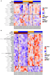
(A) The MCC dataset (GSE39612) was analysed and an expression heatmap generated for five p38 upstream genes across MCPyV-negative and MCPyV-positive MCC samples. (B) Downstream targets of p38 were identified via analysis of differentially expressed genes following inhibition of p38 signalling with SB203580 (GSE29587). The expression of these p38 downstream targets was subsequently analysed in the MCC dataset (GSE39612) and a heatmap generated across MCPyV-negative and MCPyV-positive MCC samples, showing probes that were inversely regulated compared with their expression following p38 inhibition in GSE29587. Within the heatmaps, red colour indicates high expression and blue indicates low expression.
Figure 4.
Figure 4.. Inhibition of p38 leads to loss of MCPyV ST induced cell migration.
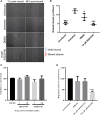
(A) i293-ST cells were induced prior to a scratch-wound. Cells were treated with vehicle control and 10 μM SB202190 to evaluate inhibitor effects upon wound closure. Wells were imaged using an EVOS II microscope 1 and 48 h post wound. (B) Wound closure rate (μm/h) of n = 3 was calculated using ImageJ software. Each point represents the average wound closure of each experimental repeat. (C) MTS viability assays were performed using 1 and 10 µM SB202474 and SB202190 to determine inhibitor effects upon viability and proliferation of the MCPyV-positive PeTa cell line. DMSO contained a comparable volume of solvent present in 10 μM inhibitor. (D) Transwell assays were performed to determine the effect of 10 µM SB202474 and SB202190 upon the migratory potential of PeTa cells. Cells were incubated with inhibitor for 24 h before addition to transwell plates. The percentage of migrated cells relative to untreated control was evaluated using a Countess II FL Automated Counter following a 48 h incubation. Significance values presented in (B) and (D) were calculated using a one-way ANOVA, with multiple comparisons to untreated displayed, where *P < 0.05, ***P < 0.0005, ****P < 0.0001. Error bars show the standard deviation of three experimental repeats.
Figure 5.
Figure 5.. MCPyV ST induced activation of p38 is via MKK4 and is independent of extracellular stimuli.
(A) HEK 293 cells were transfected with vectors that express MCPyV ST or empty vector for 48 h. Immunoblotting was performed to observe phosphorylation of MKK4 and MKK3/6. ST specific antibodies (2T2) were used to detect MCPyV ST and antibodies specific for GAPDH and endogenous levels of MKK4 used as loading controls. (B) HEK 293 cells were transfected with vectors that express MCPyV ST or empty vector for 48 h, with serum starvation or 10% (v/v) FBS containing DMEM applied for the final 2 h. Immunoblotting was performed to observe the effect of serum starvation upon phosphorylation of MK2 and MKK4. ST specific antibodies (2T2) were used to detect MCPyV ST and antibodies specific for GAPDH used as a loading control. Immunoblots are representative of experimental n = 3.
Figure 6.
Figure 6.. MCPyV ST interacts with PP4C to perturb pathway dephosphorylation.
(A) HEK 293 cells were cotransfected with vectors that express MCPyV ST and WT PP4C-Flag or TDN PP4C-HA for 48 h. For sole expression of MCPyV ST, an empty vector of equivalent mass to PP4C vectors was included during transfection. Immunoblotting was performed using specific antibodies to determine phosphorylation and endogenous levels of MK2. Specific antibodies targeting Flag and HA tags were used to detect WT and TDN PP4C, respectively. ST specific antibodies (2T2) were used to detect MCPyV ST and antibodies specific for GAPDH used as a loading control. (B) Evaluation of MK2 phosphorylation by densitometry, normalised to total MK2 expression. Significance values were calculated using a one-way ANOVA, with multiple comparisons to ST displayed, where **P < 0.005 and ***P < 0.0005. Error bars show the standard deviation of three experimental repeats.
Figure 7.
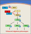
Figure 7.. Proposed mechanism via which MCPyV ST activated the p38 pathway to regulate actin dynamics.
MCPyV ST interacts with PP4C to dysregulate its role in regulating the phosphorylation of either MKK4 or an upstream kinase. Following MCPyV ST binding of PP4C, MKK4 activation, in turn, phosphorylates p38, which in turn phosphorylates substrates including MK2, ATF2 and MSK1. The activation of downstream targets of p38 subsequently enhance migratory phenotypes associated with MCPyV ST.

Similar articles

Cited by

References

    1. Becker J.C., Schrama D. and Houben R. (2008) Visions & reflections (minireview) Merkel cell carcinoma. Cell. Mol. Life Sci. 66, 1–8 10.1007/s00018-008-8483-6 - DOI - PMC - PubMed
    1. Kaae J., Hansen A.V., Biggar R.J., Boyd H.A., Moore P.S., Wohlfahrt J. et al. (2010) Merkel cell carcinoma: incidence, mortality, and risk of other cancers. J. Natl. Cancer Inst. 102, 793–801 10.1093/jnci/djq120 - DOI - PubMed
    1. Lemos B. and Nghiem P. (2007) Merkel cell carcinoma: more deaths but still no pathway to blame. J. Invest. Dermatol. 127, 2100–2103 10.1038/sj.jid.5700925 - DOI - PubMed
    1. Heath M., Jaimes N., Lemos B., Mostaghimi A., Wang L.C., Peñas P.F. et al. (2008) Clinical characteristics of Merkel cell carcinoma at diagnosis in 195 patients: the AEIOU features. J. Am. Acad. Dermatol. 58, 375–381 10.1016/j.jaad.2007.11.020 - DOI - PMC - PubMed
    1. Feng H., Shuda M., Chang Y. and Moore P.S. (2008) Clonal integration of a polyomavirus in human Merkel cell carcinoma. Science 319, 1096–1100 10.1126/science.1152586 - DOI - PMC - PubMed

Publication types

MeSH terms