

Decoding the differentiation of mesenchymal stem cells into mesangial cells at the transcriptomic level
- PMID: 32635896
- PMCID: PMC7339572
- DOI: 10.1186/s12864-020-06868-5
Decoding the differentiation of mesenchymal stem cells into mesangial cells at the transcriptomic level
Abstract
Background: Mesangial cells play an important role in the glomerulus to provide mechanical support and maintaine efficient ultrafiltration of renal plasma. Loss of mesangial cells due to pathologic conditions may lead to impaired renal function. Mesenchymal stem cells (MSC) can differentiate into many cell types, including mesangial cells. However transcriptomic profiling during MSC differentiation into mesangial cells had not been studied yet. The aim of this study is to examine the pattern of transcriptomic changes during MSC differentiation into mesangial cells, to understand the involvement of transcription factor (TF) along the differentiation process, and finally to elucidate the relationship among TF-TF and TF-key gene or biomarkers during the differentiation of MSC into mesangial cells.
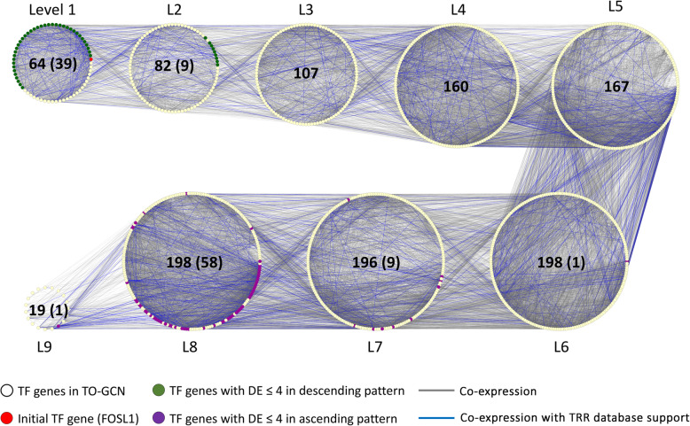
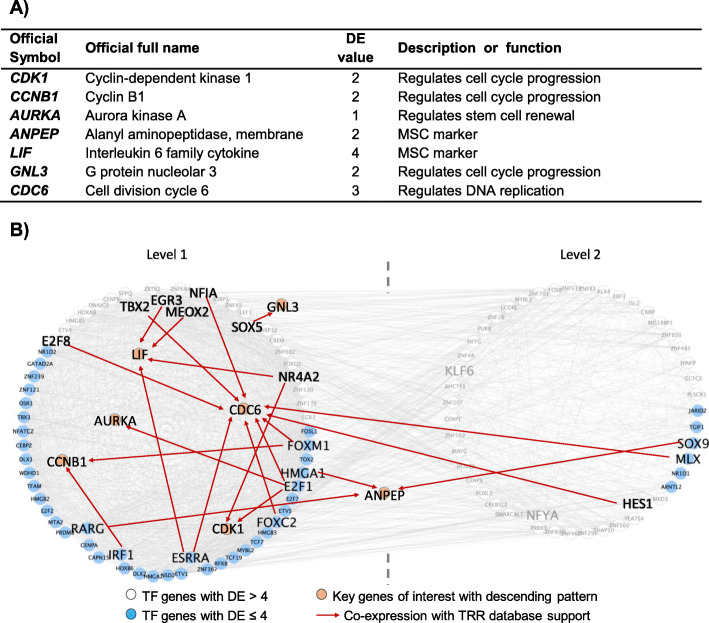
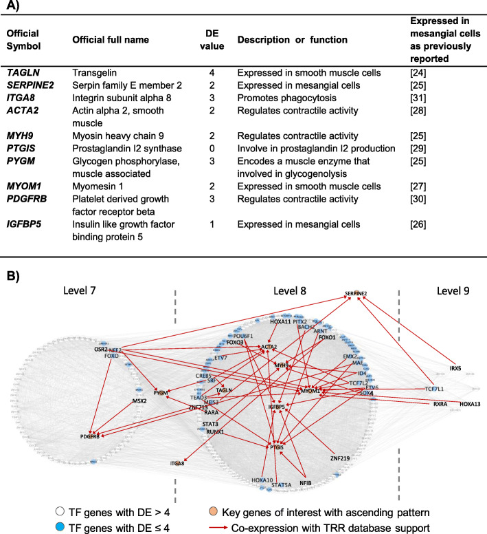
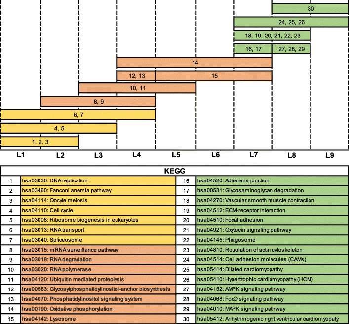
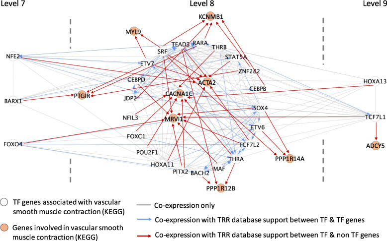
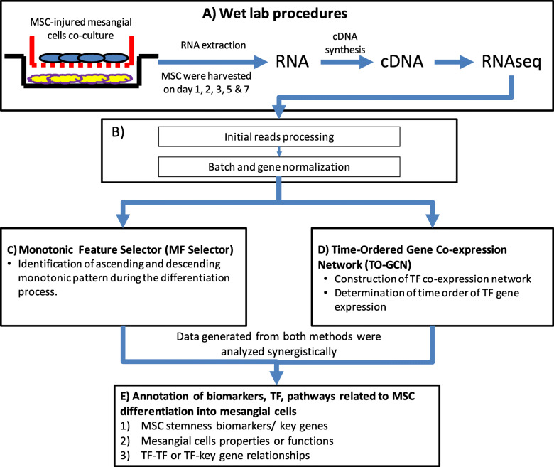
Results: Several ascending and descending monotonic key genes were identified by Monotonic Feature Selector. The identified descending monotonic key genes are related to stemness or regulation of cell cycle while ascending monotonic key genes are associated with the functions of mesangial cells. The TFs were arranged in a co-expression network in order of time by Time-Ordered Gene Co-expression Network (TO-GCN) analysis. TO-GCN analysis can classify the differentiation process into three stages: differentiation preparation, differentiation initiation and maturation. Furthermore, it can also explore TF-TF-key genes regulatory relationships in the muscle contraction process.
Conclusions: A systematic analysis for transcriptomic profiling of MSC differentiation into mesangial cells has been established. Key genes or biomarkers, TFs and pathways involved in differentiation of MSC-mesangial cells have been identified and the related biological implications have been discussed. Finally, we further elucidated for the first time the three main stages of mesangial cell differentiation, and the regulatory relationships between TF-TF-key genes involved in the muscle contraction process. Through this study, we have increased fundamental understanding of the gene transcripts during the differentiation of MSC into mesangial cells.
Keywords: Differentiation; Mesangial cell; Mesenchymal stem cell; Monotonic feature selector; Time-ordered gene co-expression network; Transcriptomic.
Conflict of interest statement
The authors declare that they have no competing interests.
Figures
Similar articles
-
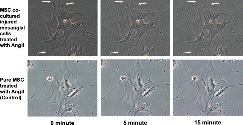
In vitro differentiation of mesenchymal stem cells into mesangial cells when co-cultured with injured mesangial cells.Cell Biol Int. 2014 Apr;38(4):497-501. doi: 10.1002/cbin.10231. Epub 2014 Jan 10. Cell Biol Int. 2014. PMID: 24375917
-
Co-expression network analysis identified key genes in association with mesenchymal stem cell osteogenic differentiation.Cell Tissue Res. 2019 Dec;378(3):513-529. doi: 10.1007/s00441-019-03071-1. Epub 2019 Aug 15. Cell Tissue Res. 2019. PMID: 31418071
-
Microenvironment in neuroblastoma: isolation and characterization of tumor-derived mesenchymal stromal cells.BMC Cancer. 2018 Nov 27;18(1):1176. doi: 10.1186/s12885-018-5082-2. BMC Cancer. 2018. PMID: 30482160 Free PMC article.
-
Transcriptional networks in the human epididymis.Andrology. 2019 Sep;7(5):741-747. doi: 10.1111/andr.12629. Epub 2019 May 2. Andrology. 2019. PMID: 31050198 Free PMC article. Review.
-
Transcriptional networks controlling stromal cell differentiation.Nat Rev Mol Cell Biol. 2021 Jul;22(7):465-482. doi: 10.1038/s41580-021-00357-7. Epub 2021 Apr 9. Nat Rev Mol Cell Biol. 2021. PMID: 33837369 Review.
Cited by
-
Time-series transcriptome provides insights into the gene regulation network involved in the icariin-flavonoid metabolism during the leaf development of Epimedium pubescens.Front Plant Sci. 2023 Jun 12;14:1183481. doi: 10.3389/fpls.2023.1183481. eCollection 2023. Front Plant Sci. 2023. PMID: 37377796 Free PMC article.
-
Generating transcriptional regulatory networks from time-ordered stem cell differentiation RNA sequencing data.STAR Protoc. 2022 Aug 19;3(3):101541. doi: 10.1016/j.xpro.2022.101541. eCollection 2022 Sep 16. STAR Protoc. 2022. PMID: 36042881 Free PMC article.
-
Current advances of stem cell-based therapy for kidney diseases.World J Stem Cells. 2021 Jul 26;13(7):914-933. doi: 10.4252/wjsc.v13.i7.914. World J Stem Cells. 2021. PMID: 34367484 Free PMC article. Review.
-
Synergistic effects of intracoronary infusion of autologous bone marrow-derived mesenchymal stem cells and revascularization procedure on improvement of cardiac function in patients with severe ischemic cardiomyopathy.Stem Cell Investig. 2021 Jan 22;8:2. doi: 10.21037/sci-2020-026. eCollection 2021. Stem Cell Investig. 2021. PMID: 33575315 Free PMC article.
-
Combining single-cell transcriptomics and CellTagging to identify differentiation trajectories of human adipose-derived mesenchymal stem cells.Stem Cell Res Ther. 2023 Feb 1;14(1):14. doi: 10.1186/s13287-023-03237-3. Stem Cell Res Ther. 2023. PMID: 36721241 Free PMC article.
References
-
- Shaw I, Rider S, Mullins J, Hughes J, Péault B. Pericytes in the renal vasculature: roles in health and disease. Nat Rev Nephrol. 2018;14(8):521–534. - PubMed
-
- Schlondorff D. The glomerular mesangial cell: an expanding role for a specialized pericyte. FASEB J. 1987;1(4):272–281. - PubMed
-
- Jefferson JA, Johnson RJ. Experimental mesangial proliferative glomerulonephritis (the anti-Thy-1.1 model) J Nephrol. 1999;12(5):297–307. - PubMed
-
- Wong CY, Cheong SK, Mok PL, Leong CF. Differentiation of human mesenchymal stem cells into mesangial cells in post-glomerular injury murine model. Pathology. 2008;40(1):52–57. - PubMed
-
- Singaravelu K, Padanilam BJ. In vitro differentiation of MSC into cells with a renal tubular epithelial-like phenotype. Ren Fail. 2009;31(6):492–502. - PubMed
MeSH terms
Substances
Grants and funding
LinkOut - more resources
Full Text Sources
Molecular Biology Databases
Miscellaneous
