. 2020 Aug 14;147(15):dev185421.
doi: 10.1242/dev.185421.

Ykt6-dependent endosomal recycling is required for Wnt secretion in the Drosophila wing epithelium

Affiliations

Ykt6-dependent endosomal recycling is required for Wnt secretion in the Drosophila wing epithelium

Karen Linnemannstöns et al. Development. .

Abstract

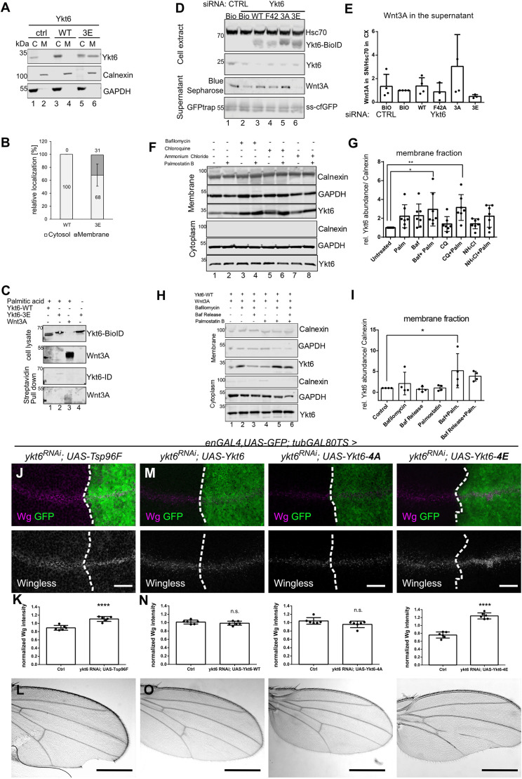
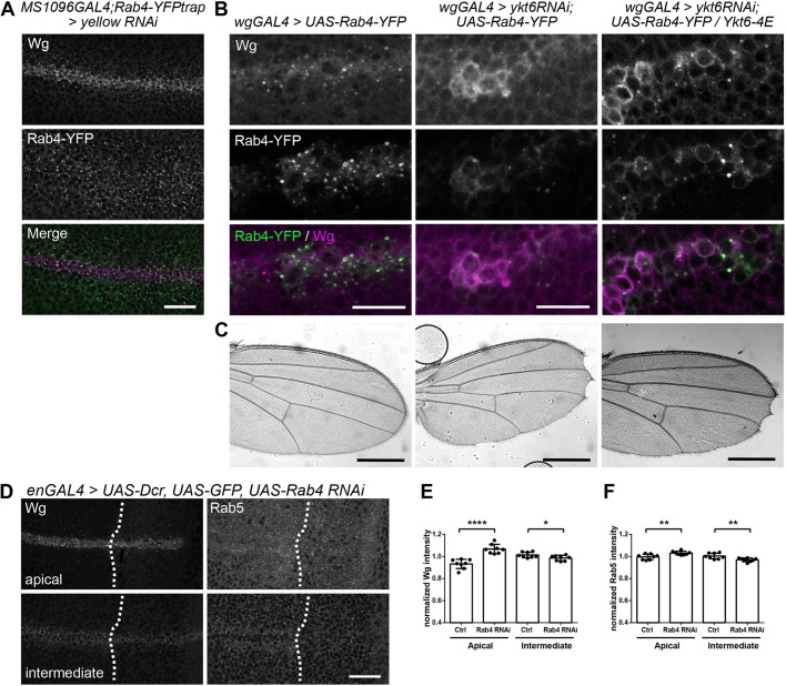
Morphogens are important signalling molecules for tissue development and their secretion requires tight regulation. In the wing imaginal disc of flies, the morphogen Wnt/Wingless is apically presented by the secreting cell and re-internalized before final long-range secretion. Why Wnt molecules undergo these trafficking steps and the nature of the regulatory control within the endosomal compartment remain unclear. Here, we have investigated how Wnts are sorted at the level of endosomes by the versatile v-SNARE Ykt6. Using in vivo genetics, proximity-dependent proteomics and in vitro biochemical analyses, we show that most Ykt6 is present in the cytosol, but can be recruited to de-acidified compartments and recycle Wnts to the plasma membrane via Rab4-positive recycling endosomes. Thus, we propose a molecular mechanism by which producing cells integrate and leverage endocytosis and recycling via Ykt6 to coordinate extracellular Wnt levels.

Keywords: Endosomal sorting; Morphogen trafficking; Wnt secretion; Wnt signalling.


Conflict of interest statement

Competing interestsThe authors declare no competing or financial interests.

Figures

Fig. 1.
Fig. 1.
Loss of Ykt6 blocks Wnt secretion. (A) RNAi-knockdown of Ykt6 in the posterior compartment of third instar WIDs marked by co-expression of GFP (engrailed-Gal4, UAS-GFP/UAS-ykt6RNAi) causes extracellular Wingless reduction. The GFP-negative anterior compartment serves as an internal control. Maximum intensity projection of 20 sections (distance 0.5 µm) depicted for visualization. (B) Scheme of Drosophila ykt6 mutant alleles. (C) Wingless protein accumulates in ykt6A clones marked by the absence of GFP. The lower panels depict enlarged images of the areas outlined in the upper panels. (D) Western blot analysis of intracellular Wnt accumulation in Hek293T cells transfected with control or Ykt6-1 or Ykt6-2 siRNA. (E) RNAi against 25 Drosophila SNAREs was screened for Wnt secretion defects in adult wings (wgGAL4) and third instar WIDs (enGAL4) (see also Table S1). (F) Knockdown of Ykt6 and Syb by RNAi in the posterior compartment of third instar WIDs marked by co-expression of GFP leads to intracellular Wg accumulation, whereas Sec22 does not affect Wg distribution. The GFP-negative compartment serves as an internal control. Left panels in F show maximum intensity projections of six (Ykt6), 15 (Syb) and two (Sec22) sections (distance 1 µm) depicted for visualization. Right panels in F show optical transverse sections. Scale bars: 20 µm.
Fig. 2.
Fig. 2.
Ykt6 acts on endosomal compartments after apical presentation. (A) Scheme of BioID labelling in Hek293T cells: Ykt6 was N-terminally tagged with a BioID domain. Upon addition of biotin, a streptavidin pull-down was performed, and control and Ykt6-WT samples were subjected to proteomics identification. (B) Western blot of biotin labelling of Ykt6-BioID and control in the presence of 50 µM biotin. All proteins identified by mass spectrometry in two independent experiments (significance level P=0.003 and 1.3-fold over BioID control samples) are listed in Table S2. (C) Enrichment scores for BioID-identified proteins of the endocytic pathway. (D) Western blot of Ykt6-WT-mediated BioID labelling of AP2A1/2 and Chmp2B. (E) Overview of Wnt secretion components involved after initial apical plasma membrane presentation of Wg. (F) Wg accumulation phenotypes of different factors required for Wg secretion. RNAi against Evi, Snx3 and AP2α expressed with enGAL4, and RNAi against Hrs and Dcr expressed with UAS-Dcr; enGAL4. Images represent a single confocal section and are representative of more than six WIDs per RNAi from three independent experiments. Scale bars: 20 µm.
Fig. 3.
Fig. 3.
Ykt6 knockdown is not sufficient to block Evi recycling. (A-C) RNAi against Ykt6 and AP2α was expressed with enGAL4,UAS-GFP, and stained for Evi. (A) The upper panels depict a maximum intensity projection of 15 apical xy sections (distance 1 µm); the lower panels are a transverse xz section of 20 pixels. (B) Comparison of Evi fluorescence intensity in n=5 biologically independent samples. Data are mean±s.d., *P=0.0425, ****P<0.0001. (C) Quantification of Evi puncta in n=5 biologically independent samples. Data are mean±s.d., ****P<0.0001. (D-F) RNAi against Ykt6 and AP2α was expressed with enGAL4 and stained for extracellular Wg. (D) A maximum intensity projection of all sections covering the entire apico-basal axis is depicted for visualization. (E) Profile of the extracellular Wg staining in the ROI depicted in D of the corresponding average intensity projection. This compares exWg in the anterior (control, no GFP) with the posterior (RNAi, GFP-positive) region for this one representative example. (F) Comparison of exWg fluorescence intensity in n=5 biologically independent samples. Data are mean±s.d., ***P=0.0007, ****P<0.0001. Scale bars: 20 µm.
Fig. 4.
Fig. 4.
Ykt6 acts on Wnt trafficking at the level of endosomes. (A) Localization of different organelle markers in wild type (left) and Ykt6 RNAi (right) from enGAL4, UAS-GFP/Ykt6 RNAi WIDs. Maximum intensity projections of 13 (Rab5), seven (Rab7, Lamp1) and nine (Hrs) sections (distance 1 µm) depicted for visualization. Scale bars: 20 µm. (B) Quantification of fluorescence intensity in n=7 (Rab5, Rab7 and Hrs) or n=8 (Lamp1) biologically independent samples from A. Data are mean±s.d., **P=0.0026, ***P=0.0004. (C) Semi-thin section of a WID (left) and electron microscopy images of MVBs in a WID (right) during time-controlled depletion of Ykt6 by RNAi (engrailed-Gal4, UAS-GFP/UAS-ykt6RNAi; tubGal80-TS/+ larvae reared for 3 days at 29°C). Scale bars: 50 μm (left); 500 nm (right). (D) Quantification of MVB size in electron microscopy images from cells in the anterior (n=17) and posterior (n=22) compartments of WIDs. (E,F) GFP-Myc-FYVE was expressed with wgGAL4 and yellow (control) or ykt6 RNAi to label PI(3)P-containing endosomes. (E) MIP of six apical sections (distance 0.5 µm). (F) Transverse xy section. Scale bars: 10 µm. (G) Quantification of E. The diameter of GFP-Myc-FYVE -positive vesicles with a clear lumen was measured. Four representative WIDs from three biological replicates with in total 394 (yellow RNAi) and 460 (ykt6 RNAi) enlarged endosomes were quantified. Data are mean±s.d., ****P<0.0001. (H) Quantification of F. The number of Wg puncta positive for GFP-Myc-2XFYVE was quantified. Data are mean±s.d., **P=0.0071. (I) Constitutively active Rab5Q88L-YFP was expressed with wgGAL4 and yellow (control) or ykt6 RNAi. Images represent a single confocal section. Scale bars: 10 µm. (J) Quantification of I. The diameter of Rab5Q88L-YFP-positive vesicles with a clear lumen was measured. Five representative WIDs from three biological replicates with, in total, 446 (yellow RNAi) and 326 (ykt6 RNAi) enlarged endosomes were quantified. Data are mean±s.d., ****P<0.0001.
Fig. 5.
Fig. 5.
A Ykt6 SNARE domain is required for cycling between compartments. (A,B) Representative blot (A) and quantification (B) of detergent fractionation of Hek293T cells transfected with Ykt6-WT and Ykt6-3E constructs. C, cytoplasmic; M, membrane fraction, n=3. (C) Click palmitoylation assay of Ykt6-WT and Ykt6-3E; Wnt3A is a positive control. Representative blot of three biological replicates. (D,E) Representative blot (D) and quantification (E) of Wnt3A secretion from Hek293T cells transfected with Ykt6-WT, phosphor-mutant Ykt6-3A and Ykt6-3E, and Longin domain mutant Ykt6-F42 constructs. n=4. One-way ANOVA (no significant differences). (F,G) Inhibiting endosomal acidification and depalmitoylation affects Ykt6 subcellular localization. (F) Representative blot of cell fractionation of untagged Ykt6 mutant constructs in Hek293T cells, treated with bafilomycin, chloroquine or ammonium chloride in combination with palmostatin B, stained for Ykt6 and fraction markers. (G) Quantification of Ykt6 in the membrane fraction, n=7; *P=0,01, **P=0.005 one-way ANOVA. (H,I) Ykt6 membrane recruitment and release. Blot of cell fractionation of untagged Ykt6 in Hek293T cells treated with bafilomycin and palmostatin B, and release from bafilomycin inhibition stained for Ykt6 and fraction markers. (I) Quantification of H from n=4; *P=0,03, one-way ANOVA. (J-L) Time-controlled depletion of Ykt6 by RNAi (engrailed-Gal4, UAS-GFP/UAS-ykt6RNAi; tubGal80-TS/UAS-Tsp96F larvae reared for 3 days at 29°C) causes intracellular Wg accumulation (J,K) and wing notches (L). (M-O) Time-controlled Ykt6 RNAi-induced block of Wg secretion and adult wing margin notches can be rescued by co-overexpression of wild-type Ykt6 and the SNARE mutant Ykt6-4A (left and middle panels), but not by Ykt6-4E (right panels). (K,N) Quantification of fluorescence intensity in n=6 biologically independent samples from J,M. Data are mean±s.d., ****P<0.0001. (J,M) Projections of six subapical sections (distance 1 µm, J) and six subapical sections (distance 0.5 µm, M). Representative images of more than 10 discs from n=3. Scale bars: 20 µm (J,M); 500 µm in adult wing images (L,O).
Fig. 6.
Fig. 6.
Ykt6 recycles Wg via Rab4 endosomes. (A) Yellow RNAi was expressed with MS1096GAL4 in the wing pouch in an endogenously tagged Rab4-YFP background. Maximum intensity projection of three sections (distance 0.5 µm) depicted for visualization. Scale bar: 20 µm. (B) UAS-Rab4-YFP was expressed with wgGAL4 alone, in combination with ykt6 RNAi or ykt6 RNAi and Ykt6-4E. A single subapical section is depicted. Scale bars: 10 µm. (C) Adult wings of the crosses from B to show adult wing notches. Scale bars: 500 µm. These wings are representative of more than 10 wings from three independent experiments. (D) RNAi against Rab4 was expressed with enGAL4, UAS-GFP,UAS-Dcr and stained for Wg and Rab5. Maximum intensity projection of three apical (upper panels) and intermediate (lower panels) sections is depicted for visualization. Scale bar: 20 μm. (E) Quantification of Wg apical versus intermediate fluorescence intensity in n=8 biologically independent samples from D. Data are mean±s.d., ****P<0.0001, *P=0.04. (F) Quantification of Rab5 apical versus intermediate fluorescence intensity in n=8 biologically independent samples from D. Data are mean±s.d., **P=0.006.

Similar articles

Cited by

References

    1. Abe M., Setoguchi Y., Tanaka T., Awano W., Takahashi K., Ueda R., Nakamura A. and Goto S. (2009). Membrane protein location-dependent regulation by PI3K (III) and Rabenosyn-5 in Drosophila wing cells. PLoS ONE 4, e7306 10.1371/journal.pone.0007306 - DOI - PMC - PubMed
    1. Acebron S. P., Karaulanov E., Berger B. S., Huang Y.-L. and Niehrs C. (2014). Mitotic Wnt signaling promotes protein stabilization and regulates cell size. Mol. Cell 54, 663-674. 10.1016/j.molcel.2014.04.014 - DOI - PubMed
    1. Albrecht L. V., Ploper D., Tejeda-Muñoz N. and De Robertis E. M. (2018). Arginine methylation is required for canonical Wnt signaling and endolysosomal trafficking. Proc. Natl. Acad. Sci. USA 115, E5317-E5325. 10.1073/pnas.1804091115 - DOI - PMC - PubMed
    1. Atanassov I. and Urlaub H. (2013). Increased proteome coverage by combining PAGE and peptide isoelectric focusing: comparative study of gel-based separation approaches. Proteomics 13, 2947-2955. 10.1002/pmic.201300035 - DOI - PMC - PubMed
    1. Baeg G. H., Lin X., Khare N., Baumgartner S. and Perrimon N. (2001). Heparan sulfate proteoglycans are critical for the organization of the extracellular distribution of Wingless. Development 128, 87-94. - PubMed

Publication types

LinkOut - more resources