Review
. 2020 Jun 29;21(13):4618.
doi: 10.3390/ijms21134618.

An Overview of Coumarin as a Versatile and Readily Accessible Scaffold with Broad-Ranging Biological Activities

Affiliations
Review

An Overview of Coumarin as a Versatile and Readily Accessible Scaffold with Broad-Ranging Biological Activities

Francesca Annunziata et al. Int J Mol Sci. .

Abstract

Privileged structures have been widely used as an effective template for the research and discovery of high value chemicals. Coumarin is a simple scaffold widespread in Nature and it can be found in a considerable number of plants as well as in some fungi and bacteria. In the last years, these natural compounds have been gaining an increasing attention from the scientific community for their wide range of biological activities, mainly due to their ability to interact with diverse enzymes and receptors in living organisms. In addition, coumarin nucleus has proved to be easily synthetized and decorated, giving the possibility of designing new coumarin-based compounds and investigating their potential in the treatment of various diseases. The versatility of coumarin scaffold finds applications not only in medicinal chemistry but also in the agrochemical field as well as in the cosmetic and fragrances industry. This review is intended to be a critical overview on coumarins, comprehensive of natural sources, metabolites, biological evaluations and synthetic approaches.

Keywords: biological activity; coumarins; metabolites; natural sources; synthesis.


Conflict of interest statement

The authors declare no conflict of interest.

Figures

Figure 1
Figure 1
Structure of coumarin nucleus.
Figure 2
Figure 2
Coumarin metabolism.
Figure 3
Figure 3
Coumarin structures reported by Couttolenc et al. [30].
Figure 4
Figure 4
Chemical structure of coumarin-fused coumarins 49.
Figure 5
Figure 5
Chitosan derivatives 10ad.
Figure 6
Figure 6
Chemical structures of tert-butylcoumarins 11 and 12 and terpenylcoumarins 1317.
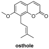
Figure 7
Figure 7
Structure of the most active O-prenylated coumarins.
Figure 8
Figure 8
Chemical structure of 5-farnesyloxycoumarin (18) and 8-farnesyloxycoumarin (19).
Figure 9
Figure 9
4-arylamino-3-nitrocoumarin derivative 20.
Figure 10
Figure 10
Chemical structure of styrylcoumarin 21.
Figure 11
Figure 11
General structure of diethylene glycol tethered isatin-1,2,3-triazole-coumarin derivatives 22a-l.
Figure 12
Figure 12
Chemical structure of compound 23.
Figure 13
Figure 13
Chemical structure of umbelliprenin (24).
Figure 14
Figure 14
Chemical structure of compound 25 and piperazine derivatives 2637.
Figure 15
Figure 15
Chemical structure and minimal inhibitory concentration (MIC) values of antibacterial coumarins proposed by Nagamallu and colleagues [79].
Figure 16
Figure 16
Coumarin dimer scaffolds proposed by Chougala and colleagues and the corresponding MIC values [80].
Figure 17
Figure 17
3,4-dihydropyrimidinone-coumarin analogues and their MIC values against S. aureus.
Figure 18
Figure 18
Coumarin-pyrazole hybrids and their MIC values against S. aureus.
Figure 19
Figure 19
Coumarins isolated from Rutacea species.
Figure 20
Figure 20
Linear coumarins isolated from the fruits of Peucedanum luxurians Tamamsch, zone of inhibition (mm) and minimum inhibitory concentration (MIC) values.
Figure 21
Figure 21
Selected coumarin-pyrazole carboxamide derivatives developed by Liu et al. and their MIC values [87].
Figure 22
Figure 22
Antifungal coumarins.
Figure 23
Figure 23
Antifungal coumarins synthesized under solvent-free conditions and exploiting ionic liquid as catalyst.
Figure 24
Figure 24
Coumarin derivatives conjugated to an imidazole-containing moiety.
Figure 25
Figure 25
Coumarins from Ferulago trachycarpa.
Figure 26
Figure 26
Coumarin-based 1,4-disubstituted 1,2,3-triazole derivatives.
Figure 27
Figure 27
New prenylated coumarins from Clausena lenis.
Figure 28
Figure 28
Prenylated coumarins from Manilkara zapota.
Figure 29
Figure 29
Coumarin derivatives synthesized by Jesumoroti and co-workers [117].
Figure 30
Figure 30
Hybrid pharmacophores proposed by Osman and co-workers [125].
Figure 31
Figure 31
Bis coumarinyl bis triazolothiadiazinyl ethane derivative.
Figure 32
Figure 32
Antiviral coumarins from Bizzarri et al. [129].
Figure 33
Figure 33
Anti-hepatitis coumarins.
Figure 34
Figure 34
Anti-inflammatory coumarin derivatives.
Figure 35
Figure 35
Anti-inflammatory 7-substituted coumarins proposed by Srivastava [146].
Figure 36
Figure 36
One of the coumarin derivatives isolated from M. exotica.
Figure 37
Figure 37
Structure of the natural coumarin osthole.
Figure 38
Figure 38
7-substituted coumarin as blockers of NF-κB signaling pathway.
Figure 39
Figure 39
Coumarins extracted from Ferulago cassia.
Figure 40
Figure 40
(a) Chemical structure of compounds 105 and 106; panel (b) SAR studies.
Figure 41
Figure 41
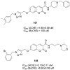
Chemical structure and IC50 values of compounds 107 and 108.
Figure 42
Figure 42
Chemical structures of alkylamino coumarins 109ad, 110a–c and 111ac.
Figure 43
Figure 43
General structure of compounds 112117.
Figure 44
Figure 44
(a) General structure of compounds 118146; (b) chemical structure of compounds 120 and 136.
Figure 45
Figure 45
Chemical structure of compounds 147ad.
Figure 46
Figure 46
Chemical structure of compounds 148151.
Figure 47
Figure 47
Chemical structure of compounds 152af.
Figure 48
Figure 48
Chemical structure of compounds 153ae.
Figure 49
Figure 49
Chemical structure of the most common anticoagulant drugs.
Figure 50
Figure 50
Structure of Tecarfarin.
Figure 51
Figure 51
Chemical structure of coumarins conjugated with salicylic acid.
Figure 52
Figure 52
C-3 alkyl coumarin derivatives.
Figure 53
Figure 53
Coumarin derivative from Ainsliaea fragrans.
Figure 54
Figure 54
General structures of compounds 158ae and 159ae.
Figure 55
Figure 55
Chemical structure of compounds 160167.
Figure 56
Figure 56
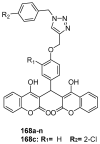
Chemical structure of biscoumarin-1,2,3-triazole hybrids. Substituents for compound 168c are reported.
Figure 57
Figure 57
Chemical structure of compounds 169, 170 and 171.
Figure 58
Figure 58
Structure of several coumarin photocleavable protecting groups.
Figure 59
Figure 59
Coumarin florescent probes for hypochlorite ion detection.
Figure 60
Figure 60
Coumarin fluorescent probes for copper (II) detection.
Figure 61
Figure 61
Chemical structure of different coumarin fluorescent probes for the detection of ions.
Figure 62
Figure 62
Chemical structure of probe 196.
Figure 63
Figure 63
Chemical structure of CG and CA probes.
Figure 64
Figure 64
Chemical structure of coumarins umbelliferone, esculetin, daphnetin and xanthotol.
Figure 65
Figure 65
Structures of compounds isolated from Citrus peel.
Figure 66
Figure 66
Chemical structure of compounds 203 and 204.
Figure 67
Figure 67
Chemical structure of coumarin-functionalized chitosan 205ad.
Figure 68
Figure 68
Chemical structure of coumarin-3-carboxilate-copper (II) complex (206).
Figure 69
Figure 69
Chemical structure of complexes 207213.
Figure 70
Figure 70
Chemical structure of derivatives 214224.
Figure 71
Figure 71
Chemical structure of Zn(II)-coumarin-hydrazone complex 225.
Scheme 1
Scheme 1
Set-up for the synthesis of coumarins via O-acetylation of salicylaldehyde in a continuous flow reactor.
Scheme 2
Scheme 2
Coumarin synthesis with polymer-supported reagent.
Scheme 3
Scheme 3
(a) Photocatalyzed synthesis of coumarins; (b) Chemical structure of (−) Riboflavin.
Scheme 4
Scheme 4
Photoredox-catalyzed synthesis of 3-fluoroalkylated coumarins.
Scheme 5
Scheme 5
(a) Bichromatic synthesis of coumarins; (b) chemical structure of catalyst A.
Scheme 6
Scheme 6
Solvent-free synthesis of 7-hydroxy-4-methylcoumarin.
Scheme 7
Scheme 7
Solvent-free Pechmann condensation with Zr-TMS-TFA as catalyst.
Scheme 8
Scheme 8
Solvent-free synthesis of hetero-annulated coumarins.
Scheme 9
Scheme 9
Lewis acid grafted sulfonated carbon@titania composite as catalyst for the Pechmann condensation.
Scheme 10
Scheme 10
Ionic liquids as an acidic catalyst in the regioselective synthesis of pyrano[3,2-c] coumarins under solvent-free conditions.
Scheme 11
Scheme 11
Imidazolium-based phosphinite ionic liquid (IL-OPPh2) for the microwave assisted synthesis of coumarins.
Scheme 12
Scheme 12
Microwave promoted Witting reaction and total synthesis of osthole. (a) general synthetic methodology; (b) osthole synthesis.

Similar articles

Cited by

References

    1. Borges F., Roleira F., Milhanzes N., Santana L., Uriarte E. Simple coumarins and analogues in medicinal chemistry: Occurrence, synthesis and biological activity. Curr. Med. Chem. 2005;12:887–916. doi: 10.2174/0929867053507315. - DOI - PubMed
    1. Hoult J.R.S., Payá M. Pharmacological and biochemical actions of simple coumarins: Natural products with therapeutic potential. Gen. Pharmacol. 1996;27:713–722. doi: 10.1016/0306-3623(95)02112-4. - DOI - PubMed
    1. Kummerle A.E., Vitorio F., Franco D.P., Pereira T.M. Coumarin Compounds in Medicinal Chemistry: Some Important Examples from the Last Year. Curr. Top. Med. Chem. 2018;18:124–128. doi: 10.2174/1568026618666180329115523. - DOI - PubMed
    1. Barot K.P., Jain S.V., Kremer L., Singh S., Ghate M.D. Recent advances and therapeutic journey of coumarins: Current status and perspectives. Med. Chem. Res. 2015;24:2771–2798. doi: 10.1007/s00044-015-1350-8. - DOI
    1. IUPAC . Nomenclature of Organic Chemistry. Pergamon Press; Oxford, UK: 1979.