Class switching and high-affinity immunoglobulin G production by B cells is dispensable for the development of hypertension in mice
- PMID: 32609312
- PMCID: PMC7983008
- DOI: 10.1093/cvr/cvaa187
Class switching and high-affinity immunoglobulin G production by B cells is dispensable for the development of hypertension in mice
Abstract
Aims: Elevated serum immunoglobulins have been associated with experimental and human hypertension for decades but whether immunoglobulins and B cells play a causal role in hypertension pathology is unclear. In this study, we sought to determine the role of B cells and high-affinity class-switched immunoglobulins on hypertension and hypertensive end-organ damage to determine if they might represent viable therapeutic targets for this disease.
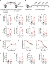
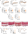
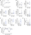
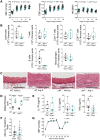
Methods and results: We purified serum immunoglobulin G (IgG) from mice exposed to vehicle or angiotensin (Ang) II to induce hypertension and adoptively transferred these to wild type (WT) recipient mice receiving a subpressor dose of Ang II. We found that transfer of IgG from hypertensive animals does not affect blood pressure, endothelial function, renal inflammation, albuminuria, or T cell-derived cytokine production compared with transfer of IgG from vehicle infused animals. As an alternative approach to investigate the role of high-affinity, class-switched immunoglobulins, we studied mice with genetic deletion of activation-induced deaminase (Aicda-/-). These mice have elevated levels of IgM but virtual absence of class-switched immunoglobulins such as IgG subclasses and IgA. Neither male nor female Aicda-/- mice were protected from Ang II-induced hypertension and renal/vascular damage. To determine if IgM or non-immunoglobulin-dependent innate functions of B cells play a role in hypertension, we studied mice with severe global B-cell deficiency due to deletion of the membrane exon of the IgM heavy chain (µMT-/-). µMT-/- mice were also not protected from hypertension or end-organ damage induced by Ang II infusion or deoxycorticosterone acetate-salt treatment.
Conclusions: These results suggest that B cells and serum immunoglobulins do not play a causal role in hypertension pathology.
Keywords: B cells; Blood pressure; Hypertension; Immunity; Inflammation.
Published on behalf of the European Society of Cardiology. All rights reserved. © The Author(s) 2020. For permissions, please email: journals.permissions@oup.com.
Figures


Similar articles
-
T-cell senescence accelerates angiotensin II-induced target organ damage.Cardiovasc Res. 2021 Jan 1;117(1):271-283. doi: 10.1093/cvr/cvaa032. Cardiovasc Res. 2021. PMID: 32049355
-
AID dysregulation in lupus-prone MRL/Fas(lpr/lpr) mice increases class switch DNA recombination and promotes interchromosomal c-Myc/IgH loci translocations: modulation by HoxC4.Autoimmunity. 2011 Dec;44(8):585-98. doi: 10.3109/08916934.2011.577128. Epub 2011 May 18. Autoimmunity. 2011. PMID: 21585311 Free PMC article.
-
CD4+ T-Cell Legumain Deficiency Attenuates Hypertensive Damage via Preservation of TRAF6.Circ Res. 2024 Jan 5;134(1):9-29. doi: 10.1161/CIRCRESAHA.123.322835. Epub 2023 Dec 4. Circ Res. 2024. PMID: 38047378
-
Absence of Activation-induced Cytidine Deaminase, a Regulator of Class Switch Recombination and Hypermutation in B Cells, Suppresses Aorta Allograft Vasculopathy in Mice.Transplantation. 2015 Aug;99(8):1598-605. doi: 10.1097/TP.0000000000000688. Transplantation. 2015. PMID: 25769064
-
Immune mechanisms in arterial hypertension. Recent advances.Cell Tissue Res. 2021 Aug;385(2):393-404. doi: 10.1007/s00441-020-03409-0. Epub 2021 Jan 4. Cell Tissue Res. 2021. PMID: 33394136 Free PMC article. Review.
Cited by
-
Therapeutic targeting of inflammation in hypertension: from novel mechanisms to translational perspective.Cardiovasc Res. 2021 Nov 22;117(13):2589-2609. doi: 10.1093/cvr/cvab330. Cardiovasc Res. 2021. PMID: 34698811 Free PMC article. Review.
-
Immune mechanisms in the pathophysiology of hypertension.Nat Rev Nephrol. 2024 Aug;20(8):530-540. doi: 10.1038/s41581-024-00838-w. Epub 2024 Apr 24. Nat Rev Nephrol. 2024. PMID: 38658669 Review.
-
Immune and inflammatory mechanisms in hypertension.Nat Rev Cardiol. 2024 Jun;21(6):396-416. doi: 10.1038/s41569-023-00964-1. Epub 2024 Jan 3. Nat Rev Cardiol. 2024. PMID: 38172242 Review.
-
Proteasome inhibition reduces plasma cell and antibody secretion, but not angiotensin II-induced hypertension.Front Cardiovasc Med. 2023 Jun 2;10:1184982. doi: 10.3389/fcvm.2023.1184982. eCollection 2023. Front Cardiovasc Med. 2023. PMID: 37332591 Free PMC article.
-
Macrophage Dectin-1 mediates Ang II renal injury through neutrophil migration and TGF-β1 secretion.Cell Mol Life Sci. 2023 Jun 20;80(7):184. doi: 10.1007/s00018-023-04826-4. Cell Mol Life Sci. 2023. PMID: 37340199 Free PMC article.
References
-
- GBD 2015 Risk Factors Collaborators. Global, regional, and national comparative risk assessment of 79 behavioural, environmental and occupational, and metabolic risks or clusters of risks, 1990-2015: a systematic analysis for the Global Burden of Disease Study 2015. Lancet 2016;388:1659–1724. - PMC - PubMed
-
- Whelton PK, Carey RM, Aronow WS, Casey DE Jr, Collins KJ, Dennison Himmelfarb C, DePalma SM, Gidding S, Jamerson KA, Jones DW, MacLaughlin EJ, Muntner P, Ovbiagele B, Smith SC Jr, Spencer CC, Stafford RS, Taler SJ, Thomas RJ, Williams KA Sr, Williamson JD, Wright JT Jr.. 2017 ACC/AHA/AAPA/ABC/ACPM/AGS/APhA/ASH/ASPC/NMA/PCNA Guideline for the prevention, detection, evaluation, and management of high blood pressure in adults: a report of the American College of Cardiology/American Heart Association Task Force on Clinical Practice Guidelines. Hypertension 2018;71:1269–1324. - PubMed
-
- Blacher J, Evans A, Arveiler D, Amouyel P, Ferrieres J, Bingham A, Yarnell J, Haas B, Montaye M, Ruidavets JB, Ducimetiere P; on behalf of the PRIME Study Group. Residual cardiovascular risk in treated hypertension and hyperlipidaemia: the PRIME Study. J Hum Hypertens 2010;24:19–26. - PubMed
-
- Struthers AD. A new approach to residual risk in treated hypertension—3P screening. Hypertension 2013;62:236–239. - PubMed
Publication types
MeSH terms
Substances
Grants and funding
LinkOut - more resources
Full Text Sources
Medical
Research Materials
Miscellaneous

