. 2020 Dec;9(12):1604-1616.
doi: 10.1002/sctm.20-0129. Epub 2020 Jun 29.

Coinhibition of activated p38 MAPKα and mTORC1 potentiates stemness maintenance of HSCs from SR1-expanded human cord blood CD34+ cells via inhibition of senescence

Affiliations

Coinhibition of activated p38 MAPKα and mTORC1 potentiates stemness maintenance of HSCs from SR1-expanded human cord blood CD34+ cells via inhibition of senescence

Xiaoyi Li et al. Stem Cells Transl Med. 2020 Dec.

Abstract

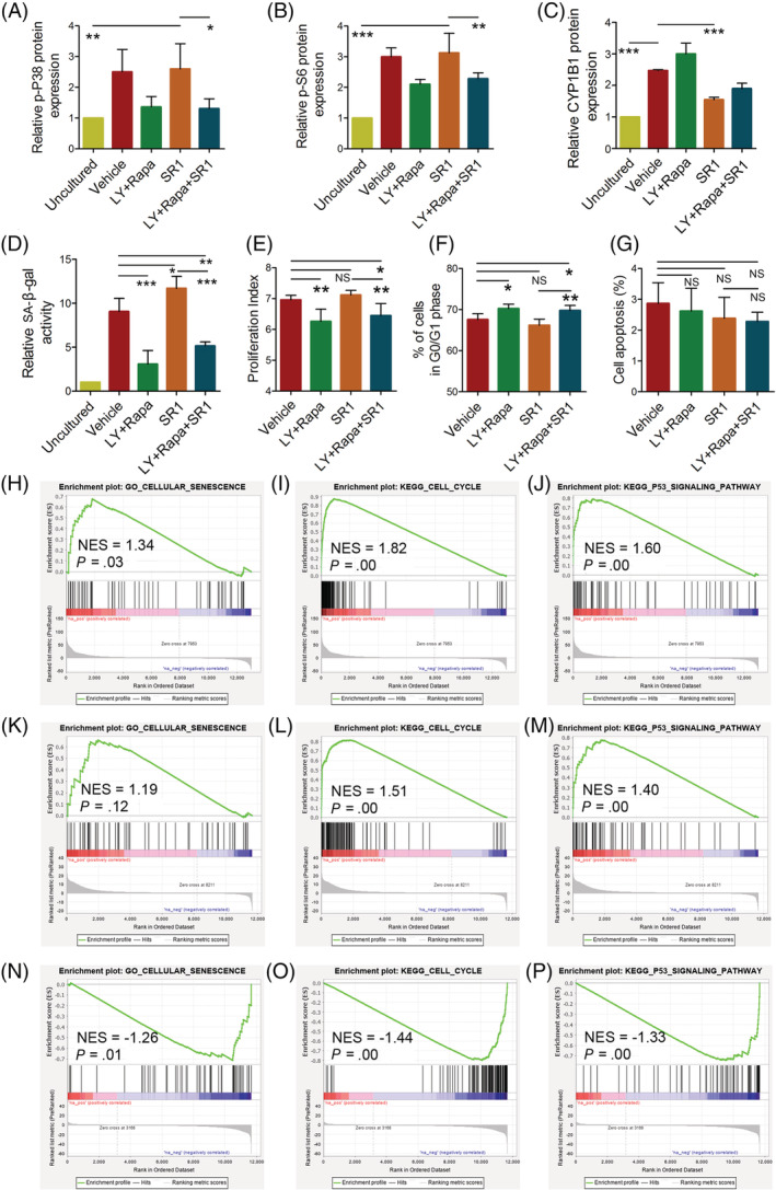
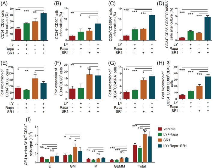
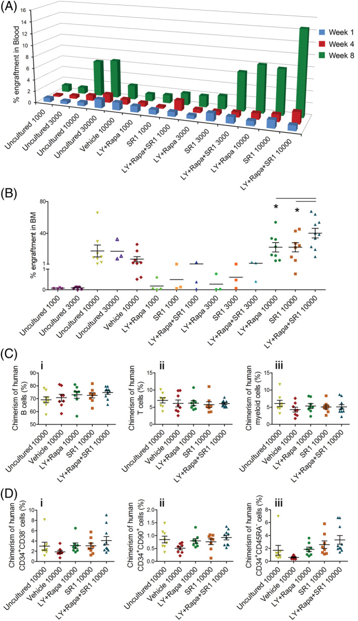
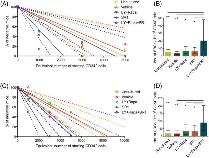
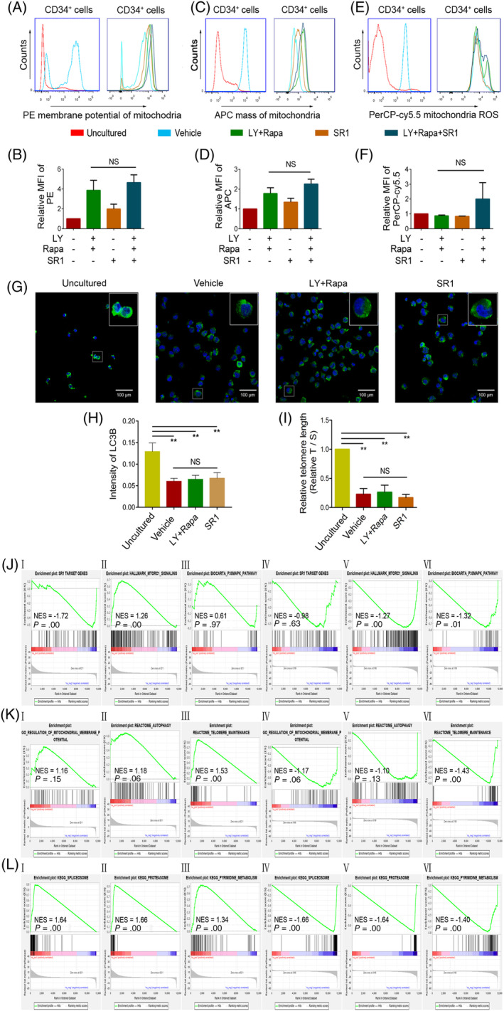
The stemness of ex vivo expanded hematopoietic stem cells (HSCs) is usually compromised by current methods. To explore the failure mechanism of stemness maintenance of human HSCs, which were expanded from human umbilical cord blood (hUCB) CD34+ cells, by differentiation inhibitor Stem Regenin 1 (SR1), an antagonist of aryl hydrocarbon receptor, we investigated the activity of p38 mitogen-activated protein kinase α (p38 MAPKα, p38α) and mammalian target of rapamycin complex 1 (mTORC1), and their effect on SR1-expanded hUCB CD34+ cells. Our results showed that cellular senescence occurred in the SR1-expanded hUCB CD34+ cells in which p38α and mTORC1 were successively activated. Furthermore, their coinhibition resulted in a further decrease in hUCB CD34+ cell senescence without an effect on apoptosis, promoted the maintenance of expanded phenotypic HSCs without differentiation inhibition, increased the hematopoietic reconstitution ability of multiple lineages, and potentiated the long-term self-renewal capability of HSCs from SR1-expanded hUCB CD34+ cells in NOD/Shi-scid/IL-2Rγnull mice. Our mechanistic study revealed that senescence inhibition by our strategy was mainly attributed to downregulation of the splicesome, proteasome formation, and pyrimidine metabolism signaling pathways. These results suggest that coinhibition of activated p38α and mTORC1 potentiates stemness maintenance of HSCs from SR1-expanded hUCB CD34+ cells via senescence inhibition. Thus, we established a new strategy to maintain the stemness of ex vivo differentiation inhibitor-expanded human HSCs via coinhibition of multiple independent senescence initiating signal pathways. This senescence inhibition-induced stemness maintenance of ex vivo expanded HSCs could also have an important role in other HSC expansion systems.

Keywords: HSC stemness maintenance; Stem Regenin 1; cellular senescence; ex vivo expansion; human cord blood CD34+ cells; mammalian target of rapamycin complex 1; p38 mitogen-activated protein kinase α.


Conflict of interest statement

The authors declared no potential conflicts of interest.

Figures

FIGURE 1
FIGURE 1
Coinhibition of activated p38α and mTORC1 prevents SR1‐expanded hUCB CD34+ cells from undergoing senescence without affecting apoptosis. hUCB CD34+ cells were cultured ex vivo for 7 days with only HGFs along with 0.1% DMSO (vehicle) or/and SR1 or/and inhibitors of p38α (LY) and mTORC1 (Rapa). The expression of p‐P38 (A) (n = 3), p‐S6 (B) (n = 3), CYP1B1 (C) (n = 4), and SA‐β‐gal activity (D) detected using C12FDG staining (n = 3) in uncultured and expanded CD34+ cells were determined by flow cytometry. The proliferation (E) of hUCB CD34+ cells was determined based on CFSE staining and flow cytometry (n = 4). The cell cycle distribution (F) of hUCB CD34+ cells was determined based on PI staining and flow cytometry (n = 4). The cell apoptosis (G) of hUCB CD34+ cells was analyzed by Annexin V and PI staining and flow cytometry (n = 4). For transcriptome sequencing and gene set enrichment analysis (GSEA), the hUCB CD34+ cells of unculture or culture with HGFs or HGFs plus SR1 (SR1) or HGFs plus both p38α and mTORC1 inhibitors (LR) treatment for 4 days were collected, their transcriptome sequences were detected and the gene NEST score based on the expression level of interacting genes in the biological network was determined using the NEST method to precisely evaluate the differences in gene expression among these different groups. Next, the GSEA was carried out. The GSEA data for cellular senescence, cell cycle, and p53 pathways from the comparison between HGF‐expanded hUCB CD34+ cells and uncultured hUCB CD34+ cells (H‐J) or between SR1‐expanded hUCB CD34+ cells (K‐M) or LR‐expanded hUCB CD34+ cells (N‐P) and HGF‐expanded hUCB CD34+ cells are shown. ***P < .001, **P < .01, and *P < .05; NS, no significance. CFSE, carboxyfluorescein diacetate succinimidyl ester; HGF, hematopoietic growth factor; hUCB, human umbilical cord blood; SR1, Stem Regenin 1
FIGURE 2
FIGURE 2
Coinhibition of activated p38α and mTORC1 promotes the maintenance of phenotypic HSCs without differentiation inhibition in ex vivo SR1‐expanded hUCB CD34+ cells. hUCB CD34+ cells were cultured ex vivo for 7 days with only HGFs along with 0.1% DMSO (vehicle) or/and SR1 or/and LY and Rapa. The proportions (A‐D) and fold expansions (E‐H) of CD34+CD38, CD34+CD90+, CD34+CD45RA, and CD34+CD38CD90+CD45RA subpopulations were determined by flow cytometry and manual calculation, respectively (n = 3). The numbers (I) of CFU‐E, CFU‐GM, CFU‐M, CFU‐GEMM and total CFUs produced by the cultured progeny of 3 × 104 hUCB CD34+ cells under different conditions for 7 days (n = 6). The fold expansion of subpopulations was calculated by the following method: fold expansion = total cells × percentage of subpopulations after culture/input cells (uncultured hUCB CD34+ cells) (3 × 104) × percentage of subpopulations before culture. ***P < .001, **P < .01, and *P < .05; NS, no significance. HGF, hematopoietic growth factor; hUCB, human umbilical cord blood; SR1, Stem Regenin 1
FIGURE 3
FIGURE 3
Coinhibition of activated p38α and mTORC1 increases the multiple lineages hematopoietic reconstitution ability and the in vivo maintenance of phenotype HSCs from ex vivo SR1 expanded hUCB CD34+ cells. A total of 1000, 3000, 10 000, and 30 000 uncultured hUCB CD34+ cells or a fraction of the final culture equivalent to 1000, 3000, and 10 000 starting hUCB CD34+ cells cultured ex vivo for 7 days with only HGFs along with 0.1% DMSO (vehicle) or/and SR1 or/and LY and Rapa were transplanted into sublethally irradiated (240 cGy) 6‐10‐week‐old female NOD/Shi‐scid/IL‐2Rγnull (NOG) mice via the tail vein within 24 hours after irradiation. The average percentage engraftment of human donor cells (hCD45+mCD45) in the blood (A) at weeks 1, 4, and 8 and in the bone marrow (B) at week 13 are shown. The chimerism of human B cells (CD19+CD3), T cells (CD3+CD19), and myeloid cells (CD11b+CD33+) (C‐i‐iii), and the human CD34+CD38, CD34+CD90+, and CD34+CD45RA subpopulations (D‐i‐iii) in the bone marrow at week 13 are shown. Each data point represents an individual mouse, and the short line for each data set represents the mean engraftment. *P < .05. HGF, hematopoietic growth factor; hUCB, human umbilical cord blood; SR1, Stem Regenin 1
FIGURE 4
FIGURE 4
Coinhibition of activated p38α and mTORC1 increases the long‐term self‐renewal capability of HSCs from ex vivo SR1‐expanded hUCB CD34+ cells. A total of 1000, 3000, and 5000 uncultured hUCB CD34+ cells or a fraction of the final culture equivalent to 1000, 3000, and 5000 starting hUCB CD34+ cells cultured ex vivo for 7 days with only HGFs along with 0.1% DMSO (vehicle) or/and SR1 or/and LY and Rapa were transplanted into sublethally irradiated (230 cGy) NOG mice via the tail vein within 24 hours after irradiation. After 16 weeks, the mice were sacrificed, and the engraftment of donor cells in bone marrow was determined by flow cytometry. For secondary engraftment, half of the bone marrow cells of both femurs and tibiae from primary recipient mice were collected and transplanted into sublethally irradiated secondary recipient NOG mice for another 16 weeks. Then, the mice were sacrificed, and the engraftment of donor cells in bone marrow was determined by flow cytometry. Linear‐regression for the percentage of negative mice and infused cell dose about primary (A) and secondary (C) transplantation. Solid lines represent the best‐fit linear regression model for each data set. Dotted lines indicate the 95% confidence intervals. The different open shapes denote the percentage of negative animals for each dose of cells. Determination of the number of SRCs produced by 1 × 105 uncultured hUCB CD34+ cells or the progeny of 1 × 105 hUCB CD34+ cells expanded ex vivo in primary (B) and secondary (D) recipient mice. Data are presented as the means ±95% confidence intervals. **P < .01 and *P < .05. HGF, hematopoietic growth factor; hUCB, human umbilical cord blood; SR1, Stem Regenin 1
FIGURE 5
FIGURE 5
Coinhibition of activated p38α and mTORC1 prevents ex vivo expanded hUCB CD34+ cells from undergoing senescence mainly via downregulation of the splicesome, proteasome formation, and pyrimidine metabolism signaling pathways. The hUCB CD34+ cells were cultured ex vivo for 7 days with only HGFs along with 0.1% DMSO (vehicle) or HGFs plus SR1 (SR1) or HGFs plus coinhibition of activated p38α and mTORC1 (LY + Rapa). Then, the uncultured hUCB CD34+ cells (uncultured) and the progeny of cultured hUCB CD34+ cells were used for detection. Representative fluorescence line diagrams of mitochondrial membrane potential (A), mass (C), and reactive oxygen species (ROS) production (E) in these hUCB CD34+ cells under different treatments. Quantitative analysis of the mean fluorescence intensity of mitochondrial membrane potential (B), mitochondrial mass (D), and mitochondrial ROS production (F) in hUCB CD34+ cells cultured under various conditions relative to HGF‐cultured hUCB CD34+ cells (n = 5). The representative immunofluorescence for LC3B (green) and DAPI (blue) (G) of the uncultured hUCB CD34+ cells or the progeny of cultured hUCB CD34+ cells under different treatments. The percentage of these cells with autophagosomes indicated by quantification of LC3B‐positive puncta (H) (n = 3). (I) The telomere lengths in these hUCB CD34+ cells under different treatments were determined by qPCR (n = 3). For transcriptome sequencing and GSEA, the hUCB CD34+ cells cultured with only HGFs, with HGFs plus SR1 (SR1) or with HGFs plus inhibitors of both p38α and mTORC1 (LR) for 4 days were collected, their transcriptome sequences were detected, and the gene NEST score based on the expression level of interacting genes in the biological network was determined using the NEST method. Next, the GSEA was carried out. The GSEA results of the signaling pathways of SR1 target genes, mTORC1 and p38α in SR1‐cultured (J‐I‐III) and LR‐cultured (J‐IV‐VI) hUCB CD34+ cells relative to those in HGF‐cultured hUCB CD34+ cells. The GSEA results of the signaling pathways of mitochondrial membrane potential, autophagy and telomere maintenance in SR1‐cultured (K‐I‐III) and LR‐cultured (K‐IV‐VI) hUCB CD34+ cells relative to those in HGF‐cultured hUCB CD34+ cells. The GSEA data for the splicesome, proteasome and pyrimidine metabolism signaling pathways in SR1‐cultured (L‐I‐III) and LR‐cultured (L‐IV‐VI) hUCB CD34+ cells relative to those in HGF‐cultured hUCB CD34+ cells (n = 3).** P < .01. HGF, hematopoietic growth factor; hUCB, human umbilical cord blood; SR1, Stem Regenin 1

Similar articles

Cited by

References

    1. Baron F, Ruggeri A, Nagler A. Methods of ex vivo expansion of human cord blood cells: challenges, successes and clinical implications. Expert Rev Hematol. 2016;9(3):297‐314. - PubMed
    1. Xie J, Zhang C. Ex vivo expansion of hematopoietic stem cells. Sci China Life Sci. 2015;58(9):839‐853. - PubMed
    1. Boelens JJ. The power of cord blood cells. Blood. 2016;127(26):3302‐3303. - PubMed
    1. Doan PL, Chute JP. Ex vivo expansion of murine and human hematopoietic stem cells. Methods Mol Biol. 2014;1185:211‐221. - PubMed
    1. Waskow C. Maintaining what is already there: strategies to rectify HSC transplantation dilemmas. Cell Stem Cell. 2015;17(3):258‐259. - PubMed

Publication types

MeSH terms

Substances