. 2020 Jun 29;10(1):10563.
doi: 10.1038/s41598-020-67578-2.

A novel FRET peptide assay reveals efficient Helicobacter pylori HtrA inhibition through zinc and copper binding

Affiliations

A novel FRET peptide assay reveals efficient Helicobacter pylori HtrA inhibition through zinc and copper binding

Sabine Bernegger et al. Sci Rep. .

Abstract

Helicobacter pylori (H. pylori) secretes the chaperone and serine protease high temperature requirement A (HtrA) that cleaves gastric epithelial cell surface proteins to disrupt the epithelial integrity and barrier function. First inhibitory lead structures have demonstrated the essential role of HtrA in H. pylori physiology and pathogenesis. Comprehensive drug discovery techniques allowing high-throughput screening are now required to develop effective compounds. Here, we designed a novel fluorescence resonance energy transfer (FRET) peptide derived from a gel-based label-free proteomic approach (direct in-gel profiling of protease specificity) as a valuable substrate for H. pylori HtrA. Since serine proteases are often sensitive to metal ions, we investigated the influence of different divalent ions on the activity of HtrA. We identified Zn++ and Cu++ ions as inhibitors of H. pylori HtrA activity, as monitored by in vitro cleavage experiments using casein or E-cadherin as substrates and in the FRET peptide assay. Putative binding sites for Zn++ and Cu++ were then analyzed in thermal shift and microscale thermophoresis assays. The findings of this study will contribute to the development of novel metal ion-dependent protease inhibitors, which might help to fight bacterial infections.


Conflict of interest statement

The authors declare no competing interests.

Figures
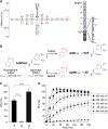
Figure 1
Figure 1
A novel FRET peptide assay to detect H. pylori HtrA activity. (A) The cleavage specificity profile of HpHtrA obtained from DIPPS profiling is represented as an iceLogo, with significantly enriched and under-represented amino acids above and below the x-axis, respectively. The scissile peptide bond between P1 and P1′ is shown as a gray dashed line in the iceLogo. The model on the right represents the domain structure of human E-cadherin (hCdh1), which is composed of the extracellular domain (EC1–EC5), a transmembrane domain (TMD) and an intracellular domain (IC). HtrA cleavage sites have been identified (red arrows) in the Ca++-binding sites located between the individual EC regions. According to the iceLogo, an additional cleavage site for HtrA is present in the linker region between the EC5 domain and TMD. (B) The sequence AQRVAF harboring 2-aminobenzoyl (2-Abz) as fluorophore and 3-nitro-tyrosine Y(NO2) as a quencher is hydrolyzed by trypsin with arginine (R) at position P1 and by HpHtrA with valine (V) at position P1. (C) 5 µM of the FRET peptide were incubated with 250 nM of HpHtrA wild type (wt), its isogenic inactive mutant (SA), or 125 nM trypsin for 180 min in 50 mM HEPES buffer (pH 7.4) at 37 °C. The data represent the relative fluorescent units (RFU) ± S.D. with the fluorescent signal obtained from trypsin-treated FRET peptide set as 100%. Asterisks indicate statistically significant differences (****p < 0.0001). (D) 4 µM of the FRET peptide were incubated with indicated concentrations of HpHtrA wt or SA for 180 min at 37 °C in 50 mM HEPES buffer (pH 7.4). The data represent the RFU ± S.D. with the fluorescent signals obtained from FRET peptide treated with 400 nM HpHtrA wt for 180 min set as 100%.
Figure 2
Figure 2
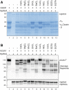
Divalent cations modulate the activity of HpHtrA. (A) 10 µg casein composed of αS1-, αS2- and β-casein were incubated with 250 ng HpHtrA for 16 h at 37 °C in 50 mM HEPES buffer (pH 7.4). Where indicated, 1 mM of the different divalent ions, EDTA, or EGTA was added. Proteins were separated by SDS-PAGE and stained with Coomassie Brilliant Blue G250. (B) 50 ng hCdh1 were incubated with 250 ng HpHtrA for 16 h at 37 °C in 50 mM HEPES buffer (pH 7.4). Where indicated, 1 mM of the different divalent ions, EDTA, or EGTA was added. Full length hCdh1 (Cdh1FL, 125 kDa) and hCdh1 cleavage fragments were detected by Western blot using an antibody recognizing the EC5 domain of hCdh1. HpHtrA and the auto-processed HpHtrA (HpHtrAs) were detected using a polyclonal HpHtrA antibody.
Figure 3
Figure 3
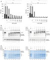
ZnCl2 and CuCl2 inhibit HpHtrA activity in a concentration-dependent manner. (A) 5 µM FRET peptide were incubated with 250 nM HpHtrA and increasing concentrations of ZnCl2 (left panel) or CuCl2 (right panel) for 15 min (black bars) and 180 min (grey bars) at 37 °C in 50 mM HEPES buffer (pH 7.4). The data represent the relative fluorescence units (RFU) ± S.D. with fluorescent signals obtained from FRET peptide treated with HpHtrA wt set as 100%. Asterisks indicate statistically significant differences (****p < 0.0001; ns, non-significant). (B) 50 ng hCdh1 were incubated with 250 ng HpHtrA wt or inactive mutant (SA) and increasing concentrations of ZnCl2 (left panel) or CuCl2 (right panel) for 16 h at 37 °C in 50 mM HEPES buffer (pH 7.4). Full length hCdh1 (Cdh1FL) and cleavage fragments were detected by Western blot using an antibody recognizing the EC5 domain of hCdh1. HpHtrA and the auto-processed short HpHtrA (HpHtrAs) were detected using a polyclonal antibody. (C) 10 µg casein composed of αS1-, αS2- and β-casein were incubated with 250 ng HpHtrA wt or inactive mutant (SA) and with increasing concentrations of ZnCl2 (left panel) or CuCl2 (right panel). After incubation at 37 °C for 16 h proteins were separated by SDS-PAGE and visualized by staining with Coomassie Brilliant Blue G250.
Figure 4
Figure 4
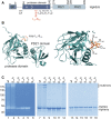
The allosteric ligand-binding loop is important for HtrA oligomer stabilization. (A) Model of HtrA showing the domain structure showing the signal peptide (SP), the extended linker region containing the LA loop, the protease domain with the catalytic triad histidine (H), aspartic acid (D), and serine (S), and the PDZ1 and PDZ2 domains. The protease domain also contains the regulatory LD, L1, L2, and L3 loops. The position of the ligand-binding loop is highlighted in red. (B) X-ray structure of the protease domain of HpHtrA with S164, D165, S166, and D168 indicated in the ligand-binding loop. S221 in the active center is highlighted in magenta. (C) Oligomerization and activity of HpHtrA wt, HpHtrA S164A, HpHtrA D165A, HpHtrA S166A, HpHtrA D168A, and HpHtrA S221A analyzed by casein zymography (lanes 1–6), non-reducing SDS-PAGE (lanes 7–12), and reducing SDS-PAGE (lanes 13–18).
Figure 5
Figure 5
Proteolytic activity of HpHtrA loop mutants. To analyze the proteolytic activity of HtrA wt and its isogenic mutants HpHtrA S164A, HpHtrA D165A, HpHtrA S166A, HpHtrA D168A, and the inactive HpHtrA S221A, 10 µg casein (A) or 50 ng E-cadherin (hCdh1) (B) were incubated with 250 ng HtrA variants as indicated for 16 h at 37 °C in 50 mM HEPES buffer (pH 7.4). Cleavage of casein was analyzed by Coomassie-stained SDS-PAGE and hCdh1 cleavage was determined by Western blot analysis using an antibody recognizing the EC5 domain of hCdh1. HpHtrA and the auto-processed HpHtrAs were detected as indicated. (C) The activity of the ligand-binding loop mutants was analyzed using the FRET peptide as a substrate. 5 µM FRET peptide were incubated with 250 nM HpHtrA wt (black circle), HpHtrA S164A (red square), HpHtrA D165A (green triangle), HpHtrA S166A (yellow inverted triangles), and HpHtrA D168A (blue rhombus) for 180 min at 37 °C in 50 mM HEPES buffer (pH 7.4). The data represent the relative fluorescent units (RFU) ± S.D. with fluorescent signals obtained from FRET peptide treated with HpHtrA wt for 180 min set as 100%.
Figure 6
Figure 6
Zn++ and Cu++ ions interfere with the stability of HpHtrA via an allosteric loop. To investigate the effect of ZnCl2 and CuCl2 on the stability and activity of HpHtrA oligomers, HpHtrA wt was incubated with increasing concentrations of ZnCl2 (A) and CuCl2 (B) and then analyzed by a Coomassie-stained SDS-PAGE under non-reducing conditions (left panels). 4 µM recombinant HpHtrA wt (black circles), HpHtrA S164A (red squares), HpHtrA D165A (green inverted triangles), HpHtrA S166A (yellow triangles), and HpHtrA D168A (blue rhombus) were incubated with SYPRO Orange and increasing concentrations of ZnCl2 and CuCl2 at a temperature ramp from 25–95 °C (increase of 0.5 °C per minute). Changes in melting temperature ΔTm are presented normalized to the intrinsic Tm of the individual HtrA mutant (right panels).

Similar articles

Cited by

References

    1. Global Burden of Disease Cancer Collaboration Global, regional, and national cancer incidence, mortality, years of life lost, years lived with disability, and disability-adjusted life-years for 32 cancer groups, 1990 to 2015: a systematic analysis for the global burden of disease study. JAMA Oncol. 2017;3:524–548. doi: 10.1001/jamaoncol.2016.5688. - DOI - PMC - PubMed
    1. Peek RM, Crabtree JE. Helicobacter infection and gastric neoplasia. J. Pathol. 2006;208:233–248. doi: 10.1002/path.1868. - DOI - PubMed
    1. Blaser MJ, Atherton JC. Helicobacter pylori persistence: biology and disease. J. Clin. Investig. 2004;113:321–333. doi: 10.1172/JCI200420925. - DOI - PMC - PubMed
    1. Hoy B, et al. Helicobacter pylori HtrA is a new secreted virulence factor that cleaves E-cadherin to disrupt intercellular adhesion. EMBO Rep. 2010;11:798–804. doi: 10.1038/embor.2010.114. - DOI - PMC - PubMed
    1. Niessen CM, Leckband D, Yap AS. Tissue organization by cadherin adhesion molecules: dynamic molecular and cellular mechanisms of morphogenetic regulation. Physiol. Rev. 2011;91:691–731. doi: 10.1152/physrev.00004.2010. - DOI - PMC - PubMed

Publication types

MeSH terms