

Regeneration in the spiny mouse, Acomys, a new mammalian model
- PMID: 32599302
- PMCID: PMC9724456
- DOI: 10.1016/j.gde.2020.05.019
Regeneration in the spiny mouse, Acomys, a new mammalian model
Abstract
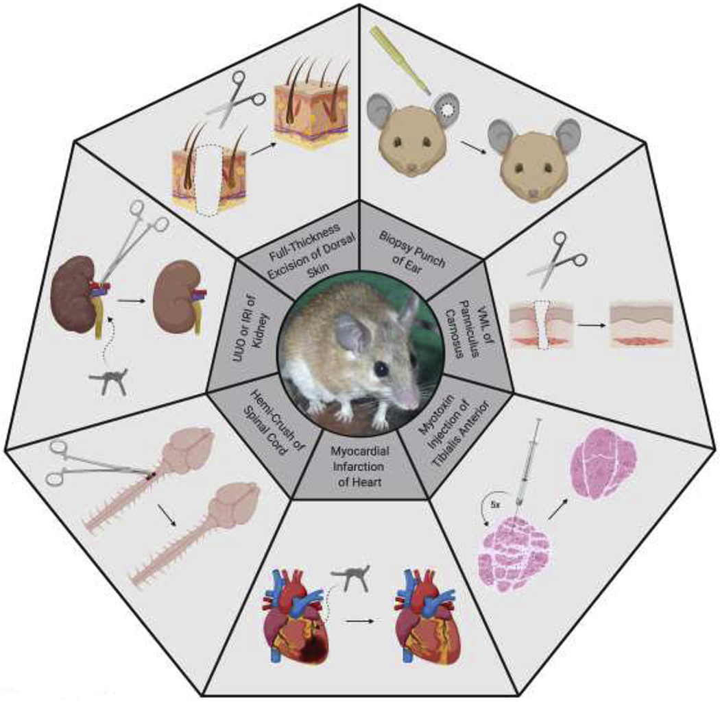
We describe the tissues and organs that show exceptional regenerative ability following injury in the spiny mouse, Acomys. The skin and ear regenerate: hair and its associated stem cell niches, sebaceous glands, dermis, adipocytes, cartilage, smooth muscle, and skeletal muscle. Internal tissues such as the heart, kidney, muscle, and spinal cord respond to damage by showing significantly reduced inflammation and improved regeneration responses. The reason for this improved ability may lie in the immune system which shows a blunted inflammatory response to injury compared to that of the typical mammal, but we also show that there are distinct biomechanical properties of Acomys tissues. Examining the regenerative behavior of closely related mammals may provide insights into the evolution of this remarkable property.
Copyright © 2020 Elsevier Ltd. All rights reserved.
Conflict of interest statement
Disclosure
The authors declare no conflict of interest.
Figures
Similar articles
-
Model systems for regeneration: the spiny mouse, Acomys cahirinus.Development. 2020 Feb 25;147(4):dev167718. doi: 10.1242/dev.167718. Development. 2020. PMID: 32098790 Free PMC article. Review.
-
Insights into the regeneration of skin from Acomys, the spiny mouse.Exp Dermatol. 2019 Apr;28(4):436-441. doi: 10.1111/exd.13847. Epub 2019 Jan 15. Exp Dermatol. 2019. PMID: 30457673 Review.
-
Spiny mice are primed but fail to regenerate volumetric skeletal muscle loss injuries.Skelet Muscle. 2024 Oct 29;14(1):26. doi: 10.1186/s13395-024-00358-y. Skelet Muscle. 2024. PMID: 39468576 Free PMC article.
-
Optimal skin regeneration after full thickness thermal burn injury in the spiny mouse, Acomys cahirinus.Burns. 2018 Sep;44(6):1509-1520. doi: 10.1016/j.burns.2018.05.018. Epub 2018 Jun 11. Burns. 2018. PMID: 29903601
-
Skin shedding and tissue regeneration in African spiny mice (Acomys).Nature. 2012 Sep 27;489(7417):561-5. doi: 10.1038/nature11499. Nature. 2012. PMID: 23018966 Free PMC article.
Cited by
-
Revelations About Aging and Disease from Unconventional Vertebrate Model Organisms.Annu Rev Genet. 2021 Nov 23;55:135-159. doi: 10.1146/annurev-genet-071719-021009. Epub 2021 Aug 20. Annu Rev Genet. 2021. PMID: 34416119 Free PMC article. Review.
-
Function and Evolution of Nuclear Receptors in Environmental-Dependent Postembryonic Development.Front Cell Dev Biol. 2021 Jun 10;9:653792. doi: 10.3389/fcell.2021.653792. eCollection 2021. Front Cell Dev Biol. 2021. PMID: 34178983 Free PMC article. Review.
-
Preclinical Models and Promising Pharmacotherapeutic Strategies in Liver Fibrosis: An Update.Curr Issues Mol Biol. 2023 May 11;45(5):4246-4260. doi: 10.3390/cimb45050270. Curr Issues Mol Biol. 2023. PMID: 37232739 Free PMC article. Review.
-
Comparative biology and non-traditional approaches for basic aging research for facilitating translational studies.Geroscience. 2024 Jun;46(3):2803-2813. doi: 10.1007/s11357-023-00992-2. Epub 2023 Nov 9. Geroscience. 2024. PMID: 37940788 Free PMC article.
-
Osteoderms in a mammal the spiny mouse Acomys and the independent evolution of dermal armor.iScience. 2023 May 24;26(6):106779. doi: 10.1016/j.isci.2023.106779. eCollection 2023 Jun 16. iScience. 2023. PMID: 37378333 Free PMC article.
References
-
- Musarò A: The Basis of Muscle Regeneration. Kuang S, ed. Adv Biol 2014, 612471. doi:10.1155/2014/612471 - DOI
-
- Goss R: Deer Antlers: Regeneration, Function and Evolution. 1st Edition. Academic Press; 1983.
-
- Wang H, Paulsen JM, Hironaka EC, Shin HS, Farry JM, Thakore AD, Jung J, Lucian HJ, Eskandari A, Anilkumar S, et al. Natural Heart Regeneration in a Neonatal Rat Myocardial Infarction Model. Cells 2020, 9:229. doi:10.3390/cells9010229 - DOI - PMC - PubMed
-
A demonstration that the neonatal rat heart can regenerate after a myocardial infarction. Increased levels of cardiomyocyte proliferation are observed and by 3 weeks the ejection fraction was the same as controls.
Publication types
MeSH terms
Grants and funding
LinkOut - more resources
Full Text Sources
Medical
