. 2020 Oct;117(10):3066-3080.
doi: 10.1002/bit.27479. Epub 2020 Jul 9.

Immobilization rapidly selects for chemoresistant ovarian cancer cells with enhanced ability to enter dormancy

Affiliations

Immobilization rapidly selects for chemoresistant ovarian cancer cells with enhanced ability to enter dormancy

Tiffany Lam et al. Biotechnol Bioeng. 2020 Oct.

Abstract

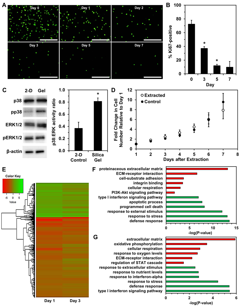
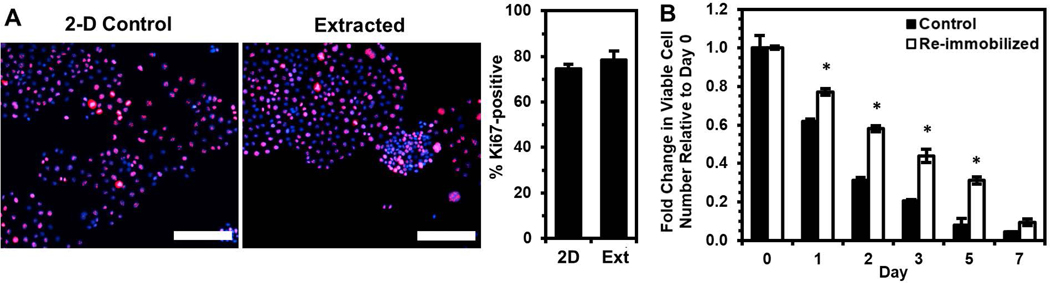
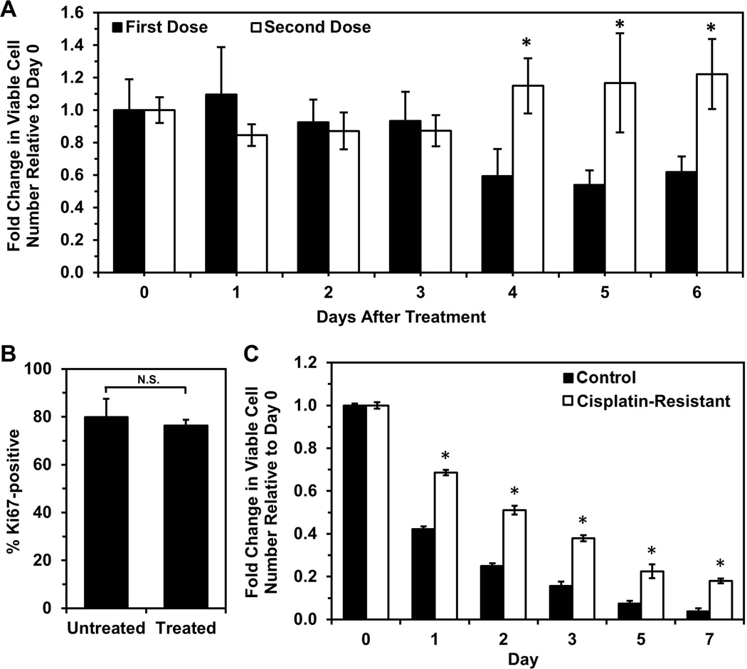
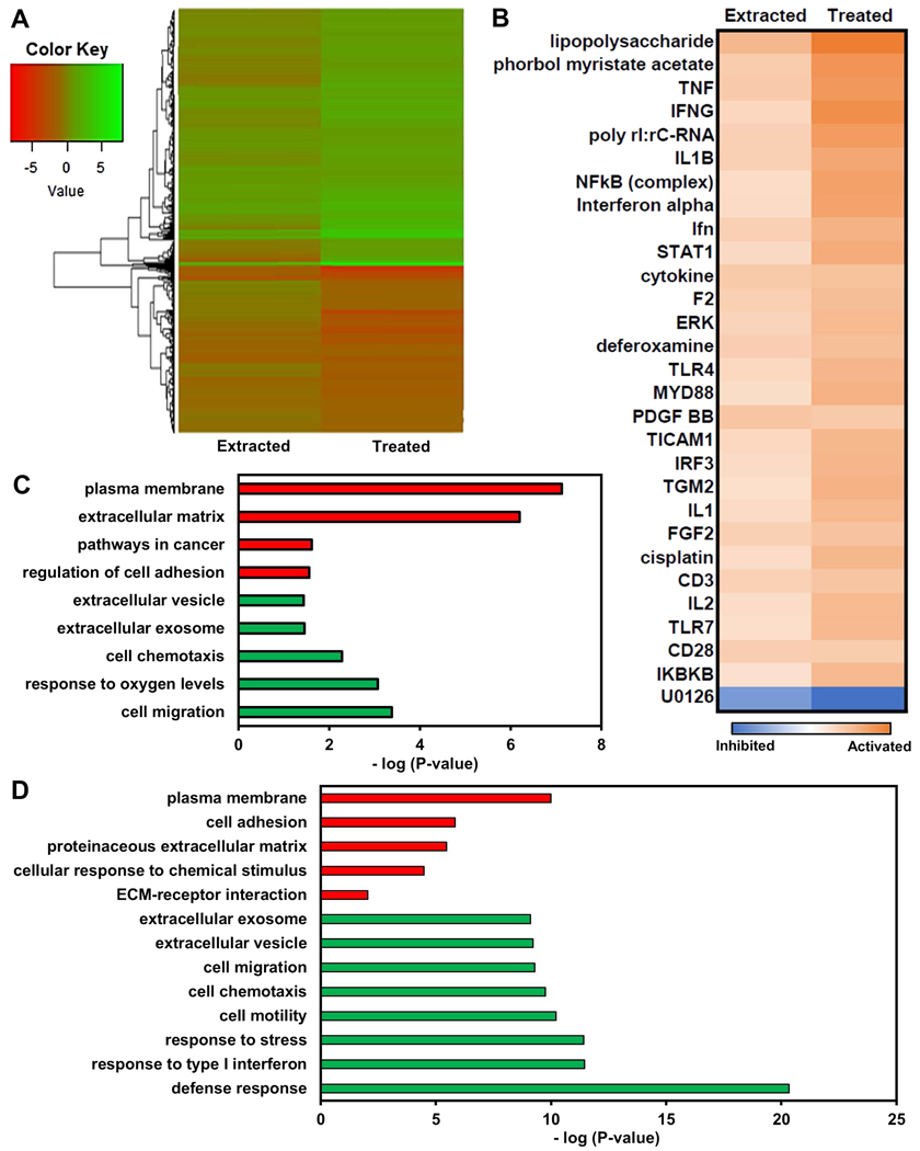
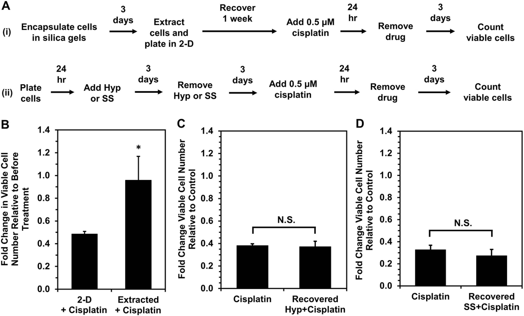
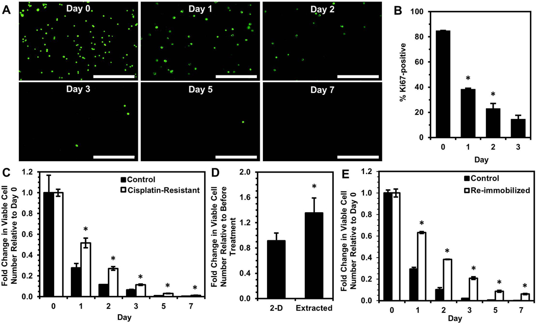
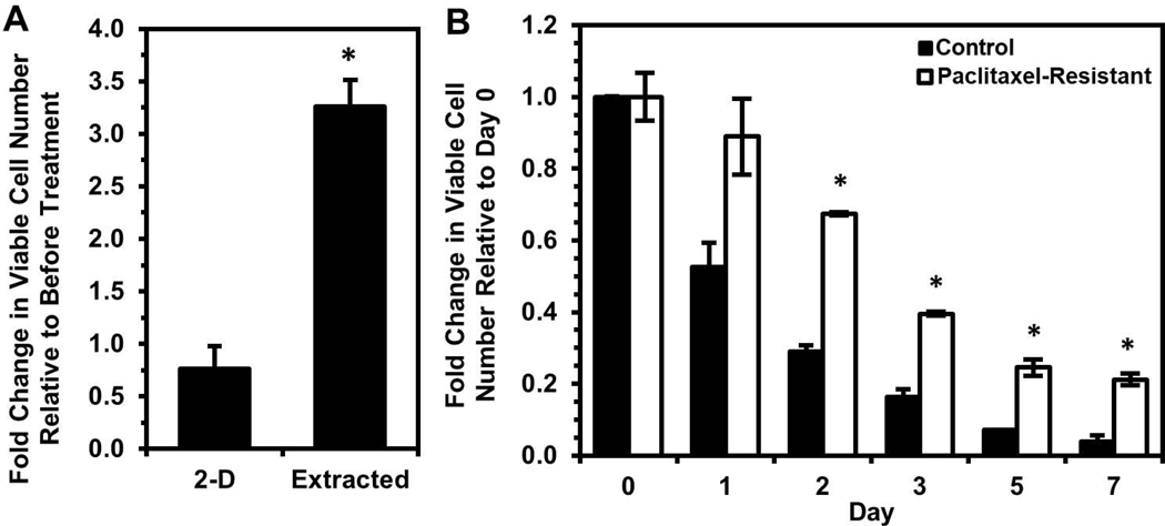
Around 20-30% of ovarian cancer patients exhibit chemoresistance, but there are currently no methods to predict whether a patient will respond to chemotherapy. Here, we discovered that chemoresistant ovarian cancer cells exhibit enhanced survival in a quiescent state upon experiencing the stress of physical confinement. When immobilized in stiff silica gels, most ovarian cancer cells die within days, but surviving cells exhibit hallmarks of single-cell dormancy. Upon extraction from gels, the cells resume proliferation but demonstrate enhanced viability upon reimmobilization, indicating that initial immobilization selects for cells with a higher propensity to enter dormancy. RNA-seq analysis of the extracted cells shows they have signaling responses similar to cells surviving cisplatin treatment, and in comparison to chemoresistant patient cohorts, they share differentially expressed genes that are associated with platinum-resistance pathways. Furthermore, these extracted cells demonstrate greater resistance to cisplatin and paclitaxel, despite being proliferative. In contrast, serum starvation and hypoxia could not effectively select for chemoresistant cells upon removal of the environmental stress. These findings demonstrate that ovarian cancer chemoresistance and the ability to enter dormancy are linked, and immobilization rapidly distinguishes chemoresistant cells. This platform could be suitable for mechanistic studies, drug development, or as a clinical diagnostic tool.

Keywords: chemoresistance; immobilization; ovarian cancer; quiescence; silica gel.


Conflict of interest statement

Conflict of Interest Statement

The authors declare that there are no conflicts of interests.

Figures

Figure 1.
Figure 1.. Immobilization of OVCAR-3 cells in silica gels results in survival of cells exhibiting quiescence.
(A) Fluorescence images of viable cells stained with calcein AM (green) at Days 0, 1, 2, 3, 5, and 7 of immobilization. Scale bar indicates 400 μm. (B) Percentage of Ki67-positive cells (number of Ki67-expressing cells normalized to number of DAPI-labeled cells) within silica gels over a one-week period quantified from fluorescence images (*P < 0.05 compared to previous timepoint). (C) Western blotting of p38 MAPK protein (“p38”), ERK1/2 MAPK proteins (“ERK1/2”), and their phosphorylated forms (“pp38” and “pERK1/2”) in cells immobilized for three days and control cells grown in standard 2-D culture conditions (*P < 0.05 compared to “2-D Control”). (D) Growth of cells extracted from silica gels after immobilization for 3 days and control cells cultured in 2-D. (E) Heatmap of log2FC of gene expression in cells immobilized for one or three days relative to control cells. (F,G) Gene ontology enrichment analysis using genes significantly upregulated (green) or downregulated (red) in “Day 1” (F) or “Day 3” (G) of immobilization relative to 2-D controls.
Figure 2.
Figure 2.. Cells surviving silica gel immobilization demonstrate enhanced survival upon re-immobilization relative to the original control population.
Cells surviving immobilization for three days were extracted and cultured in 2-D for 1 week prior to immunostaining or re-immobilization. (A) Percentage of Ki67-positive cells (number of Ki67-expressing cells (red) normalized to number of DAPI-labeled cells (blue)) from immunofluorescence images (merged images shown) compared to 2-D control cells. Scale bar indicates 200 μm. (B) Fold change in viable cell number relative to Day 0 after re-immobilizing extracted cells in silica gels relative to cells immobilized for the first time (Control) (*P < 0.05 compared to “Control” for each timepoint).
Figure 3.
Figure 3.. Cisplatin-resistant OVCAR-3 cells have higher tolerance for immobilization and silica gel environment.
(A) Cells retreated with a second dose of cisplatin are less susceptible to treatment than cells undergoing primary treatment, indicating the first dose selected for cisplatin-resistant cells. Each treatment dose was 0.5 μM cisplatin for 24 hours, and cells receiving a second dose were allowed a 2-week recovery period before treatment with the second dose. Viable cell number at Day 0 is the number of live cells counted immediately after drug removal. (*P < 0.05 compared to “First Dose”). (B) Cisplatin-resistant cells (“Treated”, i.e. treated with cisplatin and allowed to recover for 2 weeks) have similar Ki67 expression level as untreated cells. (C) Fold change in viable cell number relative to Day 0 of immobilized cisplatin-resistant cells and untreated control cells (*P < 0.05 compared to “Control” at each timepoint).
Figure 4.
Figure 4.. OVCAR-3 cells selected by immobilization regulate genes similarly to cells surviving cisplatin treatment.
(A) Heatmap of log2FC of gene expression in cells immobilized for three days, extracted, and cultured in standard 2-D conditions for two weeks (“Extracted”) and cells surviving treatment with 0.5 μM cisplatin for 24 hours and allowed to recover for 2 weeks in standard 2-D culture conditions (“Treated”) relative to control cells. (B) List of top 30 common upstream regulators (determined by average Z-score with P < 0.05) in “Extracted” and “Treated” cells based upon differentially expressed genes relative to control cells using Ingenuity Pathway Analysis. (C,D) Gene ontology enrichment analysis using genes significantly upregulated (green) or downregulated (red) in “Extracted” (C) or “Treated” (D) samples relative to 2-D controls.
Figure 5.
Figure 5.. Silica gel immobilization selects for cells that are more chemoresistant even while proliferative.
(A) Timeline for cisplatin treatment studies of cells either (i) surviving silica gel immobilization or (ii) recovered from hypoxia treatment or serum starvation. Recovery periods of three days after hypoxia or serum starvation and one week post-extraction allowed for similar cell densities and return to a proliferative state at the start of cisplatin treatment. (B) Response to cisplatin treatment of cells surviving silica gel immobilization (“Extracted+Cisplatin”) relative to cells maintained in 2-D culture conditions (“2-D+Cisplatin”). Fold change in viable cell number indicates cell number at Day 3 post-cisplatin treatment relative to cell number before drug treatment (*P < 0.05 relative to 2-D). (C,D) Fold change in viable cell number for cells recovered from (C) hypoxia (“Recovered Hyp+Cisplatin”) or (D) serum starvation (“Recovered SS+Cisplatin”) and treated with 0.5 μM cisplatin for 24 hours relative to proliferating cells grown in standard culture conditions (“Cisplatin”) after treatment. Fold change in viable cell number indicates cell number at Day 3 post-cisplatin treatment relative to untreated controls for each respective condition (*P < 0.05 compared to “Cisplatin”).
Figure 6.
Figure 6.. Selection of unique subpopulation via silica gel immobilization is also observed with SKOV-3 ovarian cancer cell line.
(A) Calcein AM staining of viable SKOV-3 cells (green) immobilized for 0, 1, 2, 3, 5, and 7 days within silica gels. Scale bar indicates 400 μm. (B) Percentage of Ki67-positive SKOV-3 cells within silica gels over a three-day period (*P < 0.05 compared to previous timepoint). (C) Fold change in viable cell number relative to Day 0 of immobilized cisplatin-resistant SKOV-3 cells and untreated control cells (*P < 0.05 compared to “Control” at each timepoint). (D,E) SKOV-3 cells surviving immobilization for 24 hours were extracted and cultured in 2-D for 1 week and used to find (D) the fold change in viable cell number three days after 5 μM cisplatin treatment for 24 hours relative to before treatment (*P < 0.05 compared to “2-D”) and (E) the fold change in viable cell number relative to Day 0 after re-immobilizing extracted SKOV-3 cells in silica gels relative to proliferating cells immobilized for the first time (Control) (*P < 0.05 compared to “Control” at each timepoint).
Figure 7.
Figure 7.. Extracted cells are less sensitive to paclitaxel, and paclitaxel-resistant cells exhibit enhanced survival upon immobilization.
(A) Fold change in viable OVCAR-3 cell number three days after 30 nM paclitaxel for 24 hours relative to before treatment (*P < 0.05 compared to “2-D”). (B) Fold change in viable cell number relative to Day 0 of immobilized paclitaxel-resistant OVCAR-3 cells and untreated control cells (*P < 0.05 compared to “Control” at each timepoint).

Similar articles

Cited by

References

    1. Abubaker K, Latifi A, Chan E, Luwor RB, Burns CJ, Thompson EW, … Ahmed N (2015). Enhanced activation of STAT3 in ascites-derived recurrent ovarian tumors: inhibition of cisplatin-induced STAT3 activation reduced tumorigenicity of ovarian cancer by a loss of cancer stem cell-like characteristics. Journal of Cancer Stem Cell Research.
    1. Agarwal R, & Kaye SB (2003). Ovarian cancer: strategies for overcoming resistance to chemotherapy. Nature Reviews Cancer, 3(7), 502–516. 10.1038/nrc1123 - DOI - PubMed
    1. Aguirre-Ghiso JA (2007). Models, mechanisms and clinical evidence for cancer dormancy. Nature Reviews Cancer, 7(11), 834–846. 10.1038/nrc2256 - DOI - PMC - PubMed
    1. Aguirre-ghiso JA, Estrada Y, Liu D, & Ossowski L (2003). ERK MAPK Activity as a Determinant of Tumor Growth and Dormancy ; Regulation by p38 SAPK. Cancer Research, 63(7), 1684–1695. - PubMed
    1. Aguirre-ghiso JA, Liu D, Mignatti A, Kovalski K, & Ossowski L (2001). Urokinase Receptor and Fibronectin Regulate the ERK MAPK to p38 MAPK Activity Ratios That Determine Carcinoma Cell Proliferation or Dormancy In Vivo. Molecular Biology of the Cell, 12, 863–879. - PMC - PubMed

Publication types