. 2020 Dec;69(12):2571-2587.
doi: 10.1007/s00262-020-02638-0. Epub 2020 Jun 25.

TIM-3 blockade combined with bispecific antibody MT110 enhances the anti-tumor effect of γδ T cells

Affiliations

TIM-3 blockade combined with bispecific antibody MT110 enhances the anti-tumor effect of γδ T cells

Qingming Guo et al. Cancer Immunol Immunother. 2020 Dec.

Abstract

As ideal cells that can be used for adoptive cell therapy, γδ T cells are a group of homogeneous cells with high proliferative and tumor killing ability. However, γδ T cells are apt to apoptosis and show decreased cytotoxicity under persistent stimulation in vitro and cannot aggregate at tumor sites efficiently in vivo, both of which are two main obstacles to tumor adoptive immunotherapy. In this study, we found that the immune checkpoint T-cell immunoglobulin domain and mucin domain 3 (TIM-3) were up-regulated significantly on γδ T cells during their ex vivo expansion and this up-regulation contributed to the dysfunction of γδ T cells. Although the killing ability of γδ T cells against breast cancer cells which exhibited a high level of epithelial cell adhesion molecule (EpCAM) was enhanced, the level of TIM-3 on γδ T cells was also further up-regulated under the application of the bispecific antibody MT110 (anti-CD3 × anti-EpCAM) which can redirect T cells to target cells. Besides, these γδ T cells with up-regulated TIM-3 exhibited an increased susceptibility to apoptosis. By reinvigorating dysfunctional γδ T cells and promoting them to accumulate at tumor sites, the combined use of TIM-3 inhibitor and MT110 could further enhance the anti-tumor effect of the adoptively transfused γδ T cells. These results may have clinical implications for the design of new translational anti-tumor regimens aimed at combining checkpoint blockade and immune cell redirection.

Keywords: Apoptosis; Bispecific antibody; Cytotoxicity; Epithelial cell adhesion molecule; T-cell immunoglobulin domain and mucin domain 3; γδ T cells.


Conflict of interest statement

The authors declare that they have no potential conflict of interest.

Figures

Fig. 1
Fig. 1
γδ T cells became dysfunctional and had a robust up-regulation of TIM-3 during ex vivo induction and expansion. Cells in the γδ T-cell culture system were collected at day 8, day 14, and day 21 and the proportion of CD3+Vδ2+T cells a and their activation markers such as CD25 b, CD69 and HLA-DR c at the corresponding time were determined by flow cytometry. d Vδ2+T cells (isolated at day 10, day 15, day 20 and day 25 from the γδ T-cell culture system) were co-cultured with K562, SH-SY5Y and MDA-MB-231 at different ratios (1:1, 5:1 and 10:1), respectively, for 12 h, and then, their cytotoxicity was determined by flow cytometry. e Representative annexin V/PI staining demonstrates increasing frequencies of the apoptosis of the isolated Vδ2+T cells in the γδ T-cell culture system at day 10, day 14, day 18, and day 22. The apoptosis rate of γδ T cells expanded from 2 healthy donors and 3 breast cancer patients was calculated as the numbers of the first plus fourth quadrants divided by those of all the four quadrants of the dot plot diagrams. Each symbol represents one individual donor (HD: Healthy donor; BCP: breast cancer patient). f The expression level of CTLA-4, PD-1, and TIM-3 on Vδ2+T cells in the γδ T-cell culture system at day 4 (red line), day 9 (green line), day 14 (pink line), and day 18 (blue line) was detected by flow cytometry. The filled histograms represent isotype controls staining and the open ones are specific staining. Similar results were obtained in three independent experiments and shown is representative data
Fig. 2
Fig. 2
Interaction of TIM-3 and TIM-3 ligands induced γδ T-cell dysfunction. a Intracellular detection of galectin-9 on γδ T cells, SH-SY5Y cells, and MDA-MB-231 cells. Shown is one representative data out of three. b The level of galectin-9 in the plasma of breast cancer patients (BCP, n = 28) and healthy donors (HD, n = 20) and the supernatant of MDA-MB-231 was determined by ELISA. Data are the mean ± SD (n = 5) of one representative experiment (**p < 0.01). c The isolated Vδ2 + T cells from the γδ T-cell culture system with 24 h of galectin-9 or agonistic TIM-3 antibody treatment were stained with Annexin V-FITC and PI and the apoptosis rate of Vδ2 + T cells were analyzed by flow cytometry. Shown is one representative experiment out of three. Columns represent the mean ± SD (n = 3) of one representative experiment out of three independent experiments (*p < 0.05). d After being treated by galectin-9 and agonistic TIM-3 antibody for 96 h, the proliferation ability of CFSE labeled Vδ2 + T cells was determined by flow cytometry. The value (inset) for the percentage of cells that divided at least once (top left corner) and the average number of cell divisions (bottom left corner) are indicated for each sample. Similar results were obtained in three independent experiments
Fig. 3
Fig. 3
TIM-3 blockade could reverse γδ T-cell dysfunction. a TIM-3 inhibitor (α-TIM-3), fusion protein TIM-3-Fc, or isotype control antibody was added to the γδ T-cell culture system (expanded from breast cancer patients) which have already supplied with galectin-9. After culturing for 24 h, the cells were stained with Annexin V-FITC and PI and analyzed by flow cytometry. Shown is one representative experiment out of three. b Western blot analyses. Galectin-9 activated caspase 3, while α-TIM-3 or TIM-3-Fc inhibits the activation of caspase 3 in γδ T cells. Galectin-9, α-TIM-3 or TIM-3-Fc could not influence the expression of cyclin B1 or cyclin D1 in γδ T cells. c γδ T cells (expanded from one breast cancer patient) with or without TIM-3 blockade (TIM-3 inhibitor or TIM-3-Fc) were co-cultured with different cell lines such as K562, SH-SY5Y and MDA-MB-231 at an E:T ratio of 1:1 for 12 h, then their cytotoxicity was determined by flow cytometry. d The levels of IFN-γ and TNF-α in each co-culture system mentioned above were determined by ELISA. Data are the mean ± SD (n = 3) of one representative experiment out of three independent experiments (*p < 0.05)
Fig. 4
Fig. 4
MT110 enhanced γδ T-cell-mediated cytotoxicity and up-regulated TIM-3 in vitro. a Surface detection of EpCAM on the neuroblastoma cell line SH-SY5Y and the breast cancer cell line MDA-MB-231. b A series dose of MT110 was added to the γδ T cell and SH-SY5Y or MDA-MB-231 co-culture system (E:T = 5:1) and the cytotoxicity of γδ T cell was determined by flow cytometry after 12 h culture. c Intracellular detection of IFN-γ and perforin on γδ T cells co-cultured with SH-SY5Y or MDA-MB-231 at an E:T ratio of 5:1 supplying MT110 (30 ng/ml) or not. d The expression level of TIM-3 on γδ T cells in the co-culture system with anti-CD3 antibody or MT110 treatment or without any treatment was determined by flow cytometry after 24 h co-culturing. Shown is one representative experiment out of three. e The levels of IFN-γ and TNF-α in each co-culture system mentioned above were determined by ELISA. f γδ T cells were treated with or without TNF-α inhibitor (α-TNFRI) or NF-κB inhibitor (MG132) in the presence of 3 ng/mL TNF-α for 24 h. g The γδ T and MDA-MB-231 co-culture system was treated with or without TNF-α inhibitor or NF-κB inhibitor for 24 h. The expression of TIM-3 was measured by flow cytometry. Shown is one representative experiment out of three. h TIM-3highγδ T cells separated in the co-culture system were treated by galectin-9 (40 pg/ml) in the presence or absence of TIM-3 inhibitor (5 μg/ml) for 24 h, and then, the cells were stained with Annexin V-FITC and PI and the apoptosis was analyzed by flow cytometry. Shown is one representative experiment out of three. All data mentioned above are the mean ± SD (n = 3) of one representative experiment out of three independent experiments (*p < 0.05, **p < 0.01)
Fig. 5
Fig. 5
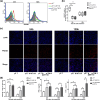
Combination of TIM-3 inhibitor and MT110 enhanced the survival and infiltration ability of adoptively transfused γδ T cells in vivo. a 24 or 120 h after breast cancer mice in γδ T group (n = 6), γδ T and MT110 group (n = 6) and γδ T and α-TIM-3 and MT110 group (n = 6) receiving transfusion of PKH26 labeled γδ T cell (expanded from a breast cancer patient) with or without α-TIM-3 and MT110, respectively, the proportions of PKH26 positive cells in the peripheral leukocytes of breast cancer mice in each group were determined by flow cytometry (gray: blank; Red: γδ T group; green: γδ T and MT110 group; blue: γδ T and α-TIM-3 and MT110 group). Shown is one representative experiment. b Comparison of proportions of PKH-positive cells in the peripheral leukocytes of breast cancer mice between the three groups mentioned above at the time point of 24 or 120 h after transfusion. (**p < 0.01). Whiskers in box plots indicate maximum and minimum values measured. Line indicates the median. c Accumulation of transfused γδ T cells (purple) in tumor tissues of three different groups mentioned above as visualized by DAPI blue and PKH26 red double labeling at the time point of 24 or 120 h after γδ T-cell transfusion (×200). Shown is one representative data out of six. d Number of DAPI blue and PKH26 red double-labeled transfused γδ T-cell count for each microscopic viewing field in shown. Bars indicate mean count for each group mentioned above and error bars are indicated. (*p < 0.05; **p < 0.01). e The plasma of breast cancer mice in each group was collected and IFN-γ and TNF-α levels were determined by ELISA. Data are the mean ± SD (n = 5) of one representative experiment (*p < 0.05; **p < 0.01)
Fig. 6
Fig. 6
Combined application of TIM-3 inhibitor and MT110 further enhanced anti-tumor effect of the adoptively transfused γδ T cells in vivo. a PBS or γδ T cells (expanded from a breast cancer patient) with or without α-TIM-3 or/and MT110 was intravenously injected into the tail of mice in the appropriate group, respectively, at day 12, day 15, and day 18. Shown is tumor growth curve of breast cancer mice in each group. Tumor sizes are plotted as mean ± SD for each group (n = 5). Experiments were repeated two to three times with similar results. (*P < 0.05). b The median survival periods of mice in γδ T and α-TIM-3 and MT110 group were 60 days, while PBS group, γδ T group, γδ T and α-TIM-3, and γδ T and MT110 group were 35, 45, 50, and 52 days, respectively. The differences were statistically significant. (*P < 0.05)

Similar articles

Cited by

References

    1. Shankaran V, Ikeda H, Bruce AT, White JM, Swanson PE, Old LJ, Schreiber RD. IFNgamma and lymphocytes prevent primary tumour development and shape tumour immunogenicity. Nature. 2001;410:1107–1111. doi: 10.1038/35074122. - DOI - PubMed
    1. Bielamowicz K, Khawja S, Ahmed N. Adoptive cell therapies for glioblastoma. Front Oncol. 2013;3:275. doi: 10.3389/fonc.2013.00275. - DOI - PMC - PubMed
    1. Pennington DJ, Silva-Santos B, Hayday AC. Gammadelta T cell development—having the strength to get there. Curr Opin Immunol. 2005;17:108–115. doi: 10.1016/j.coi.2005.01.009. - DOI - PubMed
    1. Bonneville M, Scotet E. Human Vgamma9Vdelta2 T cells: promising new leads for immunotherapy of infections and tumors. Curr Opin Immunol. 2006;18:539–546. doi: 10.1016/j.coi.2006.07.002. - DOI - PubMed
    1. Das H, Wang L, Kamath A, Bukowski JF. Vgamma2Vdelta2 T-cell receptor-mediated recognition of aminobisphosphonates. Blood. 2001;98:1616–1618. doi: 10.1182/blood.V98.5.1616. - DOI - PubMed

MeSH terms

Substances