. 2020 Jun 3:8:412.
doi: 10.3389/fcell.2020.00412. eCollection 2020.

LRP-1 Promotes Colon Cancer Cell Proliferation in 3D Collagen Matrices by Mediating DDR1 Endocytosis

Affiliations

LRP-1 Promotes Colon Cancer Cell Proliferation in 3D Collagen Matrices by Mediating DDR1 Endocytosis

Cao Cuong Le et al. Front Cell Dev Biol. .

Abstract

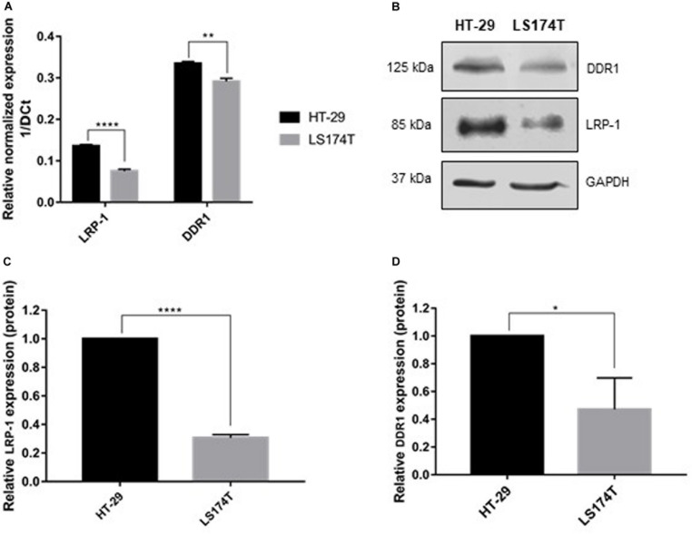
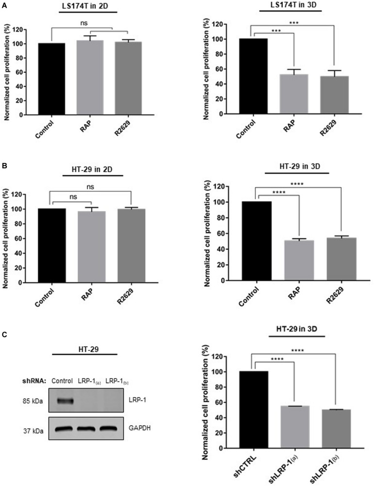
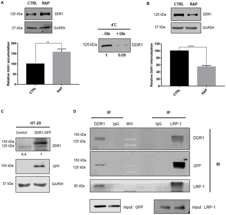
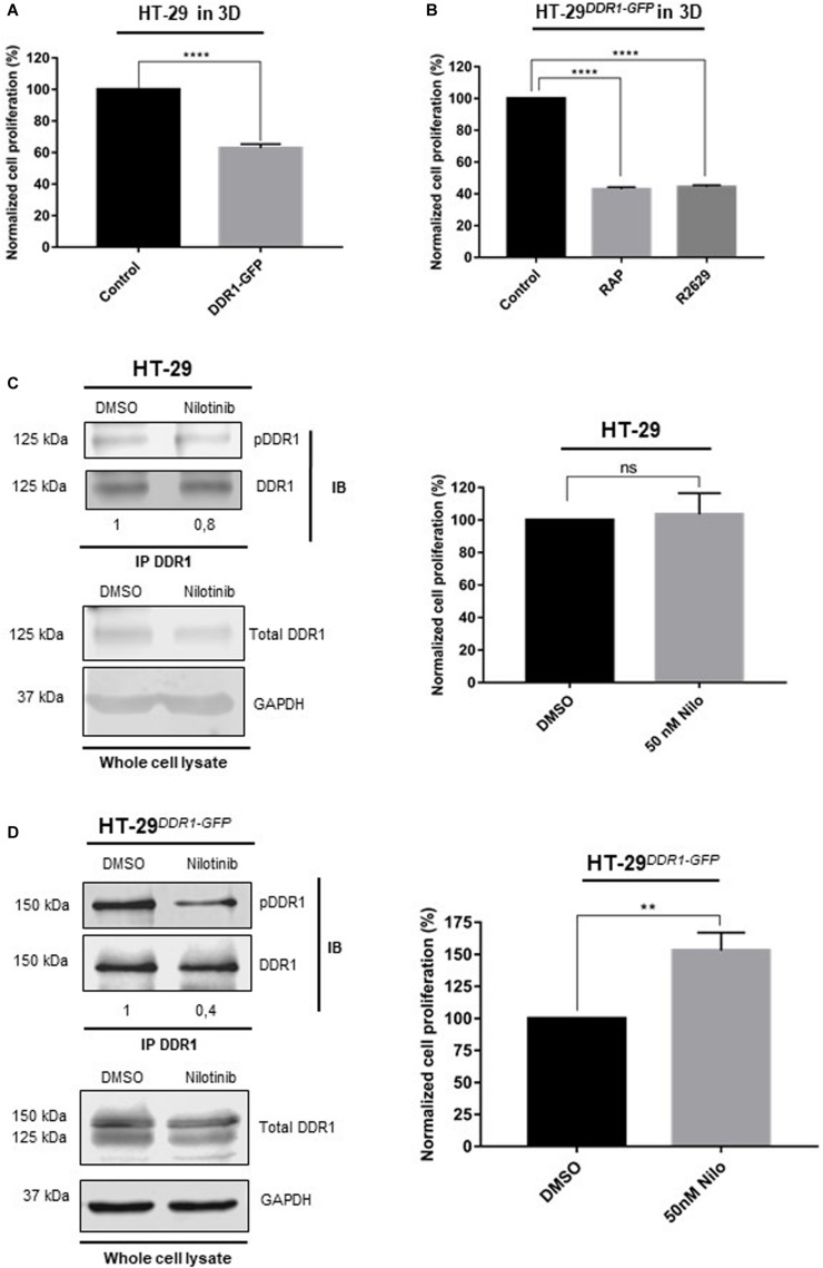
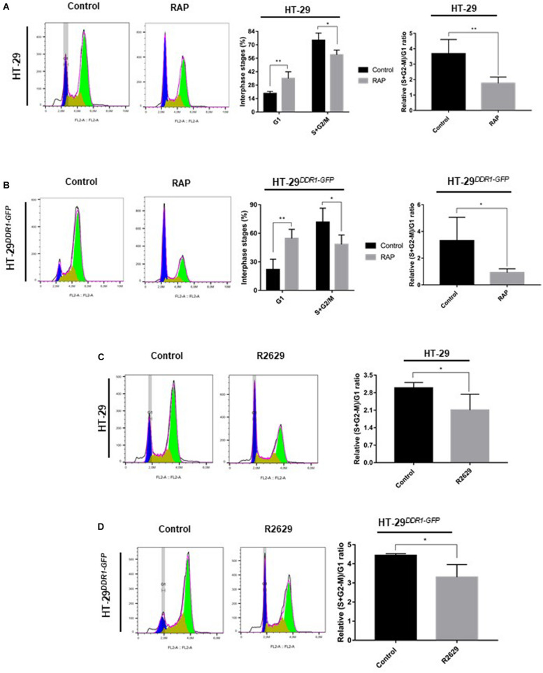
Low density lipoprotein receptor related protein-1 (LRP-1) is a large ubiquitous endocytic receptor mediating the clearance of various molecules from the extracellular matrix. Several studies have shown that LRP-1 plays crucial roles during tumorigenesis functioning as a main signal pathway regulator, especially by interacting with other cell-surface receptors. Discoïdin Domain Receptors (DDRs), type I collagen receptors with tyrosine kinase activity, have previously been associated with tumor invasion and aggressiveness in diverse tumor environments. Here, we addressed whether it could exist functional interplays between LRP-1 and DDR1 to control colon carcinoma cell behavior in three-dimensional (3D) collagen matrices. We found that LRP-1 established tight molecular connections with DDR1 at the plasma membrane in colon cancer cells. In this tumor context, we provide evidence that LRP-1 regulates by endocytosis the cell surface levels of DDR1 expression. The LRP-1 mediated endocytosis of DDR1 increased cell proliferation by promoting cell cycle progression into S phase and decreasing apoptosis. In this study, we identified a new molecular way that controls the cell-surface expression of DDR1 and consequently the colon carcinoma cell proliferation and apoptosis and highlighted an additional mechanism by which LRP-1 carries out its sensor activity of the tumor microenvironment.

Keywords: 3D collagen matrix; DDR1; LRP-1; colon cancer cell; proliferation.


Figures

FIGURE 1
FIGURE 1
Molecular characterization of colorectal carcinoma cell lines. (A) Transcriptional level of LRP-1 and DDR1 were assessed using RTqPCR. LRP-1 and DDR1 mRNA expression levels in HT-29 (black boxes) and LS174T (gray boxes) were normalized with both RPL32 and RS18 mRNA expression. (B) Whole cell extracts from HT-29 and LS174T cells were analyzed by SDS PAGE followed by western blotting using anti-DDR1, anti-LRP-1 and anti-GAPDH antibodies. Graphical representations of LRP-1 (C) and DDR1 (D) expression at protein level as normalized with GAPDH. All experiments were performed in three biological replicates. Plots are presented as the mean SD, **p < 0.01; ****p < 0.0001, n = 3, two sample t-test. *p = 0.01.
FIGURE 2
FIGURE 2
Effect of LRP-1 antagonists and LRP-1 knockdown on colorectal cancer cell proliferation. LS174T (A) and HT-29 (B) cells were cultured in 2D type I collagen coating (left panels) or 3D type I collagen matrices (right panels) without (black boxes) or with RAP (500 nM, light gray boxes) or R2629 (2.5 μg/mL, dark gray boxes) treatment. After 5 days of culture, cell growth indices were assessed using at least three separate sets of culture, all conditions were repeated at least three times. (C) HT-29 cells were transduced with lentivirus encoding non-silencing shRNA (shCTRL) or shRNA targeting LRP-1 [shLRP1(a) and shLRP1(b)] (right panel). Whole-cell extracts from each clonal cell were submitted to immunoblot analysis using anti-LRP-1 antibody (5A6). GAPDH expression level served as a loading control. shCTRL (black boxes) and shLRP-1(a) or shLRP-1(b) (gray boxes) HT-29 cells were seeded in 3D type I collagen matrix (left panel) during 5 days with or without RAP and R2629 treatment. Cell growth was evaluated by at least three separate experiments, each done in triplicate. The data are presented as the mean SD. ***p < 0.001; ****p = 0.0001; ns: not significant, One-way ANOVA test using Dunnett’s multiple comparisons.
FIGURE 3
FIGURE 3
RAP treatment inhibits DDR1 endocytosis and led to its accumulation at the plasma membrane. (A) Plasma membrane extracts from cell surface biotinylated proteins were obtained from HT-29 cells treated or not with RAP (1 μM, 1 h). Immunoblot analysis was performed using anti-DDR1 antibodies. Expression level of GAPDH in the intracellular fraction served as a loading control and for normalization. Three independent experiments were conducted, the data is represented as the mean SD. **p < 0.005, two sample t-test. (B) HT-29 cells were treated with/without RAP (1 μM) for 1 h. Plasma membrane proteins were biotinylated and endocytosis assay was carried out as reported in the experimental procedure section. DDR1 internalization was quantified by immunoblotting using DDR1 antibody (right panels including graph, ****p < 0.0001, two sample t-test). Left panel (4°C) serves to control DDR1 binding to the cell surface (-Glut, without glutathione) and glutathione efficacy for biotin stripping (+ glut, with glutathione). (C) Whole-cell extracts were obtained from HT-29 cells overexpressing GFP (control) or DDR1-GFP. Immunoblot analysis was performed using anti-DDR1 and anti-GFP antibodies and GAPDH served as a loading control. LRP-1 (D) or DDR1 containing complexes were immunoprecipitated (IP) from DDR1-GFP overexpressing HT-29 cells whole-cell extracts by using anti-LRP-1 (clone EPR3724) or anti-DDR1 (D1G6) monoclonal antibody, respectively. Immunocomplexes were then subjected to SDS-PAGE and immunoblotted (IB) by using specific antibodies for LRP-1, DDR1, and GFP.
FIGURE 4
FIGURE 4
DDR1 down-regulates colorectal cancer cell proliferation in 3D collagen matrix. (A) Wild-type HT-29 were cultured in 3D type I collagen matrix for 5 days, then cell proliferation was evaluated by three independent experiments. The data are represented as the mean SD, ****p < 0.0001, two sample t-test. (B) HT-29DDR1–GFP cells were seeded in 3D type I collagen matrix during 5 days with/without RAP or LRP-1 blocking antibodies (R2629). Cell proliferation was then evaluated by at least 3 separate sets of culture, the data are presented as the mean SD and compared to untreated cells. ****p = 0.0001, One-way ANOVA test using Dunnett’s multiple comparisons. HT-29 (C) and HT-29DDR1–GFP cells (D) were seeded in 3D type I collagen matrix and cultured with 50 nM nilotinib or DMSO (that served as a control) for 5 days. Left panels: DDR1 containing complexes were immunoprecipitated (IP) whole-cell extracts by using an anti-DDR1 monoclonal antibody (D1G6). Immunocomplexes were then subjected to SDS-PAGE and immunoblotted (IB) by using anti-DDR1 (D1G6) and anti-phospho-DDR1 (Tyr792, 4G10). Numbers under the immunoblots indicate the fold change ratio (pDDR1/DDR1), as compared to DMSO-treated cells that serve as the reference (n = 3). The bottom panel indicates the expression of DDR1 and GAPDH in whole cell lysates and served as a control. Right panels: cell proliferation was evaluated by three independent experiments, the data are presented as the mean SD. **p < 0.005; ns: not significant, two sample t-test.
FIGURE 5
FIGURE 5
Inhibition of LRP-1-mediated endocytosis induces cell cycle arrest at G1 phase. HT-29 (A,C) and HT-29DDR1–GFP (B,D) cells were grown on plastic surface and synchronized by double thymidine block. Synchronized cells were then seeded in 3D type I collagen matrix with or without 1 μM RAP (A,B) or LRP-1-blocking antibodies (R2629, 30 μg/mL) (C,D) for 24 h, followed by a cell cycle analysis. After nuclear staining with DAPI, 20.000 events were acquired and analyzed by flow cytometry. On the left colored panels, cell cycle distributions of HT-29 (A,C) and HT-29DDR1–GFP (B,D) cells treated with or without RAP or R2629 for 24 h are shown as histogram plots of the FL3 fluorescence channel. On the right panels, histograms represent the percentage of interphase stages (G1, S+G2/M) and the relative (S+G2-M)/G1 ratio of HT-29 (A,C) or HT-29DDR1–GFP (B,D) cells treated with (gray boxes) or without (black boxes) RAP or R2629. The data are presented as the mean SD. *p < 0.05; **p = 0.01, two samples t-test. Cell cycle assays were performed in four separate biological experiments for RAP treatment (A,B) and two separate experiments, each conducted in double triplicates for R2629 treatment (C,D).
FIGURE 6
FIGURE 6
Inhibition of LRP-1 results in an increase in apoptosis. HT-29 (A) and, gray boxes HT-29DDR1–GFP cells (B) were seeded in 3D type I collagen matrix and were treated without (black boxes) or with RAP (1 μM) for 3 days. The cells were then collected from digested matrix and suffered a rapid trypsinization before underwent an apoptotic assay. Apoptotic cells were stained with Annexin V and histogram (left panel), showed the percent of apoptotic. The values of treated samples were normalized to their controls, the data are represented as the mean SD, *p < 0.05, two sample t-test. (C) The plot represents the apoptotic indices of wild-type HT-29 overexpressing GFP (black boxes) and HT-29DDR1–GFP (gray boxes) cells seeded in 3D collagen matrix. The apoptosis assays were performed in two distinct experiments, each done in double triplicates. (D) Immunostaining of recombinant DDR1 (green) in untreated (left picture) or RAP-treated (right picture) HT-29DDR1–GFP cells. DNA is stained with DAPI. Scale bar: 5 μm.

Similar articles

Cited by

References

    1. Abdulhussein R., McFadden C., Fuentes-Prior P., Vogel W. F. (2004). Exploring the collagen-binding site of the DDR1 tyrosine kinase receptor. J. Biol. Chem. 279 31462–31470. 10.1074/jbc.M400651200 - DOI - PubMed
    1. Appert-Collin A., Bennasroune A., Jeannesson P., Terryn C., Fuhrmann G., Morjani H., et al. (2017). Role of LRP-1 in cancer cell migration in 3-dimensional collagen matrix. Cell Adh. Migr. 11 316–326. 10.1080/19336918.2016.1215788 - DOI - PMC - PubMed
    1. Assent D., Bourgot I., Hennuy B., Geurts P., Noel A., Foidart J. M., et al. (2015). A membrane-type-1 matrix metalloproteinase (MT1-MMP)-discoidin domain receptor 1 axis regulates collagen-induced apoptosis in breast cancer cells. PLoS One 10:e0116006. 10.1371/journal.pone.0116006 - DOI - PMC - PubMed
    1. Beaujouin M., Prebois C., Derocq D., Laurent-Matha V., Masson O., Pattingre S., et al. (2010). Pro-cathepsin D interacts with the extracellular domain of the beta chain of LRP1 and promotes LRP1-dependent fibroblast outgrowth. J. Cell. Sci. 123(Pt 19) 3336–3346. 10.1242/jcs.070938 - DOI - PMC - PubMed
    1. Boulagnon-Rombi C., Schneider C., Leandri C., Jeanne A., Grybek V., Bressenot A. M., et al. (2018). LRP1 expression in colon cancer predicts clinical outcome. Oncotarget 9 8849–8869. 10.18632/oncotarget.24225 - DOI - PMC - PubMed