. 2020 Aug 17;94(17):e01057-20.
doi: 10.1128/JVI.01057-20. Print 2020 Aug 17.

NF-κB-Interacting Long Noncoding RNA Regulates HIV-1 Replication and Latency by Repressing NF-κB Signaling

Affiliations

NF-κB-Interacting Long Noncoding RNA Regulates HIV-1 Replication and Latency by Repressing NF-κB Signaling

Hong Wang et al. J Virol. .

Abstract

NF-κB-interacting long noncoding RNA (NKILA) was recently identified as a negative regulator of NF-κB signaling and plays an important role in the development of various cancers. It is well known that NF-κB-mediated activation of human immunodeficiency virus type 1 (HIV-1) long terminal repeat (LTR)-driven gene expression is required for HIV-1 transcription and reactivation of latency. However, whether NKILA plays essential roles in HIV-1 replication and latency is unclear. Here, by ectopic expression and silencing experiments, we demonstrate that NKILA potently inhibits HIV-1 replication in an NF-κB-dependent manner by suppressing HIV-1 LTR promoter activity. Moreover, NKILA showed broad-spectrum inhibition on the replication of HIV-1 clones with different coreceptor tropisms as well as on LTR activity of various HIV-1 clinical subtypes. Chromatin immunoprecipitation (ChIP) assays revealed that NKILA expression abolishes the recruitment of p65 to the duplicated κB binding sites in the HIV-1 LTR. NKILA mutants disrupting NF-κB inhibition also lost the ability to inhibit HIV-1 replication. Notably, HIV-1 infection or reactivation significantly downregulated NKILA expression in T cells in order to facilitate viral replication. Downregulated NKILA was mainly due to reduced acetylation of histone K27 on the promoter of NKILA by HIV-1 infection, which blocks NKILA expression. Knockdown of NKILA promoted the reactivation of latent HIV-1 upon phorbol myristate acetate (PMA) stimulation, while ectopic NKILA suppressed the reactivation in a well-established clinical model of withdrawal of azidothymidine (AZT) in vitro These findings improve our understanding of the functional suppression of HIV-1 replication and latency by NKILA through NF-κB signaling.IMPORTANCE The NF-κB pathway plays key roles in HIV-1 replication and reactivation of HIV-1 latency. A regulator inhibiting NF-κB activation may be a promising therapeutic strategy against HIV-1. Recently, NF-κB-interacting long noncoding RNA (NKILA) was identified to suppress the development of different human cancers by inhibiting IκB kinase (IKK)-induced IκB phosphorylation and NF-κB pathway activation, whereas the relationship between NKILA and HIV-1 replication is still unknown. Here, our results show that NKILA inhibits HIV-1 replication and reactivation by suppressing HIV-1 long terminal repeat (LTR)-driven transcription initiation. Moreover, NKILA inhibited the replication of HIV-1 clones with different coreceptor tropisms. This project may reveal a target for the development of novel anti-HIV drugs.

Keywords: HIV-1 latency; HIV-1 replication; NF-κB; NKILA; antiviral activity; lncRNA.


Figures

FIG 1
FIG 1
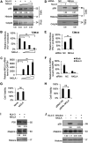
NKILA inhibits HIV-1 replication. (A to C) Overexpression of NKILA inhibits HIV-1 replication in a dose-dependent manner. (A) Multiple dose amounts of NKILA expression vector (100 ng, 300 ng, and 900 ng) or negative-control vector were transfected with the pNL4-3 viral expression vector into HEK293T cells. After 48 h, cells and supernatants were harvested and analyzed by immunoblot (IB) analysis. The densities of bands from representative immunoblotting (IB) analyses were analyzed with ImageJ software to calculate the values, for cells relative to that for histone. (B) Infectious HIV-1 production was decreased with increasing NKILA expression, as indicated in TZM-bl cells. (C) The expression levels of NKILA mRNA were measured by qRT-PCR. The mRNA level of endogenous NKILA was set as 100%. (D to F) Knockdown of NKILA increased HIV-1 replication. (D) pNL4-3 or negative-control vector was cotransfected with siRNA NKILA or siRNA NC into HEK293T cells for 48 h. Cells and supernatants were harvested for IB analysis, and the densities of bands from representative IB analyses were analyzed as described for panel A. (E) NKILA increased the infectious HIV-1 production, as indicated in TZM-bl cells. The infectious HIV-1 production of siRNA NC was set as 100%. (F) The expression levels of NKILA in cells with NKILA knockdown were measured by qRT-PCR and normalized to GAPDH expression. Overexpression (G) or knockdown (H) of NKILA had no effect on cell viability by CCK-8 detection. (I)The inhibitory effect of NKILA on HIV-1 production was not associated with altered endogenous expression of the PMEPA1 protein. NKILA or negative-control vector was cotransfected with the pNL4-3 viral vector into HEK293T cells. Forty-eight hours after transfection, cell extracts were harvested and subjected to IB analysis with anti-PMEPA1 antibody to detect the PMEPA1 protein. (J) PMEPA1 protein expression was not affected by HIV-1 infection or NKILA expression. NKILA or negative-control vector was nucleofected to Jurkat cells. Forty-eight hours posttransfection, the cells were infected with the supernatant containing NL4-3 viral particles or an equal amount of medium. After 48 h of infection, cells were harvested for IB analysis. The densities of bands were analyzed as described for panel A. All results are presented as the means ± SDs from three independent experiments. ns, not significant; **, P < 0.01.
FIG 2
FIG 2
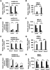
NKILA inhibits HIV-1 replication in T cells. (A to D) Knockdown of NKILA in T cells promoted infectious HIV-1 production. The pNL4-3 viral vector was transfected into HEK293T cells, and supernatants containing HIV-1 NL4-3 virus were harvested for infection of Jurkat cells, H9 cells, or primary CD4 T cells from donors for 48 h which had been transfected with siRNA NKILA or siRNA NC for 24 h. (A and C) Total RNA was extracted from the cells, and the mRNA level of NKILA was measured by qRT-PCR. Cells transfected with siRNAs were infected with NL4-3 virus for 4 h, washed twice with PBS buffer, and cultured in medium (RPMI 1640, 10% FBS). (B and D) After 48 h, the supernatants were harvested, and CAp24 expression was measured by ELISA. (E to H) Overexpression of NKILA suppressed infectious HIV-1 production. Jurkat, H9, or primary CD4+ T cells which had been transfected with NKILA or negative-control vector for 24 h were then were infected with HIV-1 NL4-3 viruses. (E and G) Forty-eight hours postinfection, cells were harvested, and the mRNA level of NKILA was measured by qRT-PCR. (F and H) The supernatants were harvested, and CAp24 expression was measured by ELISA. Expression data were normalized to those of the siRNA NC or negative vector group. All results are presented as the means ± SDs from three independent experiments. *, P < 0.05; **, P < 0.01; ***, P < 0.001.
FIG 3
FIG 3
NKILA inhibits HIV spreading replication. (A) Relative NKILA mRNA expression in MT4 cells expressing NKILA. MT4 cells were infected with lentiviruses containing NKILA or a scramble control, and at 48 h postinfection, puromycin (1 g/ml) was added to the medium for selection. (B) Two nanograms, 0.2 ng, or 0.02 ng p24 antigen of NL4-3 viral particles was added to infect the NKILA MT4 overexpression or control cell lines. Cells were washed with PBS three times after 4 h of infection. The supernatant p24 release amounts were analyzed by ELISA at 1 day, 3 days, 5 days, 7 days, and 9 days.
FIG 4
FIG 4
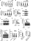
NKILA suppresses HIV-1 LTR-driven gene expression in a dose-dependent manner. (A) NKILA inhibits the transcription initiation and then inhibits the transcription elongation of HIV-1 in HEK293T cells. pNL4-3 was cotransfected with NKILA or negative-control vector into HEK293T cells, and cells were harvested 48 h posttransfection for quantitation of HIV-1 gene transcription initiation and elongation with special primers by qRT-PCR. (B and C) Effect of NKILA overexpression or knockdown on HIV 5′ LTR-driven transcription initiation and elongation in Jurkat cells. Jurkat cells transfected with NKILA vectors or siRNA NKILA were infected with HIV-luc/VSV-G for 2 days, and mRNA was extracted and analyzed by qRT-PCR. (D) NKILA inhibited NF-κB activity. Increasing amounts of NKILA or negative-control vector were cotransfected with pNF-κB-luciferase and pRenilla-luciferase reporter plasmids into HEK293T cells for 48 h. Cells were harvested for assessment of reporter gene expression by a dual luciferase reporter assay. (E) NKILA expression influenced the total and phosphorylated IĸBα protein levels, as evidenced by IB analysis, and the densities of bands were analyzed with ImageJ software to calculate the values relative to that for histone. (F) The localization of p65 protein in the cytoplasm was increased with NKILA overexpression. HEK293T cells transfected with NKILA or negative-control vector were harvested for chromatin fractionation, and protein expression was then analyzed by immunoblotting. The densities of bands were analyzed with ImageJ software to calculate the values relative to that for GAPDH. (G) Overexpression of NKILA inhibited LTR-driven gene expression. Increasing amounts of NKILA or negative-control vector were cotransfected with pHIV-1-LTR-luciferase and pRenilla-luciferase into HEK293T cells for 48 h. Cells were harvested for assessment of reporter gene expression by a dual-luciferase reporter assay. NKILA also decreased LTR-driven gene expression mediated by Tat (H) or by Tat combined with p65 (I). pHIV-1 5′ LTR plasmids were cotransfected with Tat expression vector or with both Tat and p65 expression vectors. Forty-eight hours later, cell extracts were harvested for detection of protein expression by IB analysis and quantitation of reporter activity by a dual-luciferase reporter assay. (J) NKILA repressed LTR-driven gene expression of different clinical HIV-1 strains with different NF-κB binding sites. HEK293T cells were cotransfected with the LTRs of different clinical HIV-1 strains found in China and with pRenilla for 48 h. Reporter gene expression was assessed by a luciferase assay. (K and L) Knockdown of NKILA increased LTR-driven gene expression. pHIV-1 5′ LTR plasmids were cotransfected with siRNA NKILA or siRNA NC into HEK293T cells. Forty-eight hours posttransfection, the cells were harvested and analyzed by qRT-PCR to measure the mRNA level of NKILA and were subjected to a dual-luciferase reporter assay to measure the reporter activity. The corresponding value of the control was set as 1 or 100% as appropriate. All results are representative of three independent experiments. The data are presented as the means ± SDs. **, P < 0.01; ***, P < 0.001.
FIG 5
FIG 5
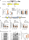
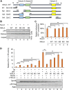
NKILA directly inhibits NF-κB-mediated HIV-1 transcription by obstructing p65 recruitment to the LTR promoter. (A) Representation of the HIV-LTR with mutations at NF-κB binding sites. (B) The effect of NKILA on the HIV-1 LTR promoter is specifically mediated by NF-κB binding sites. pHIV-1 5′ LTR WT or pHIV-1 5′ LTR mutant plasmids were cotransfected with NKILA or negative-control vector, and cells were harvested for a reporter assay 48 h posttransfection. (C) Schematic of the HIV-1 LTR primers (F1/R1, F2/R2, and F3/R3) used in the ChIP assays. (D) p65 recruitment to the LTR promoter at the NF-κB-SP1 enhancer region was disrupted by NKILA in HIV-1 NL4-3 virus-infected Jurkat cells. Jurkat cells transfected with NKILA or negative-control vector for 24 h were infected with HIV-1 NL4-3 virus for another 5 days. Fixed and isolated chromatin from HIV-1-infected Jurkat cells was immunoprecipitated with anti-p65 antibody or IgG as the negative control and was analyzed by qRT-PCR with the indicated primers spanning three different regions in the LTR promoter. The effect of NKILA on the recruitment of NFAT (E) or SP1 (F) to HIV-1 LTR. The experimental procedure is the same as for panel D. Fixed and isolated chromatin was immunoprecipitated with anti-NFAT or SP1 antibody or IgG as the negative control and then was analyzed as described for panel D. (G) The interaction of p65 and NFAT proteins with NKILA according to RNA pulldown assay. (H) NKILA interacted with NFAT and p65 proteins according to the co-IP assay. Jurkat cells were immunoprecipitated with anti-NFAT- or p65 antibody-conjugated agarose beads. NFAT and P65 proteins were detected by immunoblotting analysis. (I) The relative binding ability between NFAT or P65 with NKILA. The binding between NKILA and IgG was set as 1. **, P < 0.01; ***, P < 0.001.
FIG 6
FIG 6
NKILA mutants fail to abolish HIV-1 transcription and LTR promoter activity. (A) Schematic representation of NKILA with mutations at each of the hairpins (hairpins A, B, and C). (B) NKILA WT, the indicated mutants, or negative-control vector were cotransfected with pNL4-3ΔEnv and pVSV-G plasmids into HEK293T cells. Cells and partial supernatants were harvested for measurement of HIV-1 gene expression by IB analysis. (C) Supernatants containing virus-like particles (VLPs) were collected and used to infect TZM-bl cells. Viral infectious yield was measured by a luciferase reporter assay. (D) NKILA mutation completely abolished the ability to inhibit Tat with or without p65-mediated LTR promoter activity. NKILA WT or the indicated mutants were cotransfected with Tat or with both Tat and p65 plus pHIV-1-luciferase plasmids. Forty-eight hours posttransfection, reporter gene expression was analyzed by a luciferase reporter assay, and the expression of proteins was assessed by IB analysis. The corresponding value in the absence of Tat and p65 was set as 1 or 100% as appropriate. All results are representative of three independent experiments. The data are presented as the means ± SDs. **, P < 0.01; ***, P < 0.001.
FIG 7
FIG 7
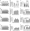
NKILA has the ability to inhibit the replication of HIV-1 clones with different coreceptor tropisms. (A) Overexpression of NKILA suppressed the replication of HIV-1 clones with different coreceptor tropisms. The NKILA or negative-control vector was cotransfected with expression plasmids for the HIV-1 clones 89.6, Yu2, or AD8. Forty-eight hours posttransfection, cells and supernatants were harvested for measurement of HIV-1 gene expression by IB analysis (A) and were analyzed by qRT-PCR to measure NKILA mRNA levels (B). (C) TZM-bl cells were infected with viral supernatants for 48 h and harvested for measurement of infectious HIV-1 yield by a luciferase assay. (D to L) NKILA mutants lost their inhibition ability toward various HIV-1 clones. NKILA WT or the indicated mutants were cotransfected with expression vectors for the HIV-1 clones AD8 (D, E, and F), 89.6 (G, H, and I), or Yu2 (J, K, and L). Cells were harvested for detection of HIV-1 gene expression by IB analysis (D, G, and J) and were analyzed by qRT-PCR to measure NKILA mRNA levels (E, H, and K). Supernatants were used to infect TZM-bl cells for measurement of the infectious yield of various HIV-1 clones (F, I, and L). The corresponding value of the control was set as 1 or 100% as appropriate. All results are representative of three independent experiments. The data are presented as the means ± SDs. **, P < 0.01; ***, P < 0.001.
FIG 8
FIG 8
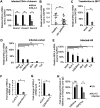
NKILA expression is inversely correlated with HIV-1 infection and production. HIV-1 infection downregulated NKILA expression in primary CD4+ T cells isolated from healthy donors (donors 1 to 3) (A), Jurkat cells (D), and H9 cells (E). Each HIV-1 strain expression vector was transfected into HEK293T cells. After 48 h, supernatants were harvested for infection of the indicated cells. The mRNA levels of NKILA in the mock- and HIV-1 NL4-3-infected primary CD4+ T (A), Jurkat (D), and H9 (E) cells were measured by qRT-PCR and normalized to the level of GAPDH. (B) The mRNA level of NKILA was decreased in primary CD4+ T cells from HIV-infected patients compared to those from healthy donors. (C) The mRNA level of NKILA was decreased by HIV-1 production. pNL4-3 or negative-control vector was transfected into HEK293T cells, and cells were harvested for analysis of NKILA expression by qRT-PCR. (F) J-Lat 6.3 cells are Jurkat cells with latent HIV-1 infection. NKILA expression in Jurkat cells with acute or latent HIV-1 infection was measured by qRT-PCR. (G) Acute infection with NL4-3 decreased the NKILA mRNA level compared to that in HIV-1 latently infected CEM cells (ACH-2), as evidenced by qRT-PCR. (H) HIV-1 infection decreased the H3K27 acetylation mark on the NKILA promoter, as revealed by anti-H3K4me and anti-H3K27ac ChIP assays in Jurkat cells showing the level of H3K27ac on the NKILA promoter following NL4-3 infection or when uninfected (UN). All results are representative of three independent experiments. The data are presented as the means ± SDs. ns, not significant; *, P < 0.05; **, P < 0.01.
FIG 9
FIG 9
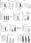
NKILA is downregulated when latent cells are activated. Reactivation of latent HIV-1 increased NKILA expression. HIV-1 latently infected J-Lat 6.3 Jurkat cells and CD4+ CEM cells (ACH-2) were utilized to measure the change in NKILA expression during viral reactivation. J-Lat 6.3 (A) or ACH-2 (C) cells were treated with or without PMA (1 μM) for 48 h and were then harvested. mRNA was extracted from the cells for measurement of NKILA expression by qRT-PCR, and the expression levels were normalized to that of GAPDH. (B) HIV-1 reactivation in J-Lat 6.3 cells was measured by detecting GFP-positive cells by flow cytometry. The corresponding value of GFP-positive cells treated with dimethyl sulfoxide (DMSO) was set as 1 or 100% as appropriate. (D) HIV-1 reactivation of ACH-2 cells was measured by IB analysis with anti-CAp24. (E) PMA treatment decreased the NKILA levels in primary CD4+ T cells from HIV-1 infected patients with cART treatment. (F to I) Knockdown of NKILA increased HIV reactivation from latency. siRNA NKILA or siRNA NC was transfected into HIV-1 latently infected C11 and J-Lat 6.3 cells. Forty-eight hours posttransfection, cells were harvested for assessment of NKILA expression by qRT-PCR (F and H) and were stimulated with PMA for 48 h for detection of the GFP-positive cells by flow cytometry (G and I). The corresponding value of GFP-positive cells treated with DMSO was set as 1 or 100% as appropriate. (J) Successful cART treatment increased NKILA expression in PBMCs of HIV-1 infected patients. PBMCs were collected from HIV-1-infected individuals pretherapy and after 48 weeks of cART therapy. mRNA was extracted from PBMCs for measurement of NKILA expression levels by qRT-PCR. (K and L) Overexpression of NKILA in CD4+ T cells caused inhibition of reactivation of HIV-1. The CD4+ cells from three patients who had undergone HAART treatment were nucleofected with NKILA or control vector. Forty-eight hours posttransfection, NKILA mRNA levels were analyzed by qRT-PCR (K), and phytohemagglutinin M (PHA-M, 5 ng/ml) was added to the cell supernatants for 7 days. (L) The supernatant p24 antigen was analyzed by ELISA. The results are representative of three independent repeats. The qRT-PCR results in panels A, C, E, F, H, J, K, and L are shown as the corresponding values, with the value in the mock group set as 100%. The data are presented as the means ± SDs. *, P < 0.05; **, P < 0.01; ***, P < 0.001 (unpaired t test).
FIG 10
FIG 10
NKILA overexpression maintains HIV-1 suppression after AZT withdrawal. (A) Experimental design for the analysis of HIV-1 replication in MT4 cells expressing NKILA or control cells. The cell lines were infected with 2 ng p24 antigen of NL4-3 viral particles. Three days postinfection, cells were treated with AZT (20 μM) or equal amounts of DMSO for 3 days and then switched to AZT-free or 20 μM AZT-containing medium for an additional 3 days. On day 9, the cells were harvested and analyzed for HIV Gag mRNA by qRT-PCR (B) and the amount of p24 antigen in the supernatant by ELISA (C).

Similar articles

Cited by

References

    1. Brooks DG, Kitchen SG, Kitchen CM, Scripture-Adams DD, Zack JA. 2001. Generation of HIV latency during thymopoiesis. Nat Med 7:459–464. doi:10.1038/86531. - DOI - PubMed
    1. Courouce AM. 1987. Latency preceding seroconversion in sexually transmitted HIV infection. Lancet 330:1025. doi:10.1016/S0140-6736(87)92587-6. - DOI - PubMed
    1. Marcello A. 2006. Latency: the hidden HIV-1 challenge. Retrovirology 3:7. doi:10.1186/1742-4690-3-7. - DOI - PMC - PubMed
    1. McCune JM. 1995. Viral latency in HIV disease. Cell 82:183–188. doi:10.1016/0092-8674(95)90305-4. - DOI - PubMed
    1. Pomerantz RJ, Trono D, Feinberg MB, Baltimore D. 1990. Cells nonproductively infected with HIV-1 exhibit an aberrant pattern of viral RNA expression: a molecular model for latency. Cell 61:1271–1276. doi:10.1016/0092-8674(90)90691-7. - DOI - PubMed

Publication types

MeSH terms

LinkOut - more resources