. 2020 Aug;13(8):e006996.
doi: 10.1161/CIRCHEARTFAILURE.120.006996. Epub 2020 Jun 24.

COP9 Signalosome Suppresses RIPK1-RIPK3-Mediated Cardiomyocyte Necroptosis in Mice

Affiliations

COP9 Signalosome Suppresses RIPK1-RIPK3-Mediated Cardiomyocyte Necroptosis in Mice

Peng Xiao et al. Circ Heart Fail. 2020 Aug.

Abstract

Background: Mechanisms governing the induction of heart failure by the impairment of autophagy and the ubiquitin-proteasome system and the molecular pathways to cardiomyocyte necrosis remain incompletely understood. COPS8 is an essential subunit of the COP9 (COnstitutive Photomorphogenesis 9) signalosome, a key regulator of ubiquitination. Mice with cardiomyocyte-restricted knockout of Cops8 (Cops8-cko) show autophagic and ubiquitin-proteasome system malfunction and massive cardiomyocyte necrosis followed by acute heart failure and premature death, providing an excellent animal model to address the mechanistic gaps specified above. This study was conducted to determine the nature and underlying mechanisms of the cardiomyocyte necrosis in Cops8-cko mice.

Methods and results: Compared with littermate control mice, myocardial protein levels of key factors in the necroptotic pathway (RIPK1 [receptor-interacting protein kinase 1], RIPK3, MLKL [mixed lineage kinase-like], the RIPK1-bound RIPK3), protein carbonyls, full-length Casp8 (caspase 8), and BCL2, as well as histochemical staining of superoxide anions were significantly higher but the cleaved Casp8 and the Casp8 activity were significantly lower in Cops8-cko mice. In vivo cardiomyocyte uptake of Evan's blue dye was used as an indicator of necrosis. Cops8-cko mice treated with a RIPK1 kinase inhibitor (Nec-1 [Necrostatin-1]) showed less Evans blue dye uptake (0.005% versus 0.20%; P<0.0001) and longer median lifespan (32.5 versus 27 days; P<0.01) than those treated with vehicle control. RIPK3 haploinsufficiency showed similar rescuing effects on Cops8-cko but Cyclophilin D deficiency did the opposite.

Conclusions: Cardiac Cops8/COP9 signalosome malfunction causes RIPK1-RIPK3 dependent, but mitochondrial permeability transition pore independent, cardiomyocyte necroptosis in mice and the COP9 signalosome plays an indispensable role in suppressing cardiomyocyte necroptosis.

Trial registration: ClinicalTrials.gov NCT04248894.

Keywords: caspase 8; mice; myocytes, cardiac; necrosis; ubiquitination.


Figures

Figure 1.
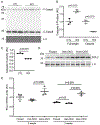
Figure 1.. Increases in myocardial RIPK1, RIPK3 and MLKL and in RIPK1-bound RIPK3 proteins in Cops8CKO mice.
A and B, Representative images (A) and pooled densitometry data (B) of western blot analysis for the indicated proteins in the myocardial extracts of 3-week-old Cops8CKO (CKO) and littermate control (CTL) mice. β-Tubulin (for RIPK1, RIPK3) and α-actinin (for MLKL) were probed as loading controls. C, Western blot (IB) analyses for RIPK1 and RIPK3 in the RIPK1 immuno-precipitates (IP) from the protein lysate of ventricular myocardium from 3-week-old CTL and Cops8CKO mice. L929 cell lysates were used as positive controls. D, RIPK1/RIPK1 ratios in the RIPK1 IP. The density of RIPK3 and RIPK1 bands for individual samples shown in panel C was used for the calculation of RIPK3 to RIPK1 ratios, the mean of the ratios of the CTL group is defined as 1 arbitrary unit (AU). The p values shown in this figure are derived from two-side unpaired t-test with Welch’s correction. Each lane and each dot represents a unique mouse.
Figure 2.
Figure 2.. Changes of myocardial reactive oxygen species (ROS) in Cops8CKO mice.
A and B, Detection of ROS in myocardial sections by dihydroethidium (DHE) staining (red). Four to six representative sections per mouse and 5 mice per genotype were analyzed. Panel A shows representative confocal fluorescence images of DHE stained myocardial sections from homozygous Cops8FL/FL, heterozygous Cops8CKO (Het-KO), and homozygous Cops8CKO (Hom-KO) mice. Scale bar=100µm. Panel B presents a scatter dot plot of individual average fluorescent intensity values for each sections, superimposed by median with 95% CI. P values are derived from the Kruskal-Walls test followed by Dunn’s pairwise comparison tests. C and D, Dot blot analyses for DNP-derivatized protein carbonyls. Equal amounts of proteins were subject to DNP-derivatization and equal proportions of the DNP-derivatized preparation were used for dot blot and subsequent immunoprobing for DNP. α-Actinin was probed as a loading control (see Supplementary Figure S1 for densitometry data). Shown are representative images (C) and pooled densitometry data (D). Mean±SEM are superimposed. Cre, Myh6-CreTG only; n = 3 mice for each group; one way ANOVA followed by Tukey’s test. E, Representative image of western blot analysis for DNP-derivatized protein carbonyls (upper image) and a-actinin (bottom). The opening curly brace demarcates the protein molecular weight range where carbonyls were increased most in the Hom-KO group. NC, negative control where equal amount of a Hom-KO myocardial protein sample that were not subject to DNP derivatization was loaded. Each lane represents a unique mouse.
Figure 3.
Figure 3.. Changes in both protein expression and activities of caspases 8 as well as BCL2 protein and mRNA levels in Cops8CKO mouse hearts.
A and B, Representative images (A) and scatter dot plots of pooled densitometry data (B) of western blot analyses for caspase 8 (Casp8). L.C., Loading control which is a portion of the image from stain-free in-gel imaging of total proteins that was used to normalize caspase 8 western blot signals. F-, full length; C-, cleaved form. C, Changes in myocardial caspase 8 activities in Cops8CKO mice at 3 weeks. CTL, littermate control; KO, homozygous Cops8CKO. D, Representative images of western blot analyses for myocardial BCL2 in homozygous Cops8FL/FL (Floxed), heterozygous Cops8CKO (Het-CKO), and homozygous Cops8CKO (Hom-CKO) mice at 3 weeks of age. E, Changes in myocardial BCL2 mRNA levels in mice at 2 and 3 weeks of age. Each scatter dot plot is superimposed by mean±SEM; each dot represents a mouse; p values are derived from unpaired t-tests with Welch’s correction (B, C) or one way ANOVA followed by Tukey’s test (E).
Figure 4.
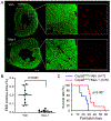
Figure 4.. Necrostatin-1 (Nec-1) treatment markedly reduces cardiomyocyte necrosis and delays premature death of Cops8CKO mice.
Cohorts of Cops8CKO mice at 2 weeks of age were treated with necrostatin-1 (Nec-1, 1.56 mg/kg/day) or vehicle (Veh) via intraperitoneal osmatic mini-pumps for 1 week (A, B) or continued for >2 weeks for the Kaplan-Meier survival analysis (C). A and B, At day 6, after min-pump implantation, mice were treated with one dose of Evan’s blue dye (EBD; 100 mg/kg, i.p.) 18 hours before they were anesthetized and perfusion-fixed in situ. Cryosections from the fixed heart were stained with Alexa488-conjugated phalloidin to identify cardiomyocytes (green) and subjected to fluorescence confocal microscopy. The images of each ventricular tissue ring were reconstructed and used for quantification of EBD-positive area (red) and total green area. Panel A shows representative reconstructed images from a pair of Cops8CKO hearts treated with Veh or Nec-1; scale bar=0.5 mm. Individual percent values of average EBD positive area in the 3 representative sections/mouse from 3 mice of each group are plotted in panel B, superimposed by median with 95% CI; Mann Whitney test. C, Kaplan-Meier survival curve of Cops8CKO mice treated with Veh or Nec-1. Nec-1 treatment significantly increased lifespan of Cops8CKO mice compared with the vehicle-treated group (median lifespan: 32.5 vs. 27 days); Log-rank Test.
Figure 5.
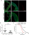
Figure 5.. RIPK3 haploinsufficiency significantly reduces cardiomyocyte necrosis and delays premature death of Cops8CKO mice.
A, Representative confocal micrographs of EBD assays. Littermate mice of the indicated genotypes at 3 weeks of age were subjected to the EBD assays in the same way as described in Figure 4. EBD positive cells display autofluorescence (red) and F-actin was stained using Alexa-488-conjugated phalloidin (green). Shown are representative composed images for the entire cross-section of the left ventricle or a higher magnification view of the marked portion of the composed image (A). Scale bar=500µm. B, Scatter dot plots of the individual percent values of EBD positive area in the 5 representative sections/mouse of 3 mice of each group, , superimposed by median with 95% CI. Mann Whitney test. C. Kaplan-Meier survival curve. The median lifespan for Cops8CKO::RIPK3+/− was significantly longer than that of Cops8CKO::RIPK3+/+ mice (31 vs. 27 days; hazard ratio=3.581). Log-Rank Test.
Figure 6.
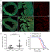
Figure 6.. Cyclophilin D knockout exacerbates cardiomyocyte necrosis and premature death of Cosp8CKO mice.
The Ppif gene encodes Cyclophilin D. Cops8CKO was induced in the Ppif wild type as well as hemizygous and homozygous null background through cross-breeding. A and B, in vivo EBD uptake assays were performed on mice at 3 weeks of age as described in Figure 4. Shown are representative confocal micrographs of myocardial sections from mice of the indicated genotypes (A) and scatter dot plot of the individual percent values of average EBD positive area in the 3 representative sections per mouse and 4 mice per group, superimposed by median with 95% CI (B). Mann Whitney test. C, Kaplan-Meier survival curves of littermate mice with the indicated genotypes. Median lifespans for the Cops8CKO::Ppif+/+ (n=13 mice), Cops8CKO::Ppif+/− (n=24), and Cops8CKO::Ppif−/− (n=24) groups were 30, 27.5 and 24.5 days, respectively. p=0.001 for all 3 groups, Log-rank test for trend; p=0.006 and hazard ratio=2.238 for Cops8CKO::Ppif−/− vs. Cops8CKO::Ppif+/+, Log-rank (Mantel-Cox) test.
Figure 7.
Figure 7.. A working model for induction of cardiomyocyte necroptosis by Cops8 deficiency.
The main interrogations of this study marked with bold black font. Loss of function of Cops8/CSN is expected to suppress the CRLs shown in green font, which in turn disrupts the respective ubiquitination events and thereby blocks NFκB-mediated survival signaling and inhibits the apoptotic pathway via increasing BCL2 and hindering the activation of caspase 8 (Casp8), steering the death receptor triggered pathway to the RIPK1-RIPK3 mediated necroptosis. The pathways in the shaded zone are likely involved but not directly examined in Cops8 deficient mice. We hypothesize that TFNα comes from autocrinal or paracrinal routes from myocardium with cardiomyocyte-restricted Cops8 knockout. Dot line denotes a potential link not tested yet. Psm, proteasomal degradation.

Similar articles

Cited by

References

    1. Wang X, Wang H. Priming the proteasome to protect against proteotoxicity. Trends Mol Med. 2020. (in press). doi: 10.1016/j.molmed.2020.02.007 - DOI - PMC - PubMed
    1. Wang X, Cui T. Autophagy modulation: A potential therapeutic approach in cardiac hypertrophy. Am J Physiol Heart Circ Physiol. 2017;313:H304–H319. - PMC - PubMed
    1. Del Re DP, Amgalan D, Linkermann A, Liu Q, Kitsis RN. Fundamental mechanisms of regulated cell death and implications for heart disease. Physiol Rev. 2019;99:1765–1817. - PMC - PubMed
    1. Choi ME, Price DR, Ryter SW, Choi AMK. Necroptosis: A crucial pathogenic mediator of human disease. JCI Insight. 2019;4:e128834. - PMC - PubMed
    1. Zhang X, Zhang H, Xu C, Li X, Li M, Wu X, Pu W, Zhou B, Wang H, Li D, Ding Q, Ying H, Wang H, Zhang H. Ubiquitination of ripk1 suppresses programmed cell death by regulating ripk1 kinase activation during embryogenesis. Nat Commun. 2019;10:4158. - PMC - PubMed

Publication types

Associated data