. 2020 Nov:93:79-94.
doi: 10.1016/j.matbio.2020.06.002. Epub 2020 Jun 17.

Mechanisms of procollagen and HSP47 sorting during ER-to-Golgi trafficking

Affiliations

Mechanisms of procollagen and HSP47 sorting during ER-to-Golgi trafficking

Shakib Omari et al. Matrix Biol. 2020 Nov.

Abstract

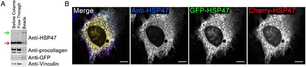
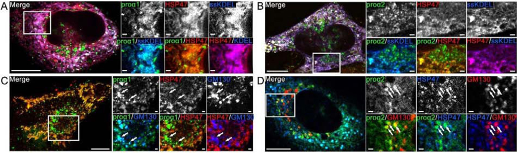
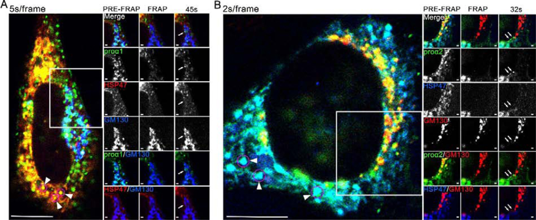
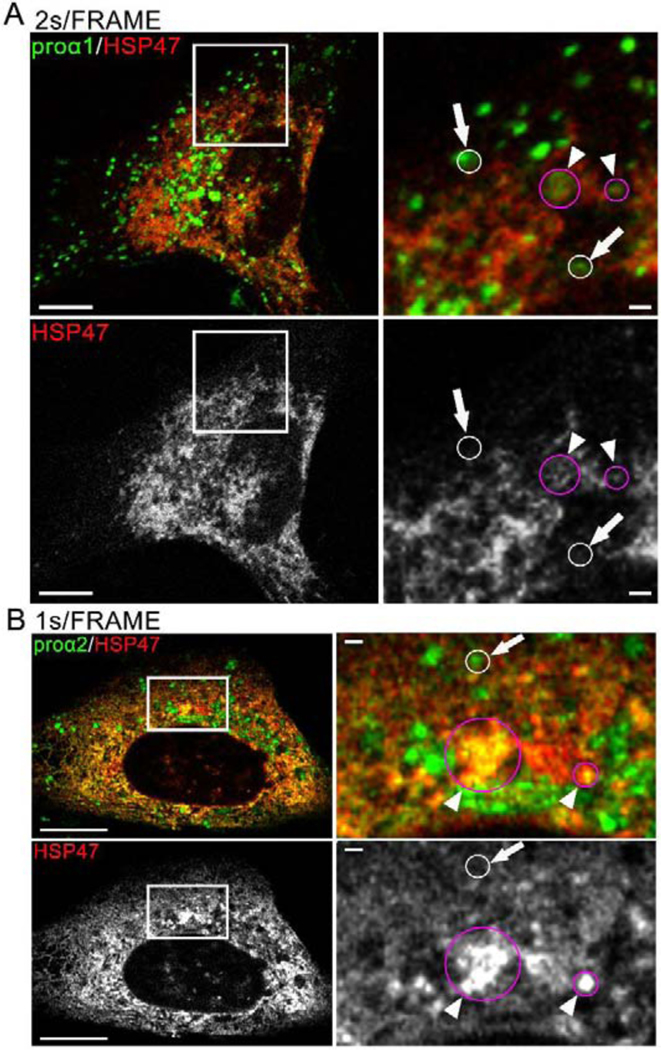
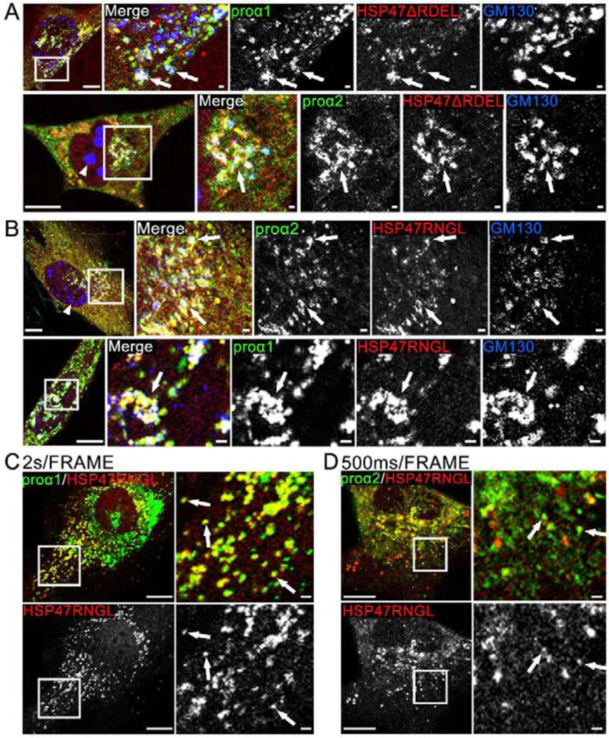
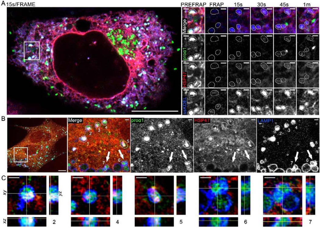
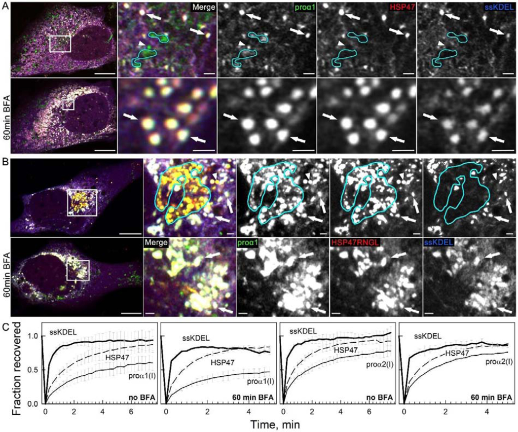
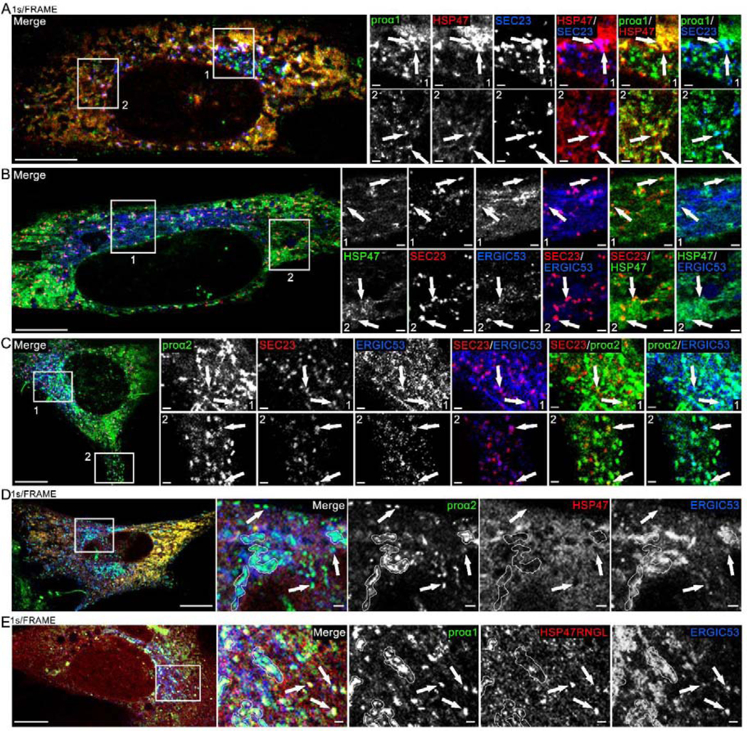
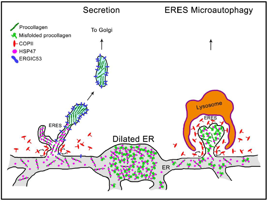
Efficient quality control and export of procollagen from the cell is crucial for extracellular matrix homeostasis, yet it is still incompletely understood. One of the debated questions is the role of a collagen-specific ER chaperone HSP47 in these processes. Most ER chaperones preferentially bind to unfolded polypeptide chains, enabling selective export of natively folded proteins from the ER after chaperone release. In contrast, HSP47 preferentially binds to the natively folded procollagen and is believed to be released only in the ER-Golgi intermediate compartment (ERGIC) or cis-Golgi. HSP47 colocalization with procollagen in punctate structures observed by immunofluorescence imaging of fixed cells has thus been interpreted as evidence for HSP47 export from the ER together with procollagen in transport vesicles destined for ERGIC or Golgi. To understand the mechanism of this co-trafficking and its physiological significance, we imaged the dynamics of fluorescently tagged type I procollagen and HSP47 punctate structures in live MC3T3 murine osteoblasts with up to 120 nm spatial and 500 ms time resolution. Contrary to the prevailing model, we discovered that most bona fide carriers delivering procollagen from ER exit sites (ERESs) to Golgi contained no HSP47, unless the RDEL signal for ER retention in HSP47 was deleted or mutated. These transport intermediates exhibited characteristic rapid, directional motion along microtubules, while puncta with colocalized HSP47 and procollagen similar to the ones described before had only limited, stochastic motion. Live cell imaging and fluorescence recovery after photobleaching revealed that the latter puncta (including the ones induced by ARF1 inhibition) were dilated regions of ER lumen, ERESs, or autophagic structures surrounded by lysosomal membranes. Procollagen was colocalized with HSP47 and ERGIC53 at ERESs. It was colocalized with ERGIC53 but not HSP47 in Golgi-bound transport intermediates. Our results suggest that procollagen and HSP47 sorting occurs at ERES before procollagen is exported from the ER in Golgi-bound transport intermediates, providing new insights into mechanisms of procollagen trafficking.

Keywords: Collagen; ER exit sites; HSP47; live cell imaging; trafficking.


Conflict of interest statement

Declaration of Competing Interests The authors have no competing interests.

Figures

Fig. 1.
Fig. 1.. Fluorescent protein-tagged HSP47 (FP-HSP47) interacts with procollagen and colocalizes with endogenous HSP47.
(A) Western blots of cell lysates before and after IP of GFP-HSP47 with Anti-GFP beads. Green arrow shows transfected GFP-HSP47; red arrow shows endogenous HSP47. Significant enrichment of procollagen relative to vinculin on the beads revealed that it co-immunoprecipitated with GFP-HSP47. Since the estimated procollagen concentration in cell lysates was lower than 1 μM and HSP47 binds procollagen with Kd ~ 1–10 μM [30], the co-immunoprecipitation indicated minimal or no disruption of HSP47-procollagen interaction by the fluorescent tag in GFP-HSP47. Because multiple endogenous HSP47 and GFP-HSP47 molecules would be bound to the same procollagen triple helix, a significant amount of endogenous HSP47 was found on the Anti-GFP beads. (B) Confocal imaging of colocalization between two FP-HSP47 constructs co-transfected into MC3T3 cells and HSP47 antibodies that label both transfected and endogenous HSP47. All spots labeled with anti-HSP47 were also positive for GFP-HSP47 and Cherry-HSP47, indicating complete colocalization of transfected and endogenous molecules in all subcellular compartments. Variations in relative fluorescence intensity of different tags are affected by antibody accessibility, uneven photobleaching during sample preparation and imaging, and other uncontrolled factors. Here and throughout the paper, different channels in multichannel images are merged using RGB pseudo color scheme and the corresponding color labels for individual channels. The scale bar is 10 μm; N=6 cells were examined.
Fig. 2.
Fig. 2.. FP-HSP47 colocalizes with procollagen in the ER but does not colocalize with the cis-Golgi marker GM130.
(A,B) MC3T3 cells transfected with Cherry-HSP47, Venus-proα1(I) (A) or Venus-proα2(I) (B), and marker of ER lumen (ssCFP-KDEL). (C,D) MC3T3 cells transfected with Cherry-HSP47 (C) or TagBFP2-HSP47 (D), Venus-proα1(I) (C) or GFP-proα2(I) (D) and marker of cis-Golgi CFP-GM130 (C) or Cherry-GM130 (D). Switching of the fluorescent tag colors in different images illustrates that each of these and subsequent experiments were performed with different combinations of fluorescent tag colors, to eliminate potential tag-specific effects. Arrows mark Golgi structures containing procollagen. All images are Airyscan (A,B) or confocal (C,D) single slices; scale bars = 10 μm (full cell) and 1 μm (zoomed areas); N≥20 (≥4 experiments with different FP combinations) for each panel.
Fig. 3,
Fig. 3,. Movies 1,2. FP-HSP47 is not found in FP-procollagen-positive transport vesicles entering cis-Golgi.
(A, Movie 1) Individual frames and time-lapse video of a cell transfected with Cherry-HSP47, Venus-proα1(I) and CFP-GM130 before and after Cherry-HSP47 and Venus-proα1(I) were photobleached from Golgi region. (B, Movie 2) Frames and time-lapse video of a cell transfected with TagBFP2-HSP47, GFP-proα2(I) and Cherry-GM130 before and after TagBFP2-HSP47 and GFP-proα2(I) were photobleached from Golgi region. Arrows in post-FRAP frames (A, B) mark Golgi-destined procollagen transport vesicles identified by time-lapse imagining (white circles in Movies 1,2). Arrowheads in pre-FRAP whole-cell images point to some of the vesicle-like procollagen/HSP47 structures (magenta circles) that exhibit limited stochastic motion. The images are confocal single slices; scale bars = 10 μm (whole cell) and = 1 μm (zoom); N≥15 (3 experiments with different FP combinations) for each panel. (Movie 2) White circle outlines the ER-to-Golgi movement of 3 procollagen transport vesicles, none of which contain HSP47.
Fig. 4,
Fig. 4,. Movies 3,4. Dynamics of vesicle-like FP-HSP47 structures resembles stochastic fluctuations of the reticular ER network but not rapid directional movement of FP-procollagen transport vesicles.
(A, Movie 3) Confocal single slice frame and 2s/frame time-lapse video of a cell transfected with GFP-proα1(I) and Apple-HSP47. (B, Movie 4) Airyscan single slice frame and 1 s/frame video of a cell transfected with Venus-proα2(I) and Cherry-HSP47. Some of procollagen transport vesicles identified by rapid directional movement are outlined by white circles in the time-lapse videos and also marked by arrows in the still frames. These transport intermediates contain procollagen but not HSP47. Stochastically moving vesicle-like structures containing procollagen and HSP47 are outlined by magenta circles in the videos and also marked by arrowheads in the still frames. Scale bars = 10 μm (whole cell) and = 1 μm (zoom); N>70 (≥14 experiments with different FP combinations) for each panel.
Fig. 5,
Fig. 5,. Movies 6,7. Deletion of RDEL and RDEL→RNGL mutation release HSP47 from the ER to cis-Golgi, causing Golgi disruption.
(A) Airyscan colocalization of Cherry-HSP47ΔRDEL with Venus-proα1(I)/α2(I) and cis-Golgi marker CFP-GM130 (white arrows); N=4 (proα1(I)) and N=4 (proα2(I)). Based on imaging of several z-slices, large CFP-GM130 spheres (arrowhead) are located inside the nuclei. At least some Golgi fragmentation was noticeable in all cells transfected with Cherry-HSP47ΔRDEL. (B) Airyscan colocalization of Cherry-HSP47RNGL with Venus-proα1(I)/α2(I) and cis-Golgi marker CFP-GM130 (white arrows); N=19 (proα1(I)) and N=7 (proα2(I)). In these cells, Golgi fragmentation was less pronounced. The nuclear CFP-GM130 spheres (arrowhead) were smaller and absent from some of the cells. (C, Movie 6) Confocal single slice frame and time-lapse video of Apple-HSP47RNGL colocalization with GFP-proα1(I)in transport vesicles marked by arrows in the still frame; N=63 (14 experiments). (D, Movie 7) Airyscan single slice frame and video of similar colocalization of Cherry-HSP47RNGL with Venus-proα2(I); N=18 (3 experiments). Scale bars = 10 μm (whole cell) and = 1 μm (zoom).
Fig. 6,
Fig. 6,. Movie 8. Vesicle-like structures containing procollagen and HSP47 inside ER lumen and lysosomal membranes.
(A, Movie 8) Airyscan single slice frames and time-lapse video of colocalization of Venus-proα1(I), Cherry-HSP47, and ssCFP-KDEL in vesicle-like structures inside ER lumen and fluorescence recovery of these markers after photobleaching (FRAP); N=18 (3 experiments). Zoomed still frames show the same area as the inset in Movie 8. In these zoomed frames, 3 vesicle-like structures marked by Venus-proα1(I), Cherry-HSP47, and ssCFP-KDEL are traced by thin white lines. One of these structures is also highlighted by a white circle in the movie inset. The bright green structure in 45 s and 1 min frames is likely a procollagen transport vesicle containing no Cherry-HSP47 and no ssCFP-KDEL, which entered the field of view between 30 and 45 s post-bleaching (the 15s/frame imaging rate in this experiment was not fast enough for more definitive identification). Quantitative analysis of FRAP kinetics in shown in Fig. 7C with and without BFA treatment. (B, C) 3D Airyscan imaging of Venus-proα1(I) and Cherry-HSP47 colocalization in lysosomes/late endosomes marked by membrane protein LAMP1-CFP; N=7. (B) A slice from the 3D z-stack shows whole cell and zoomed images of vesicle-like structures 1–7, in which Venus-proα1(I) is colocalized with LAMP1-CFP. (C) Orthogonal cross-sections of structures 2, 4, 5, 6, and 7. Structures 2, 4, and 5 were likely formed by direct lysosomal engulfment of ERES [37] or ER lumen [44], since they are surrounded by LAMP1-positive lysosomal membrane and contain internalized procollagen, HSP47, and LAMP1. Lysosomes 6 and 7 contain little or no internalized HSP47 and thereby might have a different origin, e.g. endocytosis of secreted procollagen molecules. Scale bars = 10 μm (whole cell) and = 1 μm (zoom).
Fig. 7,
Fig. 7,. Movie 9. Golgi disruption by brefeldin A (BFA) causes accumulation of vesicle-like regions of ER lumen filled with procollagen and HSP47, which were previously misinterpreted as procollagen transport vesicles.
(A) Colocalization of Venus-proα1(I), Cherry-HSP47, and ssCFP-KDEL in vesicle-like structures inside ER lumen (white puncta in merged images marked by arrows) and accumulation of these structures after 60 min treatment of the same cell with 5 μg/ml BFA (bottom panels); N=9. Golgi cisternae (outlined by cyan lines) and likely transport vesicle (arrowhead) contain Venus-proα1(I) but not Cherry-HSP47 or ssCFP-KDEL. They disappear rather than accumulate after BFA treatment. (Movie 9) Time-lapse video of similar Venus-proα1(I)/Cherry-HSP47/ssCFP-KDEL puncta in a different cell after BFA treatment shows nearly instantaneous ssCFP-KDEL fluorescence recovery after photobleaching, demonstrating that these puncta are integrated within the ER lumen network. (B) Similar accumulation of Venus-proα1(I), Cherry-HSP47RNGL, and ssCFP-KDEL in vesicle-like white puncta (arrows) inside ER lumen after 60 min treatment with 5 μg/ml BFA; N=8. Golgi cisternae (outlined in cyan) and likely transport vesicle (arrowhead) contain Venus-proα1(I) and Cherry-HSP47RNGL but not ssCFP-KDEL. They disappear after BFA treatment. (C) FRAP kinetics in vesicle-like structures containing colocalized ssCFP-KDEL, Venus-proα1(I)/proα2(I), and Cherry-HSP47; N=3. The images in (A,B) are Airyscan single slices; scale bars = 10 μm (whole cell) and = 1 μm (zoom).
Fig. 8,
Fig. 8,. Movies 10–12. HSP47 entry into ERES and release at ERES regions marked by ERGIC53.
(A, Movie 10) Airyscan single slice frame and time-lapse video of GFP-proα1(I), Cherry-HSP47, and TagBFP2-SEC23 colocalization in punctate structures representing ERES (arrows) in the Golgi region (Movie inset and zoomed still frame in top panels) and away from the Golgi region (zoomed still frame in bottom panels). Based on larger size and slower motion, procollagen carrier appearing at 00:28 s frame in the movie inset is more likely a secretory vesicle than ER-Golgi transport intermediate. N=20 (3 experiments). (B) Airyscan slice of similar colocalization of Venus-HSP47, Halo-SEC23 and Cerulean-ERGIC53 at ERES (arrows); N=6. (C) Airyscan slice of similar colocalization of Venus-proα2(I), Halo-SEC23, and Cerulean-ERGIC53 at ERES (arrows); N=22 (5 experiments). (D, Movie 11) Airyscan single slice frame and time-lapse video of Venus-proα2(I) and Cerulean-ERGIC53 colocalization without Cherry-HSP47 in procollagen transport vesicles (arrows) and Golgi (white outlines); N=4. (E, Movie 12) Airyscan single slice frame and time-lapse video of Venus-proα1(I), Cherry-HSP47RNGL, and Cerulean-ERGIC53 colocalization in procollagen transport vesicles (arrows) and Golgi (white outlines); N=17 (3 experiments). Scale bars = 10 μm (whole cell) and = 1 μm (zoom).
Fig. 9.
Fig. 9.. Model of HSP47 and procollagen sorting at ER exit sites (ERES).
Grey structure at the bottom represents interconnected network of rough ER cisternae. Procollagen is loaded into ERES together with HSP47, as indicated by colocalization of procollagen, HSP47, and SEC23 (Fig. 8A, Movie 10). Maturation of procollagen transport intermediate precursors at distal ERES is accompanied by HSP47 release and accumulation of ERGIC53, since both ERGIC53 and HSP47 colocalize with procollagen at ERES and only ERGIC53 colocalizes with procollagen in Golgi-bound transport intermediates (Fig. 8, Movies 10–12). Unless its RDEL sequence is deleted or mutated, HSP47 is found in < 1±3% ERGIC53/procollagen transport intermediates, indicating that HSP47 is primarily returned to the ER from ERES (Supp. Fig. 7C). Only few escaping HSP47 molecules are likely to be captured by cis-Golgi KDEL/RDEL receptors and returned to the ER by retrograde trafficking. As reported before, ERESs containing misfolded procollagen and HSP47 might be engulfed by lysosomes and degraded (ERES microautophagy pathway) [37]. Under a microscope, procollagen ERESs (left), dilated ER regions (middle), and ERES microautophagy intermediates (right) appear as either small puncta or larger vesicle-like structures containing procollagen and HSP47 (Figs. 6 and 7, see also Figs. 1–7 in Ref. [37]). They accumulate upon inhibition of procollagen export from the ER by brefeldin A and are easy to confuse with transport vesicles, unless their motion and/or composition is imaged.

Similar articles

Cited by

References

    1. Raote I, Malhotra V, Protein transport by vesicles and tunnels, J Cell Biol 218(3) (2019) 737–739. - PMC - PubMed
    1. McCaughey J, Stephens DJ, ER-to-Golgi Transport: A Sizeable Problem, Trends Cell Biol 29(12) (2019) 940–953. - PubMed
    1. Hutchings J, Zanetti G, Coat flexibility in the secretory pathway: a role in transport of bulky cargoes, Curr Opin Cell Biol 59 (2019) 104–111. - PMC - PubMed
    1. McGourty CA, Akopian D, Walsh C, Gorur A, Werner A, Schekman R, Bautista D, Rape M, Regulation of the CUL3 Ubiquitin Ligase by a Calcium-Dependent Co-adaptor, Cell 167(2) (2016) 525–538 e14. - PubMed
    1. Jin L, Pahuja KB, Wickliffe KE, Gorur A, Baumgartel C, Schekman R, Rape M, Ubiquitin-dependent regulation of COPII coat size and function, Nature 482(7386) (2012) 495–500. - PMC - PubMed

Publication types

LinkOut - more resources