Myasthenia Gravis: From the Viewpoint of Pathogenicity Focusing on Acetylcholine Receptor Clustering, Trans-Synaptic Homeostasis and Synaptic Stability
- PMID: 32547365
- PMCID: PMC7272578
- DOI: 10.3389/fnmol.2020.00086
Myasthenia Gravis: From the Viewpoint of Pathogenicity Focusing on Acetylcholine Receptor Clustering, Trans-Synaptic Homeostasis and Synaptic Stability
Abstract
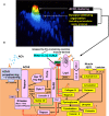
Myasthenia gravis (MG) is a disease of the postsynaptic neuromuscular junction (NMJ) where nicotinic acetylcholine (ACh) receptors (AChRs) are targeted by autoantibodies. Search for other pathogenic antigens has detected the antibodies against muscle-specific tyrosine kinase (MuSK) and low-density lipoprotein-related protein 4 (Lrp4), both causing pre- and post-synaptic impairments. Agrin is also suspected as a fourth pathogen. In a complex NMJ organization centering on MuSK: (1) the Wnt non-canonical pathway through the Wnt-Lrp4-MuSK cysteine-rich domain (CRD)-Dishevelled (Dvl, scaffold protein) signaling acts to form AChR prepatterning with axonal guidance; (2) the neural agrin-Lrp4-MuSK (Ig1/2 domains) signaling acts to form rapsyn-anchored AChR clusters at the innervated stage of muscle; (3) adaptor protein Dok-7 acts on MuSK activation for AChR clustering from "inside" and also on cytoskeleton to stabilize AChR clusters by the downstream effector Sorbs1/2; (4) the trans-synaptic retrograde signaling contributes to the presynaptic organization via: (i) Wnt-MuSK CRD-Dvl-β catenin-Slit 2 pathway; (ii) Lrp4; and (iii) laminins. The presynaptic Ca2+ homeostasis conditioning ACh release is modified by autoreceptors such as M1-type muscarinic AChR and A2A adenosine receptors. The post-synaptic structure is stabilized by: (i) laminin-network including the muscle-derived agrin; (ii) the extracellular matrix proteins (including collagen Q/perlecan and biglycan which link to MuSK Ig1 domain and CRD); and (iii) the dystrophin-associated glycoprotein complex. The study on MuSK ectodomains (Ig1/2 domains and CRD) recognized by antibodies suggested that the MuSK antibodies were pathologically heterogeneous due to their binding to multiple functional domains. Focussing one of the matrix proteins, biglycan which functions in the manner similar to collagen Q, our antibody assay showed the negative result in MG patients. However, the synaptic stability may be impaired by antibodies against MuSK ectodomains because of the linkage of biglycan with MuSK Ig1 domain and CRD. The pathogenic diversity of MG is discussed based on NMJ signaling molecules.
Keywords: Lrp4; MuSK; acetylcholine receptor; agrin; matrix proteins; myasthenia gravis; neuromuscular junction; wnts.
Copyright © 2020 Takamori.
Figures

Similar articles
-
Roles of collagen Q in MuSK antibody-positive myasthenia gravis.Chem Biol Interact. 2016 Nov 25;259(Pt B):266-270. doi: 10.1016/j.cbi.2016.04.019. Epub 2016 Apr 24. Chem Biol Interact. 2016. PMID: 27119269 Review.
-
Collagen Q and anti-MuSK autoantibody competitively suppress agrin/LRP4/MuSK signaling.Sci Rep. 2015 Sep 10;5:13928. doi: 10.1038/srep13928. Sci Rep. 2015. PMID: 26355076 Free PMC article.
-
MuSK myasthenia gravis IgG4 disrupts the interaction of LRP4 with MuSK but both IgG4 and IgG1-3 can disperse preformed agrin-independent AChR clusters.PLoS One. 2013 Nov 7;8(11):e80695. doi: 10.1371/journal.pone.0080695. eCollection 2013. PLoS One. 2013. PMID: 24244707 Free PMC article.
-
[Genetic defects and disorders at the neuromuscular junction].Brain Nerve. 2011 Jul;63(7):669-78. Brain Nerve. 2011. PMID: 21747136 Review. Japanese.
-
Antibodies against Wnt receptor of muscle-specific tyrosine kinase in myasthenia gravis.J Neuroimmunol. 2013 Jan 15;254(1-2):183-6. doi: 10.1016/j.jneuroim.2012.09.001. Epub 2012 Sep 18. J Neuroimmunol. 2013. PMID: 22999188
Cited by
-
Integrating Network Pharmacology and Component Analysis to Study the Potential Mechanisms of Qi-Fu-Yin Decoction in Treating Alzheimer's Disease.Drug Des Devel Ther. 2023 Sep 14;17:2841-2858. doi: 10.2147/DDDT.S402624. eCollection 2023. Drug Des Devel Ther. 2023. PMID: 37727255 Free PMC article.
-
The Neuromuscular Junction in Health and Disease: Molecular Mechanisms Governing Synaptic Formation and Homeostasis.Front Mol Neurosci. 2020 Dec 3;13:610964. doi: 10.3389/fnmol.2020.610964. eCollection 2020. Front Mol Neurosci. 2020. PMID: 33343299 Free PMC article. Review.
-
Pathophysiology of Childhood-Onset Myasthenia: Abnormalities of Neuromuscular Junction and Autoimmunity and Its Background.Pathophysiology. 2023 Dec 2;30(4):599-617. doi: 10.3390/pathophysiology30040043. Pathophysiology. 2023. PMID: 38133144 Free PMC article. Review.
-
Narrative review of immunotherapy in thymic malignancies.Transl Lung Cancer Res. 2021 Jun;10(6):3001-3013. doi: 10.21037/tlcr-20-1222. Transl Lung Cancer Res. 2021. PMID: 34295693 Free PMC article. Review.
-
Treating myasthenia gravis beyond the eye clinic.Eye (Lond). 2024 Aug;38(12):2422-2436. doi: 10.1038/s41433-024-03133-x. Epub 2024 May 24. Eye (Lond). 2024. PMID: 38789789 Free PMC article. Review.
References
LinkOut - more resources
Full Text Sources
Miscellaneous

