. 2020 Jul 23;5(14):e136496.
doi: 10.1172/jci.insight.136496.

Chronic-plus-binge alcohol intake induces production of proinflammatory mtDNA-enriched extracellular vesicles and steatohepatitis via ASK1/p38MAPKα-dependent mechanisms

Affiliations

Chronic-plus-binge alcohol intake induces production of proinflammatory mtDNA-enriched extracellular vesicles and steatohepatitis via ASK1/p38MAPKα-dependent mechanisms

Jing Ma et al. JCI Insight. .

Abstract

Alcohol-associated liver disease is a spectrum of liver disorders with histopathological changes ranging from simple steatosis to steatohepatitis, cirrhosis, and hepatocellular carcinoma. Recent data suggest that chronic-plus-binge ethanol intake induces steatohepatitis by promoting release by hepatocytes of proinflammatory mitochondrial DNA-enriched (mtDNA-enriched) extracellular vesicles (EVs). The aim of the present study was to investigate the role of the stress kinase apoptosis signal-regulating kinase 1 (ASK1) and p38 mitogen-activated protein kinase (p38) in chronic-plus-binge ethanol-induced steatohepatitis and mtDNA-enriched EV release. Microarray analysis revealed the greatest hepatic upregulation of metallothionein 1 and 2 (Mt1/2), which encode 2 of the most potent antioxidant proteins. Genetic deletion of the Mt1 and Mt2 genes aggravated ethanol-induced liver injury, as evidenced by elevation of serum ALT, neutrophil infiltration, oxidative stress, and ASK1/p38 activation in the liver. Inhibition or genetic deletion of Ask1 or p38 ameliorated ethanol-induced liver injury, inflammation, ROS levels, and expression of phagocytic oxidase and ER stress markers in the liver. In addition, inhibition of ASK1 or p38 also attenuated ethanol-induced mtDNA-enriched EV secretion from hepatocytes. Taken together, these findings indicate that induction of hepatic mtDNA-enriched EVs by ethanol is dependent on ASK1 and p38, thereby promoting alcoholic steatohepatitis.

Keywords: Hepatology; Toxicology.


Conflict of interest statement

Conflict of interest: The authors have declared that no conflict of interest exists.

Figures

Figure 1
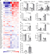
Figure 1. Mt1/2–/– mice are more susceptible to chronic-plus-binge ethanol–induced liver injury.
(A) C57BL/6N mice were fed an ethanol diet for 8 weeks (E8w) or 8 weeks plus 1 binge (E8w+1B). Liver tissues were collected 9 hours after binge in the E8w+1B group and subjected to microarray analysis. A heat map of oxidative stress–related genes is shown. (B) C57BL/6N mice were pair-fed with control diet (pair-fed, n = 4) or fed an ethanol diet for 8 weeks (E8w), or E8w+1B, and were euthanized 9 hours after binge. C57BL/6N mice were fed a control diet (pair-fed) or ethanol diet for 10 days plus 1 binge (E10d+1B) and were euthanized 3, 6, or 9 hours later. Hepatic mRNA levels of Mt1 and Mt2 were examined by RT-qPCR. (CF) WT and Mt1/2–/– mice were pair-fed or fed an ethanol diet for 10 days, followed by gavage of a single dose of maltose or ethanol, respectively and were euthanized 9 hours later. Serum ALT levels and hepatic TG levels were measured (C). Hepatic neutrophils were examined by IHC staining with an anti-MPO antibody or RT-qPCR analysis of Ly6g mRNA (D). Hepatic macrophages were examined by immunostaining with an anti-F4/80 antibody or RT-qPCR analysis of F4/80 mRNA (E). Liver tissues were also subjected to TUNEL staining, and the TUNEL+ hepatocytes were counted (F). Values represent mean ± SEM (each dot represents 1 mouse sample). Statistical evaluation was performed by Student’s t test or 1-way ANOVA with Tukey’s post hoc test for multiple comparisons. *P < 0.05, **P < 0.01, ***P < 0.001.
Figure 2
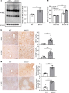
Figure 2. Deletion of the Mt1/2 gene enhances oxidative stress and ASK1/p38 MAPK activation in the E10d+1B model.
WT and Mt1/2–/– mice were fed an ethanol-containing diet for 10 days, followed by gavage of ethanol, and were euthanized 9 hours later. (A) Liver tissues from ethanol-fed mice were subjected to immunoblot analysis of 4-HNE. Quantification of 4-HNE was normalized to β-actin. (B) Liver tissues were subjected to TBARS assay for determination of MDA levels. (C) Liver tissues were subjected to IHC staining with antibodies against MDA and 4-HNE. Representative images (scale bars: 100 μm) and quantification of the area positive for each staining are shown. (D) Liver tissues were subjected to immunostaining with antibody against p-ASK1 or p-p38. Representative images (scale bars: 200 μm) and quantification of the positive area or density for each staining are shown. Values represent mean ± SEM (each dot represents 1 mouse sample). Statistical evaluation was performed by Student’s t test or 1-way ANOVA with Tukey’s post hoc test for multiple comparisons. *P < 0.05, **P < 0.01, ***P < 0.001.
Figure 3
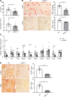
Figure 3. Deletion of ASK1 ameliorates chronic-plus-binge ethanol–induced liver injury.
WT and Ask1–/– mice were subjected to E10d+1B feeding and were euthanized at 9 hours after gavage. (A) Serum ALT levels and circulating neutrophil percentage were measured. (B) Liver tissues were subjected to IHC staining with anti-MDA and anti–4-HNE antibodies. Representative images (scale bars: 100 μm) and quantification of areas positive for staining are shown. Values represent mean ± SEM (each dot represents 1 mouse sample). Statistical evaluation was performed by Student’s t test. *P < 0.05, **P < 0.01.
Figure 4
Figure 4. Inhibition of ASK1 ameliorates chronic-plus-binge ethanol–induced hepatic inflammation.
C57BL/6J mice were subjected to E10d+1B feeding and received i.p. injection of ASK1 inhibitors (3 mg/kg GS-4997 [GS+E] or 4 mg/kg NQDI-1 ,NQ+E]) 30 minutes before gavage. Mice were euthanized 9 hours after gavage. Veh, vehicle. (A) Serum ALT levels and circulating neutrophils were quantified. (B) Hepatic neutrophils and macrophages were examined by IHC staining with anti-MPO or anti-F4/80 antibody or by RT-qPCR analysis of Ly6g and F4/80 mRNA. Representative images (scale bars: 100 μm) and quantification of the area positive for each staining are shown. Values represent mean ± SEM (each dot represents 1 mouse sample). Arrowheads indicate MPO+ cells. Statistical evaluation was performed by 1-way ANOVA with Tukey’s post hoc test for multiple comparisons. *P < 0.05, **P < 0.01, ***P < 0.001.
Figure 5
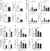
Figure 5. Inhibition of ASK1 ameliorates chronic-plus-binge ethanol–induced hepatic oxidative stress and ER stress.
C57BL/6J mice were subjected to E10d+1B feeding and received i.p. injection of ASK1 inhibitors (3 mg/kg GS-4997 or 4 mg/kg NQDI-1) 30 minutes before gavage. Mice were euthanized 9 hours after gavage. (A) Liver tissues were subjected to immunostaining with antibody against MDA or 4-HNE. Representative images (scale bars: 100 μm) and quantification of the area positive for each staining are shown. (B) Western blot analysis of CHOP, BCL2, p-JNK, JNK, and β-actin in the liver. Values represent mean ± SEM (each dot represents one mouse sample). Statistical evaluation was performed by 1-way ANOVA with Tukey’s post hoc test for multiple comparisons. **P < 0.01, ***P < 0.001.
Figure 6
Figure 6. Hepatocyte-specific deletion of the p38a gene ameliorates chronic-plus-binge ethanol–induced liver injury.
WT and p38aHep–/– mice were subjected to E10d+1B feeding and were euthanized 9 hours after gavage. (A) Serum ALT levels and circulating neutrophils were quantified. (B) Liver tissues were subjected to IHC staining with antibodies against MPO and F4/80. Representative images (scale bars: 200 μm) and quantification of MPO+ cells and F4/80+ area (%) are shown. (C) Liver tissues were subjected to RT-qPCR analysis of inflammatory cytokine genes. (D) Liver tissues were subjected to immunostaining with antibodies against MDA and 4-HNE. Representative images (scale bars: 200 μm) and quantification of the area positive for each staining are shown. Values represent mean ± SEM (each dot represents 1 mouse sample). Statistical evaluation was performed by Student’s t test. *P < 0.05, ***P < 0.001.
Figure 7
Figure 7. Treatment with p38 inhibitors ameliorates chronic-plus-binge ethanol–induced hepatic inflammation.
C57BL/6J mice were subjected to E10d+1B feeding and received i.p. injection of p38 inhibitors (3 mg/kg LY2228820 [LY+E], 12 mg/kg PH797804 [PH+E], or 10 mg/kg SB239063 [SB+E]) 30 minutes before gavage. Mice were euthanized 9 hours after gavage. (A) Serum ALT levels and circulating neutrophils were measured. (B) Liver tissues were subjected to IHC staining with antibody against MPO or F4/80. Representative images are shown (scale bars: 100 μm). (C) RT-qPCR was performed to analyze hepatic expression of proinflammatory mediators. Values represent mean ± SEM (each dot represents 1 mouse sample). Statistical evaluation was performed by 1-way ANOVA with Tukey’s post hoc test for multiple comparisons. *P < 0.05, **P < 0.01, ***P < 0.001.
Figure 8
Figure 8. Treatment with p38 inhibitors ameliorates chronic-plus-binge ethanol–induced hepatic oxidative stress and ER-stress.
C57BL/6J mice were subjected to E10d+1B feeding and received i.p. injection of p38 inhibitors (3 mg/kg LY2228820, 12 mg/kg PH797804, or 10 mg/kg SB239063) 30 minutes before gavage. Mice were euthanized 9 hours after gavage. (A) Liver tissues were subjected to immunostaining with antibody against MDA and 4-HNE. Representative images (scale bars: 100 μm) and quantification of the area positive for each staining are shown. (B) Western blot analysis of CHOP, BIP, p-eIF2α, total eIF2α, and β-actin in the liver. Values represent mean ± SEM (each dot represents 1 mouse sample). Statistical evaluation was performed by 1-way ANOVA with Tukey’s post hoc test for multiple comparisons. *P < 0.05, ***P < 0.001.
Figure 9
Figure 9. ASK1/p38 pathway activation contributes to elevated release of mtDNA enriched EVs in E10d+1B model.
(A) WT, Ask1–/–, and p38aHep–/– mice were subjected to E10+1B ethanol feeding and were euthanized 9 hours after gavage. Serum EVs were isolated and quantified by exosome quantitation assay. Cytochrome c oxidase (Cyto c ox) DNA levels in EVs were measured. (B) C57BL/6J mice were subjected to E10d+1B feeding and received i.p. injection of ASK1 or p38 inhibitors 30 minutes before gavage. Mice were euthanized 9 hours after gavage. Serum EVs were isolated and counted by exosome quantitation assay. Cyto c ox DNA levels in EVs were measured. (C) Hepatocytes were isolated from WT, Ask1–/–, or p38aHep–/– mice and treated with ethanol (100 mM) or PBS for 24 hours. EVs were isolated from culture medium and counted by exosome quantitation assay. (D and E) Hepatocytes were isolated from WT mice and treated with ethanol (100 mM) or ethanol plus ASK1 or p38 inhibitors for 24 hours. EVs were isolated from culture medium and counted by exosome quantitation assay. Cyto c ox DNA levels in the EVs were measured. Values represent mean ± SEM (each dot represents 1 mouse sample). Statistical evaluation was performed by Student’s t test or 1-way ANOVA with Tukey’s post hoc test for multiple comparisons. *P < 0.05, **P < 0.01, ***P < 0.001.
Figure 10
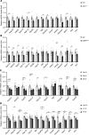
Figure 10. Inhibition of ASK1 or p38 activation attenuates expression of the genes related to EV biogenesis and secretion in the E10d+1B model.
(A and B) WT and Ask1–/– or p38aHep–/– mice were subjected to E10d+1B feeding and euthanized 9 hours after gavage. Liver tissues were subjected to RT-qPCR analysis of EV biogenesis– and secretion–related genes. (C and D) C57BL/6J mice were subjected to E10d+1B feeding and received i.p. injection of ASK1 inhibitor (3 mg/kg GS-4997 or 4 mg/kg NQDI-1) or p38 inhibitor (3 mg/kg LY2228820 or 12 mg/kg PH797804) 30 minutes before gavage. Mice were euthanized 9 hours after ethanol gavage Liver tissues were subjected to RT-qPCR analysis of EV biogenesis– and secretion– related genes. Values represent mean ± SEM (each dot represents 1 mouse sample). Statistical evaluation was performed by Student’s t test or 1-way ANOVA with Tukey’s post hoc test for multiple comparisons. *P < 0.05, **P < 0.01, ***P < 0.001.

Similar articles

Cited by

References

    1. Gao B, Bataller R. Alcoholic liver disease: pathogenesis and new therapeutic targets. Gastroenterology. 2011;141(5):1572–1585. doi: 10.1053/j.gastro.2011.09.002. - DOI - PMC - PubMed
    1. Szabo G, Kamath PS, Shah VH, Thursz M, Mathurin P, EASL-AASLD Joint Meeting Alcohol-related liver disease: areas of consensus, unmet needs and opportunities for further study. Hepatology. 2019;69(5):2271–2283. doi: 10.1002/hep.30369. - DOI - PubMed
    1. Crabb DW, Im GY, Szabo G, Mellinger JL, Lucey MR. Diagnosis and treatment of alcohol-associated liver diseases: 2019 practice guidance from the American Association for the Study of Liver Diseases. Hepatology. 2020;71(1):306–333. doi: 10.1002/hep.30866. - DOI - PubMed
    1. Bertola A, Mathews S, Ki SH, Wang H, Gao B. Mouse model of chronic and binge ethanol feeding (the NIAAA model) Nat Protoc. 2013;8(3):627–637. doi: 10.1038/nprot.2013.032. - DOI - PMC - PubMed
    1. Lazaro R, et al. Osteopontin deficiency does not prevent but promotes alcoholic neutrophilic hepatitis in mice. Hepatology. 2015;61(1):129–140. doi: 10.1002/hep.27383. - DOI - PMC - PubMed

Publication types

MeSH terms