

Molecular mechanisms of splenectomy-induced hepatocyte proliferation
- PMID: 32531779
- PMCID: PMC7292681
- DOI: 10.1371/journal.pone.0233767
Molecular mechanisms of splenectomy-induced hepatocyte proliferation
Abstract
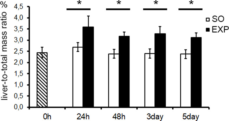
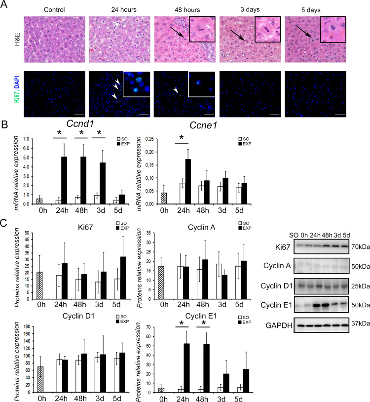
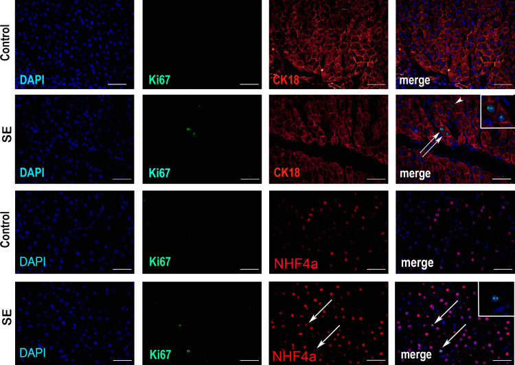
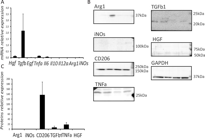
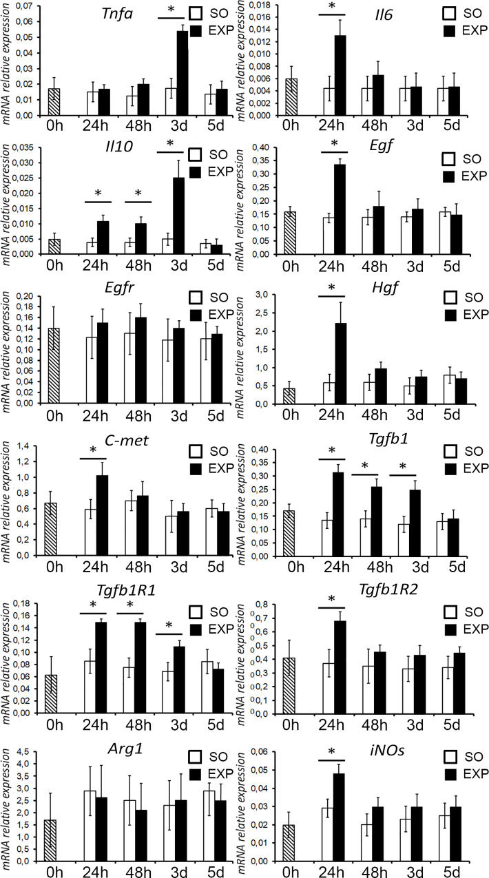
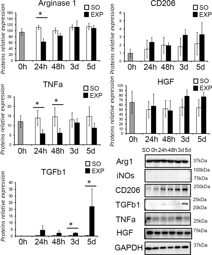
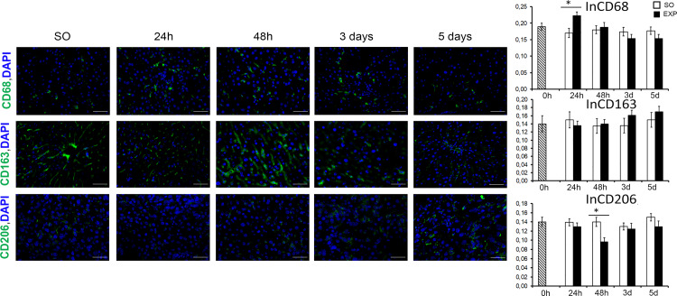
Functional and anatomical connection between the liver and the spleen is most clearly manifested in various pathological conditions of the liver (cirrhosis, hepatitis). The mechanisms of the interaction between the two organs are still poorly understood, as there have been practically no studies on the influence exerted by the spleen on the normal liver. Mature male Sprague-Dawley rats of 250-260 g body weight, 3 months old, were splenectomized. The highest numbers of Ki67+ hepatocytes in the liver of splenectomized rats were observed at 24 h after the surgery, simultaneously with the highest index of Ki67-positive hepatocytes. After surgical removal of the spleen, expression of certain genes in the liver tissues increased. A number of genes were upregulated in the liver at a single time point of 24 h, including Ccne1, Egf, Tnfa, Il6, Hgf, Met, Tgfb1r2 and Nos2. The expression of Ccnd1, Tgfb1, Tgfb1r1 and Il10 in the liver was upregulated over the course of 3 days after splenectomy. Monitoring of the liver macrophage populations in splenectomized animals revealed a statistically significant increase in the proportion of CD68-positive cells in the liver (as compared with sham-operated controls) detectable at 24 h and 48 h after the surgery. The difference in the liver content of CD68-positive cells between splenectomized and sham-operated animals evened out by day 3 after the surgery. No alterations in the liver content of CD163-positive cells were observed in the experiments. A decrease in the proportion of CD206-positive liver macrophages was observed at 48 h after splenectomy. The splenectomy-induced hepatocyte proliferation is described by us for the first time. Mechanistically, the effect is apparently induced by the removal of spleen as a major source of Tgfb1 (hepatocyte growth inhibitor) and subsequently supported by activation of proliferation factor-encoding genes in the liver.
Conflict of interest statement
The authors have declared that no competing interests exist.
Figures
Similar articles
-
Splenectomy enhances the therapeutic effect of adipose tissue-derived mesenchymal stem cell infusion on cirrhosis rats.Liver Int. 2016 Aug;36(8):1151-9. doi: 10.1111/liv.12962. Epub 2015 Oct 1. Liver Int. 2016. PMID: 26353075
-
Role of the spleen in liver fibrosis in rats may be mediated by transforming growth factor beta-1.J Gastroenterol Hepatol. 2002 Jan;17(1):59-65. doi: 10.1046/j.1440-1746.2002.02667.x. J Gastroenterol Hepatol. 2002. PMID: 11895554
-
Role of the spleen in liver regeneration in relation to transforming growth factor-β1 and hepatocyte growth factor.J Surg Res. 2015 Jun 15;196(2):270-7. doi: 10.1016/j.jss.2015.02.025. Epub 2015 Feb 19. J Surg Res. 2015. PMID: 25862490
-
The Spleen Promotes the Secretion of CCL2 and Supports an M1 Dominant Phenotype in Hepatic Macrophages During Liver Fibrosis.Cell Physiol Biochem. 2018;51(2):557-574. doi: 10.1159/000495276. Epub 2018 Nov 20. Cell Physiol Biochem. 2018. PMID: 30458454
-
Cooperation of liver cells in health and disease.Adv Anat Embryol Cell Biol. 2001;161:III-XIII, 1-151. doi: 10.1007/978-3-642-56553-3. Adv Anat Embryol Cell Biol. 2001. PMID: 11729749 Review.
Cited by
-
An Eye on Kupffer Cells: Development, Phenotype and the Macrophage Niche.Int J Mol Sci. 2022 Aug 30;23(17):9868. doi: 10.3390/ijms23179868. Int J Mol Sci. 2022. PMID: 36077265 Free PMC article. Review.
-
Spleen: Reparative Regeneration and Influence on Liver.Life (Basel). 2022 Apr 22;12(5):626. doi: 10.3390/life12050626. Life (Basel). 2022. PMID: 35629294 Free PMC article. Review.
-
Splenectomy ameliorates liver cirrhosis by restoring the gut microbiota balance.Cell Mol Life Sci. 2024 Jan 12;81(1):32. doi: 10.1007/s00018-023-05055-5. Cell Mol Life Sci. 2024. PMID: 38214780 Free PMC article.
-
Spleen regeneration after subcutaneous heterotopic autotransplantation in a mouse model.Biol Res. 2023 Mar 29;56(1):15. doi: 10.1186/s40659-023-00427-4. Biol Res. 2023. PMID: 36991509 Free PMC article.
-
Comparative Analysis of the Transcriptome, Proteome, and miRNA Profile of Kupffer Cells and Monocytes.Biomedicines. 2020 Dec 18;8(12):627. doi: 10.3390/biomedicines8120627. Biomedicines. 2020. PMID: 33352881 Free PMC article.
References
Publication types
MeSH terms
Substances
Grants and funding
LinkOut - more resources
Full Text Sources
Research Materials
Miscellaneous
