. 2020 Jun 2;10(1):8912.
doi: 10.1038/s41598-020-65914-0.

Topographic distinction in long-term value signals between presumed dopamine neurons and presumed striatal projection neurons in behaving monkeys

Affiliations

Topographic distinction in long-term value signals between presumed dopamine neurons and presumed striatal projection neurons in behaving monkeys

Kazuki Enomoto et al. Sci Rep. .

Abstract

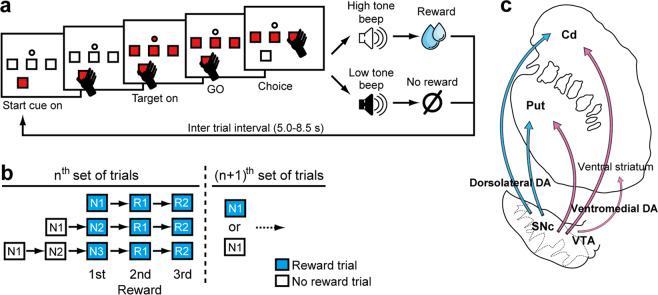
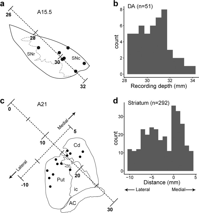
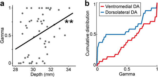
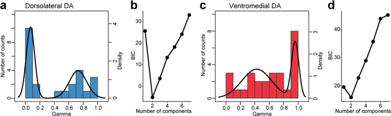
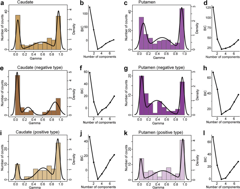
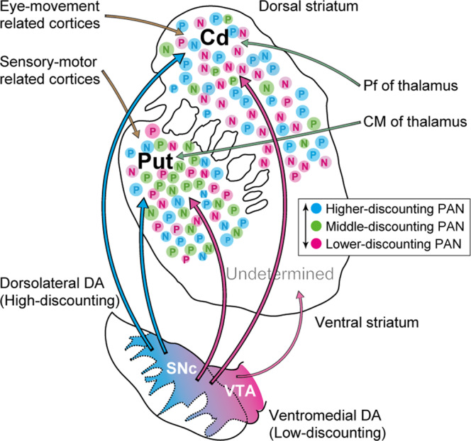
Nigrostriatal dopamine (DA) projections are anatomically organized along the dorsolateral-ventromedial axis, conveying long-term value signals to the striatum for shaping actions toward multiple future rewards. The present study examines whether the topographic organization of long-term value signals are observed upon activity of presumed DA neurons and presumed striatal projection neurons (phasically active neurons, PANs), as predicted based on anatomical literature. Our results indicate that DA neurons in the dorsolateral midbrain encode long-term value signals on a short timescale, while ventromedial midbrain DA neurons encode such signals on a relatively longer timescale. Activity of the PANs in the dorsal striatum is more heterogeneous for encoding long-term values, although significant differences in long-term value signals were observed between the caudate nucleus and putamen. These findings suggest that topographic DA signals for long-term values are not simply transferred to striatal neurons, possibly due to the contribution of other projections to the striatum.


Conflict of interest statement

The authors declare no competing interests.

Figures

Figure 1
Figure 1
Multi-step choice task. (a) Sequence of events in a single trial. Monkeys chose one of three targets in a trial to find one rewarding target. Following the ITI, monkeys chose a target again based on the reward and no-reward outcomes in the previous trials. (b) Schematic drawing of a series of choices to obtain multiple rewards. After finding a rewarding target, monkeys were required to repeat the rewarded choices in the first and second repeat trials (R1 and R2). Monkeys obtained two (monkey RO, no R2 trials) or three rewards (monkeys SK, CC, and TN) in a series of trials. (c) Schematic drawing of the nigrostriatal projection. Anatomical projections from the midbrain to the striatum based on Haber et al., 2000 are shown.
Figure 2
Figure 2
Recording locations of DA neurons and PANs. (a) Recording sites of DA neurons (black dots) in a representative coronal section reconstructed histologically at 15.5 mm anterior (A15.5). Depth from the cortical surface was represented along the electrode insertion. SNr, substantia nigra pars reticulata; SNc, substantia nigra pars compacta. (b) Numbers of DA neurons at each recording depth. (c) Recording sites of PANs in a representative coronal section reconstructed histologically at 21 mm anterior (A21). Depth from the cortical surface was represented along the electrode insertion. Mediolateral location relative to the edge of the caudate nucleus was represented along the axis orthogonal to the electrode insertion. Cd, caudate nucleus; Put, putamen; ic, internal capsule; AC, anterior commissure. (d) Numbers of PANs at each mediolateral location. See Supplementary Fig. 2 for more detail.
Figure 3
Figure 3
Representative activity of DA neurons in the ventromedial and dorsolateral parts of the midbrain. (a) Raster plots and histograms of DA neuron activity recorded from the ventromedial part of the midbrain after the start cue (left panel), reward beep (center panel), and no-reward beep (right panel). Firing rates relative to baseline activity are shown. Spikes were sorted according to trial as follows: N1 (orange), N2 (cyan), N3 (magenta), R1 (light green), and R2 (dark green). Hatched gray areas represent the time windows used to measure response amplitude in each histogram. (b) Response amplitude of the example neuron shown in (a) after the start cue, reward beep, and no-reward beep for each trial type (bar graph, mean and SE). Superimposed line plots indicate the long-term value (start cue) and the prediction errors of long-term values (reward and no-reward beeps) estimated based on the best-fit γ value (see Materials and Methods). A single best-fit γ value was estimated against responses to start cue, reward beep, and no-reward beep simultaneously. (c,d) Same as (a,b) but for a DA neuron recorded from the dorsolateral part of the midbrain.
Figure 4
Figure 4
Ventromedial DA neurons encode multiple future rewards on a longer timescale than dorsolateral ones. (a) Scatter plot of the estimated γ value against recording depth in each DA neuron. Regression line is presented in black. Asterisks indicate the significance of the regression coefficient (**p < 0.01). (b) Cumulative distributions of the estimated γ values in dorsolateral (blue) and ventromedial (red) DA neurons.
Figure 5
Figure 5
PANs in the caudate nucleus encode future rewards on shorter and longer timescales than PANs in the putamen. (a) Cumulative distributions of the estimated γ values in positive-coding (gray) and negative-coding type PANs (black). (b) Same as (a) but for PANs in the caudate nucleus (positive-coding type, yellow; negative-coding type, brown) and putamen (positive-coding type, light purple; negative-coding type, dark purple). (c) Scatter plot of the estimated γ value against mediolateral recording axis in the caudate nucleus and putamen. Regression lines are presented for positive- (gray) and negative-coding type PANs (black) in each of the caudate nucleus and putamen, respectively.
Figure 6
Figure 6
Long-term value signals encoded by DA neurons are composed of two subgroups. (a) Distribution of γ values for dorsolateral DA neurons (bar graph). The best-fitting model is indicated by the black line. (b) Plots of the estimated Bayesian information criterion (BIC) for the data in (a) for each model, which included one to seven components. (c,d) Same as (a,b) but for ventromedial DA neurons.
Figure 7
Figure 7
Long-term value signals encoded by PANs are composed of three subgroups. (a) Distribution of γ values for PANs in the caudate nucleus (bar graph). The best-fitting model is indicated by the black line. b) Plots of the estimated Bayesian information criterion (BIC) for the data in (a) for each model, which included one to seven components. (c,d) Same as (a,b) but for PANs in the putamen. (e,f) Same as (a,b) but for negative-coding type PANs in the caudate nucleus. (g,h) Same as (a,b) but for negative-coding type PANs in the putamen. i,j) Same as (a,b) but for positive-coding type PANs in the caudate nucleus. (k,l) Same as (a,b) but for positive-coding type PANs in the putamen.
Figure 8
Figure 8
Schematic drawing of the nigrostriatal projection as well as the topography of long-term reward value coding in the midbrain and striatum. Anatomical projections from the midbrain to the striatum based on Haber et al., 2000 and the discount factor observed in the present study are shown. PANs representing long-term values with high (cyan), medium (purple), and low (magenta) discounting of future rewards are shown by dots. DA neurons representing long-term values with dorsolateral-ventromedial arrangement are shown by filled colors. Discount factor in the ventral striatum was not examined. Centromedian (CM) and parafascicular (Pf) nuclei of the thalamus; Cd, caudate nucleus; Put, putamen; VTA, ventral tegmental area.

Similar articles

Cited by

References

    1. Björklund A, Dunnett SB. Dopamine neuron systems in the brain: an update. Trends Neurosci. 2007;30:194–202. - PubMed
    1. Schultz W. Neuronal Reward and Decision Signals: From Theories to Data. Physiol. Rev. 2015;95:853–951. - PMC - PubMed
    1. Watabe-Uchida M, Eshel N, Uchida N. Neural Circuitry of Reward Prediction Error. Annu. Rev. Neurosci. 2017;40:373–394. - PMC - PubMed
    1. Bromberg-Martin ES, Matsumoto M, Nakahara H, Hikosaka O. Multiple Timescales of Memory in Lateral Habenula and Dopamine Neurons. Neuron. 2010;67:499–510. - PMC - PubMed
    1. Enomoto K, et al. Dopamine neurons learn to encode the long-term value of multiple future rewards. Proc. Natl. Acad. Sci. 2011;108:15462–15467. - PMC - PubMed

Publication types