. 2020 Jun 1;10(1):8877.
doi: 10.1038/s41598-020-65469-0.

Mouse intestinal tuft cells express advillin but not villin

Affiliations

Mouse intestinal tuft cells express advillin but not villin

Amin Esmaeilniakooshkghazi et al. Sci Rep. .

Abstract

Tuft (or brush) cells are solitary chemosensory cells scattered throughout the epithelia of the respiratory and alimentary tract. The actin-binding protein villin (Vil1) is used as a marker of tuft cells and the villin promoter is frequently used to drive expression of the Cre recombinase in tuft cells. While there is widespread agreement about the expression of villin in tuft cells there are several disagreements related to tuft cell lineage commitment and function. We now show that many of these inconsistencies could be resolved by our surprising finding that intestinal tuft cells, in fact, do not express villin protein. Furthermore, we show that a related actin-binding protein, advillin which shares 75% homology with villin, has a tuft cell restricted expression in the gastrointestinal epithelium. Our study identifies advillin as a marker of tuft cells and provides a mechanism for driving gene expression in tuft cells but not in other epithelial cells of the gastrointestinal tract. Our findings fundamentally change the way we identify and study intestinal tuft cells.


Conflict of interest statement

The authors declare no competing interests.

Figures
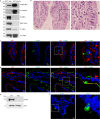
Figure 1
Figure 1
Mouse intestinal cells that express advillin do not express villin. (a) Specificity of villin and advillin antibodies was confirmed by Western analysis using recombinant full-length human villin and human advillin proteins. Two antibodies raised against the amino-terminus of villin (N-villin and N20-villin) did not cross-react with advillin compared to antibodies raised against the carboxyl-terminus of villin (C-villin) or full-length villin (FL-villin) protein. Antibodies raised against the amino-terminus of advillin (N-advillin) and those raised against the carboxyl-terminus of advillin (C-advillin) detect advillin better than they cross-react with villin. Original full length data are available in supplementary Fig S1. (b) H & E staining of mouse distal ileum identifies solitary cells with candle-like “tufted morphology” (identified by arrowhead). Right panel shows higher magnification of the boxed area. (c) Immunohistochemistry of paraffin embedded tissue from distal ileum of C57BL/6 J mice using N-villin (red) and advillin (green) antibodies. Nuclei are counter stained with DAPI. N-villin antibody does not cross-react with advillin in cells that express advillin in apical tufts and along the basolateral surface. Right panel shows higher magnification of boxed area. (d) Immunohistochemistry of cryopreserved tissue from distal ileum of C57BL/6 J mice using N-villin (red) and advillin (green) antibodies. Nuclei are counter stained with DAPI. N-villin does not co-localize with advillin in cells that express advillin in apical tufts and along the basolateral surface. Right panel shows higher magnification of boxed area. (e) Western analysis of distal ileal tissue from villin knockout (VKO) mice and their wild type (WT) littermates show the absence of villin protein in VKO mice. Original full length data are available in supplementary Fig S2. (f) Immunohistochemistry of paraffin embedded tissue from distal ileum of villin knockout mice identify advillin but not villin expressing cells. Nuclei are counter stained with DAPI. Right panel shows higher magnification of boxed area. Data shown in Western blots are representative of three independent experiments and in immunohistochemistry of n = 5 animals. Scale bars represent 10 μm.
Figure 2
Figure 2
Intestinal tuft cells express advillin. (a) Immunohistochemistry of paraffin-embedded tissue from distal ileum of C57BL/6 J mice using advillin (green) and DCLK1 (red) antibodies. Nuclei are counter stained with DAPI. Both proteins co-localize in the same cell. Note that not all DCLK1 positive cells are positive for advillin expression. Right panel shows higher magnification of boxed area and shows co-localization of DCLK1 and advillin in a tuft cell. (b) Immunohistochemistry of paraffin embedded tissue from distal ileum of C57BL/6 J mice using advillin (green) and PTGS1 (red) antibodies. Nuclei are counter stained with DAPI. Right panel shows higher magnification of boxed area and co-localization of PTGS1 and advillin in a tuft cell. (c) Immunohistochemistry of cryopreserved tissue from distal ileum of C57BL/6 J mice using Alexa Fluor 568 Phalloidin (red) and advillin (green). Nuclei are counter stained with DAPI. Right panel shows higher magnification of boxed area and co-localization of advillin and F-actin at the apical surface of a tuft cell. Data shown are representative of n = 5 animals. Scale bar represents 10 μm.
Figure 3
Figure 3
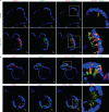
In intestinal enteroids, tuft cells express advillin. (a) Immuno-histochemistry of enteroids from distal ileum of C57BL/6 J mice shows tuft cell hyperplasia 72 hours post IL-4 and IL-13 treatment. Control refers to untreated enteroids from C57BL/6 J mice. Advillin (green) and DCLK1 (red) co-localization was used to identify tuft cells. Nuclei are counter stained with DAPI. Right panel shows higher magnification of the boxed area. (b) Immunohistochemistry of IL-4 and IL-13 treated enteroids from distal ileum of C57BL/6 J mice, show co-localization in tuft cells of advillin (green) and DCLK1 (red) in the upper panel; and advillin and PTGS1 (red) in the lower panel. Nuclei were counter stained with DAPI. Right panels show higher magnification of boxed area. Data shown are representative of n = 5 animals. Scale bar represents 10 μm.
Figure 4
Figure 4
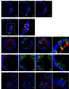
Tuft cells positive for advillin are negative for villin. (a) Immunohistochemistry and Virtual Channel confocal microscopy of IL-4 and IL-13 treated enteroids from distal ileum of C57BL/6 J mice, show co-localization of advillin (green), DCLK1 (red) and PTGS1 (magenta) within the same cells identified as tuft cells. Nuclei are counter stained with DAPI. Right panel shows higher magnification of boxed area. (b) Immunohistochemistry of IL-4 and IL-13 treated enteroids from distal ileum of C57BL/6 J mice show co-localization in tuft cells of advillin (green) with F-actin (red) in the upper panel; and co-localization of advillin (red) and tubulin (green) in the lower panel. Nuclei were counter stained with DAPI. Right panel shows higher magnification of boxed area. Note the co-localization of advillin to the apical tuft that terminates near the perinuclear region of the cells. Advillin appears in vesiculated structures that co-localize with tubulin near the lateral cell surface and in the cytoplasm. (c) Immunohistochemistry of IL-4 and IL-13 treated enteroids from distal ileum of C57BL/6 J mice show the absence of villin (red) from advillin (green) expressing tuft cells. Nuclei are counter stained with DAPI. Right panel shows higher magnification of boxed area. Data shown are representative of n = 5 animals. Scale bar represents 10 μm.
Figure 5
Figure 5
Regulation of actin dynamics by villin and advillin. (a) Villin and advillin bind F-actin. Recombinant villin and advillin proteins (60 nM) were incubated with 3 μM F-actin and centrifuged at 200,000 x g for 15 min. The supernatant (S) precipitated with 2 volumes of acetone and the pellet (P) were analyzed by SDS-PAGE and gels were stained with GelCode Blue. Control refers to assay in the absence of either protein. (b) Villin and advillin bundle F-actin. Recombinant villin and advillin proteins (1 μM) were incubated with F-actin and centrifuged at 10,000 x g for 15 min. The supernatant (S) and pellet (P) fractions were analyzed by SDS-PAGE and gels were stained with GelCode Blue. Control refers to assay in the absence of either protein. (c) Villin and advillin (60 nM each) nucleate actin filaments. Pyrene-labeled G-actin (6 μM) was polymerized in the presence of villin or advillin protein and fluorescence intensity measured over time. (d) Villin and advillin (60 nM each) cap actin filaments. Polymerization of 1.4 μM pyrene-labeled G-actin in the presence of F-actin seeds (290 nM) was measured in the presence of villin or advillin protein. (e) Villin and advillin sever actin filaments. Pyrene-labeled F-actin (1 μM) was diluted to 0.1 μM in actin-polymerizing buffer in the presence of villin or advillin protein (60 nM each) and the decrease in fluorescence intensity was followed over time. All gels are representative of three experiments with similar results. In all actin kinetic assays, values represent the mean of three independent experiments.

Similar articles

Cited by

References

    1. Von Moltke J. Intestinal tuft cells. Physiology of the Gastrointestinal Tract. 2018;Chapter 31:721–733. doi: 10.1016/B978-0-12-809954-4.00031-1. - DOI
    1. Isomaki AM. A new cell type (tuft cell) in the gastrointestinal mucosa of the rat. A transmission and scanning electron microscopic study. Acta Pathol Microbiol Scand A. 1973;Suppl 240:241–235. - PubMed
    1. Hofer D, Drenckhahn D. Cytoskeletal markers allowing discrimination between brush cells and other epithelial cells of the gut including enteroendocrine cells. Histochem Cell Biol. 1996;105:405–412. doi: 10.1007/BF01463662. - DOI - PubMed
    1. Allan EM. The ultrastructure of the brush cell in bovine lung. Res Vet Sci. 1978;25:314–317. doi: 10.1016/S0034-5288(18)32946-1. - DOI - PubMed
    1. Hofer D, Drenckhahn D. Cytoskeletal markers allowing discrimination between brush cells and other epithelial cells of the gut including enteroendocrine cells. Histochem Cell Biol. 1996;105:405–412. doi: 10.1007/BF01463662. - DOI - PubMed

Publication types