Review
. 2020 May 27;12(6):587.
doi: 10.3390/v12060587.

Structural Insights into APOBEC3-Mediated Lentiviral Restriction

Affiliations
Review

Structural Insights into APOBEC3-Mediated Lentiviral Restriction

Krista A Delviks-Frankenberry et al. Viruses. .

Abstract

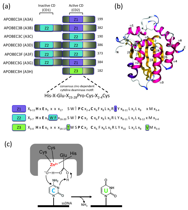
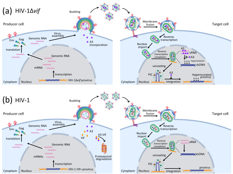
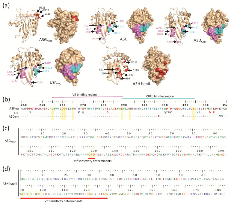
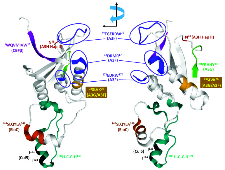
Mammals have developed clever adaptive and innate immune defense mechanisms to protect against invading bacterial and viral pathogens. Human innate immunity is continuously evolving to expand the repertoire of restriction factors and one such family of intrinsic restriction factors is the APOBEC3 (A3) family of cytidine deaminases. The coordinated expression of seven members of the A3 family of cytidine deaminases provides intrinsic immunity against numerous foreign infectious agents and protects the host from exogenous retroviruses and endogenous retroelements. Four members of the A3 proteins-A3G, A3F, A3H, and A3D-restrict HIV-1 in the absence of virion infectivity factor (Vif); their incorporation into progeny virions is a prerequisite for cytidine deaminase-dependent and -independent activities that inhibit viral replication in the host target cell. HIV-1 encodes Vif, an accessory protein that antagonizes A3 proteins by targeting them for polyubiquitination and subsequent proteasomal degradation in the virus producing cells. In this review, we summarize our current understanding of the role of human A3 proteins as barriers against HIV-1 infection, how Vif overcomes their antiviral activity, and highlight recent structural and functional insights into A3-mediated restriction of lentiviruses.

Keywords: APOBEC3G; CBFβ; Vif; cullin 5; cytidine deamination; elongin b and c; hypermutation; nucleic acid binding; proteasomal degradation; restriction factor; substrate selection.


Conflict of interest statement

The authors declare no conflicts of interest.

Figures

Figure 1
Figure 1
A3 domain organization. (a) Zinc-coordinating domains of A3A/B/C/D/F/G/H. A3 gene duplications have given rise to A3A/C/H with single Z-domains and A3B/D/F/G with two Z-domains. Numbers correspond to amino acid sequence length for each A3. Both the catalytically inactive and active Z domains contain the consensus motif H-X-E-X23-28-P-C-X2-4-C, which is structurally defined as starting at the loop3/α-helix 2 junction. The amino acid similarity between the three Z domains (Z1, Z2, Z3) is shown, each with their distinctive amino acids highlighted. X represents any amino acid. (b) Crystal structure of A3 cytidine deaminase (CD) domain. Five β strands (yellow), six α helices (pink), and zinc atom (black) of the A3G catalytic domain are shown (PDB: 3IR2). (c) Mechanism of cytidine deamination proceeds by a direct nucleophilic attack on the C4 pyrimidine ring. In the catalytic site, cysteines and histidine together coordinate a zinc atom (Zn2+), whereby a water molecule is hydrolyzed, donating a proton to the catalytic glutamate and forming a hydroxyl group. The glutamate acts as a proton shuffle during the catalysis, converting cytidine to uridine.
Figure 2
Figure 2
A3 protein-mediated HIV-1 restriction and its counteraction by Vif. (a) A3G inhibition of HIV-1Δvif. In the absence of a functional Vif protein (HIV-1Δvif), A3G is packaged into HIV-1 virions in the producer cells, exerting its antiviral activity in the target cells by inducing lethal hypermutation, inhibiting reverse transcription and blocking integration. Reverse transcription and pre-integration complex (PIC) formation occurs within mostly intact viral cores in the cytoplasm and the nucleus [48]. (b) Vif induces degradation of A3 proteins. In the presence of wild-type HIV-1, expression of Vif in the producer cells targets A3 proteins for proteasomal degradation, leading to productive HIV-1 infection of target cells. Created with BioRender.com.
Figure 3
Figure 3
Comparison of A3GNTD, A3C, A3DCTD, A3FCTD, and A3H hap II structures. (a) Cartoon and surface representations of the structure of A3GNTD (PDB: 2MZZ), A3C (PDB: 3VOW), and A3DCTD (a predicted model generated through homology modeling based on the A3FCTD structure (PDB: 6NIL) by using the Phyre2 web portal for protein modeling, prediction and analysis [140], and A3H hap II (PDB: 6BBO). The known Vif-mediated degradation determinants labeled in red (for A3G and A3H), the critical residues for Vif-binding in magenta (for A3C, A3D, and A3F), and the CBFβ binding residues in cyan (for A3C, A3D, A3F). (b) Multiple sequence alignment of A3 domains (A3FCTD, A3C, A3DCTD) and the critical Vif- and CBFβ-binding residues are highlighted in yellow bars. A3F was used as a reference and hence the numbering is based on A3F’s residue numbers. The determinants for Vif- and CBFβ-binding are in non-overlapping regions, which are indicated above the alignment. (c,d) Similar primary sequences of residues in A3GNTD and A3H hap II are shown, respectively. The residues highlighted in yellow bars are identified to be critical for Vif-mediated degradation of the respective A3 proteins. The sequence alignment was performed by a Clustal W multiple alignment in BioEdit.
Figure 4
Figure 4
Domains of Vif involved in interactions with A3 proteins and CRL5 E3 ubiquitin ligase. Only the structure of Vif, in complex with CBFβ, Cul5, and EloB/C, is shown (PDB: 4N9F). Residues involved in interactions with A3 proteins are shown using Rasmol software (www.rasmol.org). Residues involved in interaction with A3F, A3G, and A3H hap II are shown in purple, green, and red, respectively. Residues that are involved in interaction with both A3G and A3F are shown in gold. Residues involved in interactions with CBFβ, EloC, and Cul5 are shown in magenta, brown, and black, respectively.
Figure 5
Figure 5
Ternary A3FCTD-Vif-CBFβ complex and the role of CBFβ-Vif dimer in recruiting A3FCTD to the CRL5 E3 ubiquitin ligase complex. (a) Overall structure of A3FCTD-Vif–CBFβ ternary complex. Vif (magenta) and CBFβ (cyan) form a shallow wedge-like platform for A3FCTD (orange) to dock primarily via the α2-helix on Vif and the α3- and α4-helical surface on CBFβ. (b) Detailed illustrations of the A3F-CBFβ interface and interacting residues that form two distinct electrostatic interfaces; the first involves A3F (E289 and R293), CBFβ (E54), and Vif (K50); the second involves A3F (E324) and CBFβ (E35/E43). (c) Detailed illustrations of the A3F-Vif interface and interacting residues. The extensive hydrophobic and electrostatic A3F-Vif interactions are indicated with dashed lines and all critical residues involved in the interactions are shown in stick form.
Figure 6
Figure 6
A3 deamination and ssDNA interactions. (a) Co-crystal structure of A3GCTD in complex with ssDNA substrate 5′-AATCCCAAA-3′ (PDB 6BUX). Shown is the three-dimensional fold of the A3GCTD with α-helices (purple) and β-sheets (yellow) with the ssDNA (red) co-ordinated with the reactive cytosine (C0) positioned next to the catalytic zinc atom. Loops 1, 3, 5, and 7, which influence substrate binding, specificity, and dinucleotide preference selection, are labeled. (b) Co-crystal of A3H in complex with duplex RNA substrate 5′-UAAAAAAA-3′ + 5′-UUUUUUUUU-3′ with a 1 nucleotide overhang (PDB 6B0B). Shown are α-helices (purple) and β-sheets (yellow) with the duplex RNA (red and blue) positioned with zinc atom. (c) Preferred ssDNA substrates of the A3 enzymes where the underlined C is the preferred target nucleotide of the deamination reaction. (d) Summary of the interactions of the 5′-TCCCA target ssDNA substrate with amino acids in A3GCTD (PBD 6BUX). A3G loop 1 contains amino acids W211, R213, R215, H216; A3G loop 3 contains amino acids N244, H257; A3G α-helix 2 contains amino acids A258 and E259; A3G loop 7 contains amino acids Y315, D316, D317, Q318 and A3G α-helix 6 contains amino acid R374.

Similar articles

Cited by

References

    1. Conticello S.G. The AID/APOBEC family of nucleic acid mutators. Genome Biol. 2008;9:229. doi: 10.1186/gb-2008-9-6-229. - DOI - PMC - PubMed
    1. Desimmie B.A., Delviks-Frankenberrry K.A., Burdick R.C., Qi D., Izumi T., Pathak V.K. Multiple APOBEC3 restriction factors for HIV-1 and one Vif to rule them all. J. Mol. Biol. 2014;426:1220–1245. doi: 10.1016/j.jmb.2013.10.033. - DOI - PMC - PubMed
    1. Stenglein M.D., Burns M.B., Li M., Lengyel J., Harris R.S. APOBEC3 proteins mediate the clearance of foreign DNA from human cells. Nat. Struct. Mol. Biol. 2010;17:222–229. doi: 10.1038/nsmb.1744. - DOI - PMC - PubMed
    1. Jarmuz A., Chester A., Bayliss J., Gisbourne J., Dunham I., Scott J., Navaratnam N. An anthropoid-specific locus of orphan C to U RNA-editing enzymes on chromosome 22. Genomics. 2002;79:285–296. doi: 10.1006/geno.2002.6718. - DOI - PubMed
    1. Muramatsu M., Kinoshita K., Fagarasan S., Yamada S., Shinkai Y., Honjo T. Class switch recombination and hypermutation require activation-induced cytidine deaminase (AID), a potential RNA editing enzyme. Cell. 2000;102:553–563. doi: 10.1016/S0092-8674(00)00078-7. - DOI - PubMed

Publication types

LinkOut - more resources