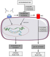
Vascular Signaling in Allogenic Solid Organ Transplantation - The Role of Endothelial Cells
- PMID: 32457653
- PMCID: PMC7227440
- DOI: 10.3389/fphys.2020.00443
Vascular Signaling in Allogenic Solid Organ Transplantation - The Role of Endothelial Cells
Abstract
Graft rejection remains the major obstacle after vascularized solid organ transplantation. Endothelial cells, which form the interface between the transplanted graft and the host's immunity, are the first target for host immune cells. During acute cellular rejection endothelial cells are directly attacked by HLA I and II-recognizing NK cells, macrophages, and T cells, and activation of the complement system leads to endothelial cell lysis. The established forms of immunosuppressive therapy provide effective treatment options, but the treatment of chronic rejection of solid organs remains challenging. Chronic rejection is mainly based on production of donor-specific antibodies that induce endothelial cell activation-a condition which phenotypically resembles chronic inflammation. Activated endothelial cells produce chemokines, and expression of adhesion molecules increases. Due to this pro-inflammatory microenvironment, leukocytes are recruited and transmigrate from the bloodstream across the endothelial monolayer into the vessel wall. This mononuclear infiltrate is a hallmark of transplant vasculopathy. Furthermore, expression profiles of different cytokines serve as clinical markers for the patient's outcome. Besides their effects on immune cells, activated endothelial cells support the migration and proliferation of vascular smooth muscle cells. In turn, muscle cell recruitment leads to neointima formation followed by reduction in organ perfusion and eventually results in tissue injury. Activation of endothelial cells involves antibody ligation to the surface of endothelial cells. Subsequently, intracellular signaling pathways are initiated. These signaling cascades may serve as targets to prevent or treat adverse effects in antibody-activated endothelial cells. Preventive or therapeutic strategies for chronic rejection can be investigated in sophisticated mouse models of transplant vasculopathy, mimicking interactions between immune cells and endothelium.
Keywords: HLA I and II; donor-specific antibodies; endothelial activation; transplant vasculopathy; vascular signaling.
Copyright © 2020 Kummer, Zaradzki, Vijayan, Arif, Weigand, Immenschuh, Wagner and Larmann.
Figures


Similar articles
-
Propagation and characterization of lymphocytes from transplant biopsies.Crit Rev Immunol. 1991;10(6):455-80. Crit Rev Immunol. 1991. PMID: 1830745 Review.
-
The Role of the Endothelium during Antibody-Mediated Rejection: From Victim to Accomplice.Front Immunol. 2018 Jan 29;9:106. doi: 10.3389/fimmu.2018.00106. eCollection 2018. Front Immunol. 2018. PMID: 29434607 Free PMC article. Review.
-
The role of the graft endothelium in transplant rejection: evidence that endothelial activation may serve as a clinical marker for the development of chronic rejection.Pediatr Transplant. 2000 Nov;4(4):252-60. doi: 10.1034/j.1399-3046.2000.00031.x. Pediatr Transplant. 2000. PMID: 11079263 Review.
-
Renal ischemia-reperfusion injury: new implications of dendritic cell-endothelial cell interactions.Transplant Proc. 2006 Apr;38(3):670-3. doi: 10.1016/j.transproceed.2006.01.059. Transplant Proc. 2006. PMID: 16647440 Review.
-
Non-HLA antibodies against endothelial targets bridging allo- and autoimmunity.Kidney Int. 2016 Aug;90(2):280-288. doi: 10.1016/j.kint.2016.03.019. Epub 2016 May 14. Kidney Int. 2016. PMID: 27188505 Review.
Cited by
-
Cellular activation pathways and interaction networks in vascularized composite allotransplantation.Front Immunol. 2023 May 17;14:1179355. doi: 10.3389/fimmu.2023.1179355. eCollection 2023. Front Immunol. 2023. PMID: 37266446 Free PMC article. Review.
-
IL-6 inhibition prevents costimulation blockade-resistant allograft rejection in T cell-depleted recipients by promoting intragraft immune regulation in mice.Nat Commun. 2024 Jun 3;15(1):4309. doi: 10.1038/s41467-024-48574-w. Nat Commun. 2024. PMID: 38830846 Free PMC article.
-
Oxygen carriers affect kidney immunogenicity during ex-vivo machine perfusion.Front Transplant. 2023 Jun 16;2:1183908. doi: 10.3389/frtra.2023.1183908. eCollection 2023. Front Transplant. 2023. PMID: 38993849 Free PMC article.
-
Endothelial cell provenance: an unclear role in transplant medicine.Front Transplant. 2023 Apr 28;2:1130941. doi: 10.3389/frtra.2023.1130941. eCollection 2023. Front Transplant. 2023. PMID: 38993867 Free PMC article. Review.
-
Diminished Immune Cell Adhesion in Hypoimmune ICAM-1 Knockout Pluripotent Stem Cells.bioRxiv [Preprint]. 2024 Jun 9:2024.06.07.597791. doi: 10.1101/2024.06.07.597791. bioRxiv. 2024. PMID: 38895244 Free PMC article. Preprint.
References
Publication types
LinkOut - more resources
Full Text Sources
Other Literature Sources
Research Materials

