Review
. 2021 Apr 6;60(13):941-955.
doi: 10.1021/acs.biochem.0c00343. Epub 2020 Jun 2.

Emerging Approaches to Functionalizing Cell Membrane-Coated Nanoparticles

Affiliations
Review

Emerging Approaches to Functionalizing Cell Membrane-Coated Nanoparticles

Xiangzhao Ai et al. Biochemistry. .

Abstract

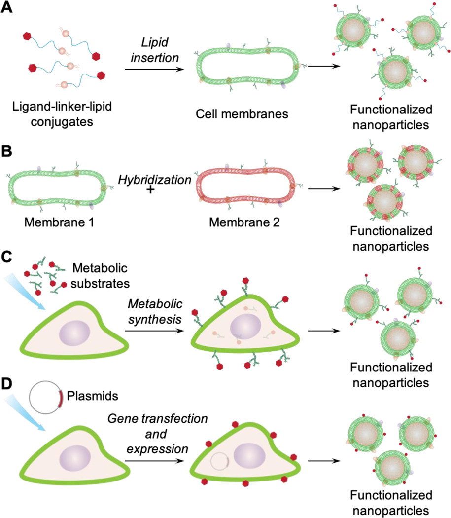
There has been significant interest in developing cell membrane-coated nanoparticles due to their unique abilities of biomimicry and biointerfacing. As the technology progresses, it becomes clear that the application of these nanoparticles can be drastically broadened if additional functions beyond those derived from the natural cell membranes can be integrated. Herein, we summarize the most recent advances in the functionalization of cell membrane-coated nanoparticles. In particular, we focus on emerging methods, including (1) lipid insertion, (2) membrane hybridization, (3) metabolic engineering, and (4) genetic modification. These approaches contribute diverse functions in a nondisruptive fashion while preserving the natural function of the cell membranes. They also improve on the multifunctional and multitasking ability of cell membrane-coated nanoparticles, making them more adaptive to the complexity of biological systems. We hope that these approaches will serve as inspiration for more strategies and innovations to advance cell membrane coating technology.


Figures

Figure 1.
Figure 1.
Schematic showing different methods for functionalizing cell membrane-coated nanoparticles. (A) lipid insertion, (B) membrane hybridization, (C) metabolic engineering, and (D) genetic modification.
Figure 2.
Figure 2.
(A) Schematic of the preparation of nanoparticles coated with DCDX-modified RBC membranes (DCDX-RBCNPs). Streptavidin-PEG-DSPE is synthesized and then inserted into RBC membranes. After coating polymeric cores, biotin-PEG-DCDX binds to the streptavidin on the surface of the resulting RBCNPs to form DCDX-modified RBCNPs. (B) Transmission electron microscope image of DCDX-RBCNPs. (C) The distribution of nanoparticles in the brain of tumor-bearing mice 14 days post-implantation. Nuclei were stained with DAPI (blue), blood vessels were labeled with anti-CD31 (red), while green represents the DiI-loaded nanoparticles. The yellow dotted lines represent the margins of the glioma and the yellow arrows point to the glioma (scale bars, 200 μm). (D) Ex vivo images and average radiant efficacy of brains and tumors in tumor-bearing mice (7 or 14 days after implantation). Bars represent means with SD, n = 3, *p < 0.05, **p < 0.005. (E) Kaplan-Meier survival curves of nude mice bearing intracranial U87 glioma. Mice (n = 10) were injected at 7, 9, 11, 13 and 15 days after glioma implantation with saline, free Dox, Dox-loaded RBC-NPs (RBCNPs/Dox), and Dox-loaded DCDX-RBCNPs (DCDX-RBCNPs/Dox). Reproduced with permission from ref 50. Copyright 2017 Elsevier.
Figure 3.
Figure 3.
(A) The near infrared light (NIR)-driven drug release of the red blood cell (RBC) membrane-coated nanoparticles (PTX-PN@DiR-RV). DiR dye was embedded in the RBC membrane (DiR-RV), and the thermosensitive lipid DPPC was added to the polymeric cores (PN). Under the 808 nm laser irradiation (+L), DiR provided strong thermal energy and then triggered the phase transition of DPPC, leading to the destruction of the cores and the release of paclitaxel (PTX). (B) The infrared thermographic images of mice after 4 h i.v. injection with PBS, free DiR, PN@DiR-RV, and PTX-PN@DiR-RV, respectively. (C) The temperature elevation profile of each group in (B). (D-E) In vivo antitumor and anti-metastasis efficacy by the synergetic chemo-photothermal therapy of PTX-PN@DiR-RV. (D) Tumor growth of mice after intravenous injection of different formulations. (E) Quantitative analysis of the lung metastatic nodules for each group. Data were presented as mean ± SD (n = 6), ** P < 0.01, *** P < 0.005. Reproduced with permission from ref 51. Copyright 2016 John Wiley and Sons.
Figure 4.
Figure 4.
Development of RBC–platelet hybrid membrane-coated nanoparticles ([RBC-P]NPs). (A) Schematic of membrane fusion and coating. Membrane material is derived from both RBCs and platelets and then fused together. The resulting fused membrane is used to coat poly(lactic-co-glycolic acid) (PLGA) polymeric cores to produce [RBC-P]NPs. (B) A representative TEM image of [RBC-P]NPs negatively stained with vanadium (scale bar = 100 nm). (C) Confocal fluorescent microscopy images of either a mixture of RBC membrane-coated nanoparticles (RBCNPs) and platelet membrane-coated nanoparticles [PNPs] or of the [RBC-P]NPs (red = RBC membrane, green = platelet membrane; scale bar = 10 μm). (D) Circulation time of fluorescently labeled RBCNPs, [RBC-P]NPs, and PNPs after intravenous administration to mice via the tail vein (n = 4; mean ± SEM; lines represent two-phase decay model) (E) Amount of free dichlorvos, a model organophosphate, remaining in solution after incubation with RBCNPs, [RBC-P]NPs, or PNPs (n = 3; mean  ± SD). UD = undetectable. (F) Imaging of aortas from ApoE knockout mice fed with a high fat western diet, after intravenous administration with dye-labeled RBCNPs, [RBC-P]NPs, and PNPs (red = nanoparticles; scale bars = 1 mm). Oil Red O staining was used to confirm the presence of atherosclerotic plaque. Reproduced with permission from ref 53. Copyright 2017 John Wiley & Sons.
Figure 5.
Figure 5.
Development of cancer-dendritic hybrid membrane-coated metal organic framework (MOF) nanoparticles as a cancer vaccine. (A) Schematic of the process for preparing MOF nanoparticles coated with the membrane of the fused cells (MOF@FM). (B) Dendritic cells (DCs, anti-MHC II-labeled, green), 4T1 cells (anti-CD44-APC labeled, magenta), and the fused cells (FC, double labeled) observed with the confocal laser scanning microscopy (CLSM). Scale bar = 10 μm. (C) TEM images of MOF@FM. Scale bar =100 nm. (D) Percentage of DC maturation based on the quantification of CD80 and CD86 expression after in vitro incubation of DCs with 4T1 cancer cell membrane (CM), DC membrane (DM), and fused cell membrane (FM) for 48 h. The mean values and s.d. were presented and measurements were taken from distinct samples (one-way ANOVA; **p<0.01, ***p<0.001, n = 3). (E) In vitro cytotoxicity of the T lymphocytes after incubation with above-pretreated DCs for 48 h to 3T3, 4T1, and CT26 cells. The mean values and s.d. were presented and measurements were taken from distinct samples (n = 5). (F) Percentage of tumor-free mice receiving immunization with MOF@FM vaccine followed by tumor challenge. Reproduced with permission from ref 56. Copyright 2019 Springer Nature.
Figure 6.
Figure 6.
Metabolic glycoengineering approach for membrane modification. (A) Scheme of glycoengineered T cell membrane extraction and N3-labeled membrane-coated nanoparticles (N3-TINPs) construction. (B) Illustration of tumor-bearing mice with BCN group expression upon Ac4ManN-BCN injection. N3-TINPs could targeted anchor in tumor region through immune recognition of T cell membrane and bioorthogonal reaction between BCN and N3 groups, and effectively eliminate tumors based on ICG-mediated photothermal effects. (C) Identification of N3 group on the surface of N3-TINPs. Tumor cells were incubated with N3-TINPs or TINPs (control) for 1 h, and then stain with anti-CD3-FITC and DBCO-Sata650. (D) In vivo photothermal therapy efficacy based on tumor growth curves of different groups in Raji tumor-bearing mice (n = 5). Reproduced with permission from ref 90. Copyright 2019 John Wiley & Sons.
Figure 7.
Figure 7.
Metabolic lipid-engineering approach for membrane modification. (A) Illustration of N3-tagged leukocyte membrane via lipid-engineering to develop biomimetic nanoplatform (MNCs) for enhanced CD8+ T cell proliferation. The T-cell stimuli conjugations were identified by immunostaining with the fluorescence-labeled secondary antibody of antiCD28 and pMHC-I. Then the N3 groups on cell membrane were confirmed by DBCO-Flour525. After incubation with CD8+ T cells for 7 days, the aAPCs presented the highest proliferation efficiency. Reproduced with permission from ref. . Copyright 2017 American Chemical Society. (B) Scheme of N3-labeled macrophage membrane-coated nanoplatform for targeted siRNA delivery. Following the modification via metabolic lipid-engineering, the N3-labeled membrane was coated onto MNC-siRNA nanocomplex and conjugated with DBCO-RGD for tumor targeting. The imaging of tumor and various organs were performed at 24 h after intravenous injection of different MNC-based nanoformulations. (i) PBS, (ii) MNC:siRNA, (iii) M-MNC:siRNA, (iv) R-M-MNC:siRNA. Reproduced with permission from ref. 92. Copyright 2018 John Wiley & Sons.
Figure 8.
Figure 8.
Schematic of bioengineered cell membrane nanovesicle coated oncolytic adenoviruses (OA@BCMNs) for OA delivery and in vivo antitumor efficacy of OA@BCMNs. (A) Design features and proposed mechanism of OA@BCMNs. The BCMNs encapsulated OA, protecting OAs from neutralizing antibodies and delivering them to tumors through receptor mediated endocytosis. Once entered tumor cells, OAs infect and amplify the tumor cells, causing the tumor cell lysis. (B) Viral genome copies in excised tumors and organs, after intravenous injection of OA and OA@BCMNs-preS1 into HepG2-NTCP bearing nude mouse model, were quantified using real time qPCR. (C) Tumor growth curve of HepG2-NTCP bearing nude mouse model after the indicated treatment. (D) Tumor growth curve of HepG2-APN bearing mouse model after the indicated treatment. Reproduced with permission from ref 113. Copyright 2019 American Chemical Society.
Figure 9.
Figure 9.
(A) Illustration of the steps involved in the synthesis of PASylated nanoghosts. A plasmid that expresses the proline-alanine-serine (PAS) peptides on the surface membrane is transfected into mammalian cells. PASylated cell membranes are then harvested and coated on PLGA nanoparticles (PLGA NPs) to form PASylated nanoghosts. (B) In vivo serum concentration of DiR dye from nanoparticle groups over 48 h. Sample groups are PLGA NPs, non-transfected nanoghosts (No PAS), and PASylated nanoghosts (PAS40). ⁎ and # denote statistical significance of PAS40 (P ≤ 0.005) in comparison to No PAS and PLGA NP. (C) Biodistribution of dye-loaded sample groups in the liver, spleen kidney, heart and lungs of mice at 48 h post treatment. * denotes statistical significance (P ≤ 0.001) in comparison to the PLGA NP control group. Reproduced with permission from ref 115. Copyright 2019 Elsevier.

Similar articles

Cited by

References

    1. Ragelle H, Danhier F, Preat V, Langer R, and Anderson DG (2017) Nanoparticle-based drug delivery systems: a commercial and regulatory outlook as the field matures, Expert Opin. Drug Deliv 14, 851–864. - PubMed
    1. Fang RH, Kroll AV, Gao W, and Zhang L. (2018) Cell Membrane Coating Nanotechnology, Adv. Mater 30, e1706759. - PMC - PubMed
    1. Hu CMJ, Fang RH, Luk BT, Chen KNH, Carpenter C, Gao W, Zhang K, and Zhang L. (2013) ‘Marker-of-self’ functionalization of nanoscale particles through a top-down cellular membrane coating approach, Nanoscale 5, 2664–2668. - PMC - PubMed
    1. Hu CMJ, Zhang L, Aryal S, Cheung C, Fang RH, and Zhang L. (2011) Erythrocyte membrane-camouflaged polymeric nanoparticles as a biomimetic delivery platform, Proc. Natl. Acad. Sci. U. S. A 108, 10980–10985. - PMC - PubMed
    1. Piao JG, Wang LM, Gao F, You YZ, Xiong YJ, and Yang LH (2014) Erythrocyte Membrane Is an Alternative Coating to Polyethylene Glycol for Prolonging the Circulation Lifetime of Gold Nanocages for Photothermal Therapy, ACS Nano 8, 10414–10425. - PubMed