. 2020 Jun 9;117(23):13117-13126.
doi: 10.1073/pnas.2000430117. Epub 2020 May 20.

DCyFIR: a high-throughput CRISPR platform for multiplexed G protein-coupled receptor profiling and ligand discovery

Affiliations

DCyFIR: a high-throughput CRISPR platform for multiplexed G protein-coupled receptor profiling and ligand discovery

N J Kapolka et al. Proc Natl Acad Sci U S A. .

Abstract

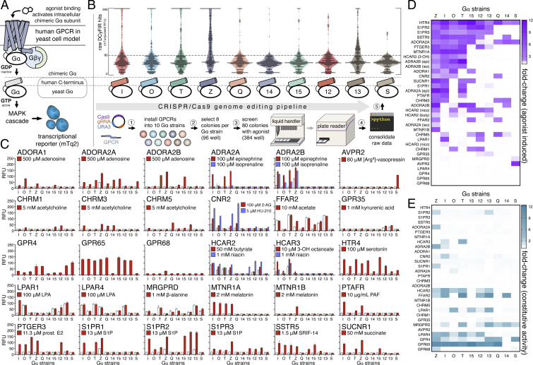
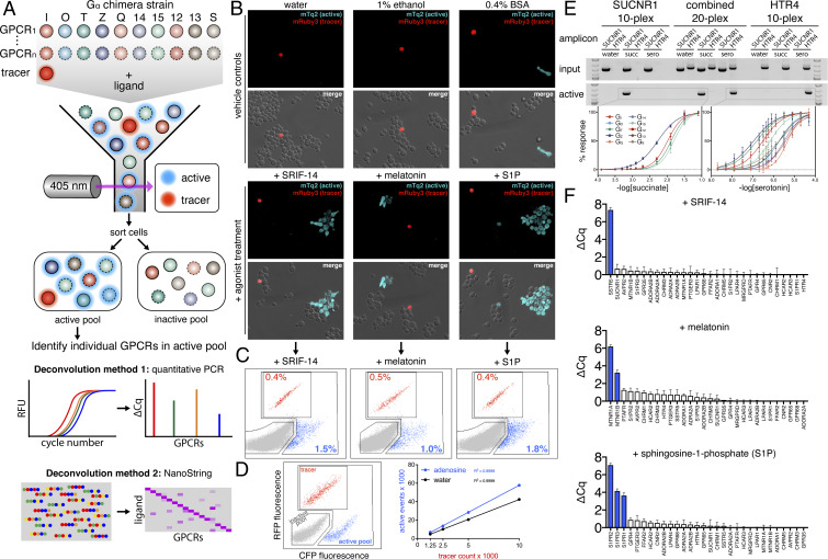
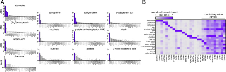
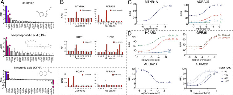
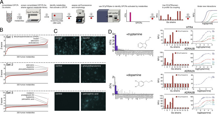
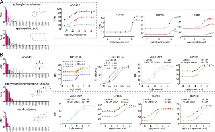
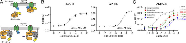
More than 800 G protein-coupled receptors (GPCRs) comprise the largest class of membrane receptors in humans. While there is ample biological understanding and many approved drugs for prototypic GPCRs, most GPCRs still lack well-defined biological ligands and drugs. Here, we report our efforts to tap the potential of understudied GPCRs by developing yeast-based technologies for high-throughput clustered regularly interspaced short palindromic repeats (CRISPR) engineering and GPCR ligand discovery. We refer to these technologies collectively as Dynamic Cyan Induction by Functional Integrated Receptors, or DCyFIR. A major advantage of DCyFIR is that GPCRs and other assay components are CRISPR-integrated directly into the yeast genome, making it possible to decode ligand specificity by profiling mixtures of GPCR-barcoded yeast strains in a single tube. To demonstrate the capabilities of DCyFIR, we engineered a yeast strain library of 30 human GPCRs and their 300 possible GPCR-Gα coupling combinations. Profiling of these 300 strains, using parallel (DCyFIRscreen) and multiplex (DCyFIRplex) DCyFIR modes, recapitulated known GPCR agonism with 100% accuracy, and identified unexpected interactions for the receptors ADRA2B, HCAR3, MTNR1A, S1PR1, and S1PR2. To demonstrate DCyFIR scalability, we profiled a library of 320 human metabolites and discovered several GPCR-metabolite interactions. Remarkably, many of these findings pertained to understudied pharmacologically dark receptors GPR4, GPR65, GPR68, and HCAR3. Experiments on select receptors in mammalian cells confirmed our yeast-based observations, including our discovery that kynurenic acid activates HCAR3 in addition to GPR35, its known receptor. Taken together, these findings demonstrate the power of DCyFIR for identifying ligand interactions with prototypic and understudied GPCRs.

Keywords: DCyFIR; G protein-coupled receptors; multiplex; pharmacologically dark GPCR; yeast.


Conflict of interest statement

The authors declare no competing interest.

Figures

Fig. 1.
Fig. 1.
High-throughput CRISPR engineering and DCyFIRscreen validation of the DCyFIR yeast strain library. (A) Simplified schematic of our yeast pheromone pathway model for studying human GPCRs (see SI Appendix, Fig. S1 for further details). (B) Workflow of our high-throughput CRISPR/Cas9 genome editing pipeline showing primary screening data (more than 5,000 mTq2 fluorescence measurements) for our exploratory panel of 30 human GPCRs. All fluorescence values are reported as RFUs. (C) DCyFIRscreen profiles for 300 GPCR–Gα strains against their known agonists, with error bars representing the SD of n = 4 experimental replicates. Untreated/treated conditions are represented by white/colored bars. All RFU measurements were quantified using the same gain setting. (D) Heat map of agonist-induced signaling in the 300 GPCR–Gα strains. (E) Heat map of constitutive activity in the 300 GPCR–Gα strains.
Fig. 2.
Fig. 2.
Developing and validating DCyFIRplex. (A) Schematic of the DCyFIRplex workflow showing strain consolidation to build the multiplex, FACS to collect active receptor strain pools, and our two primary multiplex deconvolution techniques. (B) Confocal microscopy images of treated and untreated (vehicle) samples of the GPCR–Gα 300-plex with the added mRuby3 tracer strain (maximum intensity projections, 63X magnification). (C) FACS analysis of the inactive (gray), active (cyan), and tracer (red) pools for the 300-plex shown in B. (D) FACS analysis of negative (gray), positive (cyan), and tracer (red) controls (see Methods for details); also, a representative standard curve of tracer event counts versus active pool event counts for our reference conditions of ±adenosine used to calibrate the FACS sorting procedure. Tracer event counts between 3,000 and 5,000 gave the most consistent deconvolution results. (E) PCR deconvolution of SUCNR1 and HTR4 10-plexes and combined 20-plex visualized by gel electrophoresis; also, the family of normalized titration curves that corresponded to the active GPCR–Gα strains in each 10- and 20-plex (errors bars represent the SD of n = 4 experimental replicates). (F) DCyFIRplex profiles deconvoluted via qPCR for the agonist-treated 300-plexes characterized in B and C. Expected hits are colored blue. ΔCq values correspond to the Cq difference between treated and untreated conditions, with error bars representing the SEM of n = 6 repeats derived from 3 independent 300-plex consolidations deconvoluted in technical duplicate. ΔCq values correspond to a log2 scale.
Fig. 3.
Fig. 3.
Using DCyFIRplex profiling to recapitulate known agonist interactions. (A) DCyFIRplex profiles for known GPCR agonists in our 30-receptor panel deconvoluted via qPCR. (B) Same samples as in A deconvoluted using NanoString. (A and B) ΔCq values correspond to the Cq difference between treated and untreated conditions, with error bars representing the SEM of n = 6 repeats derived from 3 independent 300-plex consolidations deconvoluted in technical duplicate. ΔCq values correspond to a log2 scale. NanoString transcript counts were collected in technical duplicate, averaged, and normalized to the maximal RNA transcript count for each GPCR gene.
Fig. 4.
Fig. 4.
Using DCyFIRplex to discover interactions for known GPCR agonists. (A) DCyFIRplex profiles identifying GPCR–ligand interactions (pink bars) discovered in the process of screening known agonists (purple bars) within our panel of 30 exploratory receptors. (B) DCyFIRscreen profiles confirming the DCyFIRplex discoveries in A, with error bars representing the SD of n = 4 experimental replicates. (C and D) Select titrations confirming the DCyFIRplex discoveries in A, with error bars representing the SD of n = 4 experimental replicates. Dashed lines and boxes indicate datasets showing that KYNA activates HCAR3 with greater potency than GPR35 and is also an endogenous negative allosteric modulator of ADRA2B. Full titration datasets are available in SI Appendix, Fig. S5.
Fig. 5.
Fig. 5.
DCyFIR profiling of a human metabolite library. (A) Step-by-step workflow used to screen a library of 320 endogenous human metabolites. (B) Z-score profiles for metabolite screens of receptor set 1 (ADORA1, ADORA2A, FFAR2, GPR4, GPR65, GPR68, HCAR2, HCAR3, LPAR1, LPAR4, MRGPRD), set 2 (ADORA2B, ADRA2A, ADRA2B, AVPR2, CHRM1, CHRM3, CHRM5, CNR2, GPR35), and set 3 (HTR4, MTNR1A, MTNR1B, PTAFR, PTGER3, S1PR1, S1PR2, S1PR3, SSTR5, SUCNR1). Gray bands indicate Z-scores between ± 1. (C) Representative fluorescence microscopy images for Z-score hits in receptor subsets 1 (110-plex), 2 (90-plex), and 3 (100-plex). (D) Discovery workflow illustrating tryptamine agonism of HTR4 and ADRA2B and dopamine agonism of ADRA2A and ADRA2B. Once tryptamine and dopamine were identified as hits (steps 1 to 3 in A), we used DCyFIRplex profiling to identify their GPCR target or targets, DCyFIRscreen profiling to identify their Gα coupling pattern or patterns, and titrations to quantify their EC50 values (steps 4 to 6 in A).
Fig. 6.
Fig. 6.
Identification and validation of GPCR–metabolite interactions. (A) DCyFIRplex profiles and titrations for metabolite agonists and (B) positive allosteric modulators. For B, full titration datasets for all GPCR–Gα coupling combinations are available in SI Appendix, Fig. S7. DCyFIRplex error bars represent the SEM of n = 6 repeats derived from 3 independent 300-plex consolidations deconvoluted in technical duplicate, and titration error bars represent the SD of n = 4 experimental replicates.
Fig. 7.
Fig. 7.
Validation of DCyFIR discoveries in mammalian cells. GPCR signaling as measured by BRET between a GPCR fused to the Renilla luciferase (Rec-Rluc8) and Venus fluorescent protein (V) fused to either a mini G protein (A, Top) or split between the βγ subunits (βγ-V) of a full G protein heterotrimer (A, Bottom). (B) Net BRET for KYNA titrations of HCAR3 (using the mini G protein V-mGsi containing an N-terminal nuclear export signal, NES-V-mGsi) and GPR35 (using full-length Gα12 with βγ-V). Error bars represent SEM of at least n = 6 independent experiments. (C) Net BRET for titrations of ADRA2B (using full-length Gαi1 with βγ-V) with a known agonist (norepinephrine) and discovered agonists (dopamine, phenylethanolamine, tryptamine, and serotonin). Error bars represent SEM of at least n = 4 independent experiments.

Similar articles

Cited by

References

    1. Sriram K., Insel P. A., G protein-coupled receptors as targets for approved drugs: How many targets and how many drugs? Mol. Pharmacol. 93, 251–258 (2018). - PMC - PubMed
    1. Rodgers G. et al. ., Glimmers in illuminating the druggable genome. Nat. Rev. Drug Discov. 17, 301–302 (2018). - PMC - PubMed
    1. Lyu J. et al. ., Ultra-large library docking for discovering new chemotypes. Nature 566, 224–229 (2019). - PMC - PubMed
    1. Dowell S. J., Brown A. J., Yeast assays for G-protein-coupled receptors. Receptors Channels 8, 343–352 (2002). - PubMed
    1. Dowell S. J., Brown A. J., Yeast assays for G protein-coupled receptors. Methods Mol. Biol. 552, 213–229 (2009). - PubMed

Publication types

MeSH terms

LinkOut - more resources