Cyclin A2 is essential for mouse gonocyte maturation
- PMID: 32420805
- PMCID: PMC7469458
- DOI: 10.1080/15384101.2020.1762314
Cyclin A2 is essential for mouse gonocyte maturation
Abstract
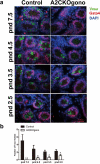
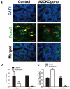
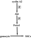
In mammals, male gonocytes are derived from primordial germ cells during embryogenesis, enter a period of mitotic proliferation, and then become quiescent until birth. After birth, the gonocytes proliferate and migrate from the center of testicular cord toward the basement membrane to form the pool of spermatogonial stem cells (SSCs) and establish the SSC niche architecture. However, the molecular mechanisms underlying gonocyte proliferation, migration and differentiation are largely unknown. Cyclin A2 is a key component of the cell cycle and required for cell proliferation. Here, we show that cyclin A2 is required in mouse male gonocyte development and the establishment of spermatogenesis in the neonatal testis. Loss of cyclin A2 function in embryonic gonocytes by targeted gene disruption affected the regulation of the male gonocytes to SSC transition, resulting in the disruption of SSC pool formation, imbalance between SSC self-renewal and differentiation, and severely abnormal spermatogenesis in the adult testis.
Keywords: Cyclin A2; gonocyte differentiation; spermatogonial stem cells.
Conflict of interest statement
The authors declare there are no conflicts of interest.
Figures


Similar articles
-
Temporal-Spatial Establishment of Initial Niche for the Primary Spermatogonial Stem Cell Formation Is Determined by an ARID4B Regulatory Network.Stem Cells. 2017 Jun;35(6):1554-1565. doi: 10.1002/stem.2597. Epub 2017 Mar 16. Stem Cells. 2017. PMID: 28207192 Free PMC article.
-
Establishment of a proteomic profile associated with gonocyte and spermatogonial stem cell maturation and differentiation in neonatal mice.Proteomics. 2014 Feb;14(2-3):274-85. doi: 10.1002/pmic.201300395. Epub 2014 Jan 10. Proteomics. 2014. PMID: 24339256
-
AIP1-mediated actin disassembly is required for postnatal germ cell migration and spermatogonial stem cell niche establishment.Cell Death Dis. 2015 Jul 16;6(7):e1818. doi: 10.1038/cddis.2015.182. Cell Death Dis. 2015. PMID: 26181199 Free PMC article.
-
Mammalian gonocyte and spermatogonia differentiation: recent advances and remaining challenges.Reproduction. 2015 Mar;149(3):R139-57. doi: 10.1530/REP-14-0431. Reproduction. 2015. PMID: 25670871 Review.
-
Gonocytes, the forgotten cells of the germ cell lineage.Birth Defects Res C Embryo Today. 2009 Mar;87(1):1-26. doi: 10.1002/bdrc.20142. Birth Defects Res C Embryo Today. 2009. PMID: 19306346 Review.
Cited by
-
Sertoli cell and spermatogonial development in pigs.J Anim Sci Biotechnol. 2022 Apr 11;13(1):45. doi: 10.1186/s40104-022-00687-2. J Anim Sci Biotechnol. 2022. PMID: 35399096 Free PMC article.
-
Understanding the Underlying Molecular Mechanisms of Meiotic Arrest during In Vitro Spermatogenesis in Rat Prepubertal Testicular Tissue.Int J Mol Sci. 2022 May 24;23(11):5893. doi: 10.3390/ijms23115893. Int J Mol Sci. 2022. PMID: 35682573 Free PMC article.
-
Roles of Spermatogonial Stem Cells in Spermatogenesis and Fertility Restoration.Front Endocrinol (Lausanne). 2022 May 12;13:895528. doi: 10.3389/fendo.2022.895528. eCollection 2022. Front Endocrinol (Lausanne). 2022. PMID: 35634498 Free PMC article. Review.
-
Molecular characterization, expression patterns and cellular localization of BCAS2 gene in male Hezuo pig.PeerJ. 2023 Oct 24;11:e16341. doi: 10.7717/peerj.16341. eCollection 2023. PeerJ. 2023. PMID: 37901468 Free PMC article.
References
-
- Blanchard JM. Cyclin A2 transcriptional regulation: modulation of cell cycle control at the G1/S transition by peripheral cues. Biochem Pharmacol. 2000;60:1179–1184. - PubMed
-
- Murphy M, Stinnakre MG, Senamaud-Beaufort C, et al. Delayed early embryonic lethality following disruption of the murine cyclin A2 gene. Nat Genet. 1997;15:83–86. - PubMed
-
- Sweeney C, Murphy M, Kubelka M, et al. A distinct cyclin A is expressed in germ cells in the mouse. Development. 1996;122:53–64. - PubMed
-
- Liu D, Matzuk MM, Sung WK, et al. Cyclin A1 is required for meiosis in the male mouse. Nat Genet. 1998;20:377–380. - PubMed
Publication types
MeSH terms
Substances
Grants and funding
LinkOut - more resources
Full Text Sources
Molecular Biology Databases
