

KSHV infection skews macrophage polarisation towards M2-like/TAM and activates Ire1 α-XBP1 axis up-regulating pro-tumorigenic cytokine release and PD-L1 expression
- PMID: 32418990
- PMCID: PMC7374093
- DOI: 10.1038/s41416-020-0872-0
KSHV infection skews macrophage polarisation towards M2-like/TAM and activates Ire1 α-XBP1 axis up-regulating pro-tumorigenic cytokine release and PD-L1 expression
Abstract
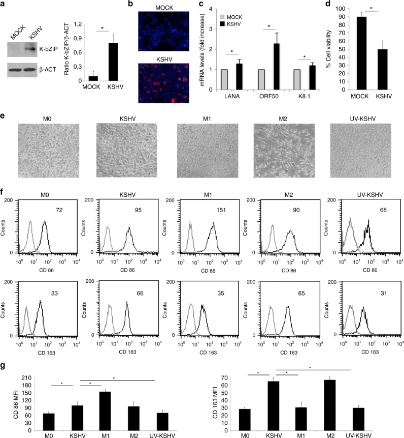
Background: Kaposi's Sarcoma Herpesvirus (KSHV) is a gammaherpesvirus strongly linked to human cancer. The virus is also able to induce immune suppression, effect that contributes to onset/progression of the viral-associated malignancies. As KSHV may infect macrophages and these cells abundantly infiltrate Kaposi's sarcoma lesions, in this study we investigated whether KSHV-infection could affect macrophage polarisation to promote tumorigenesis.
Methods: FACS analysis was used to detect macrophage markers and PD-L1 expression. KSHV infection and the molecular pathways activated were investigated by western blot analysis and by qRT-PCR while cytokine release was assessed by Multi-analyte Kit.
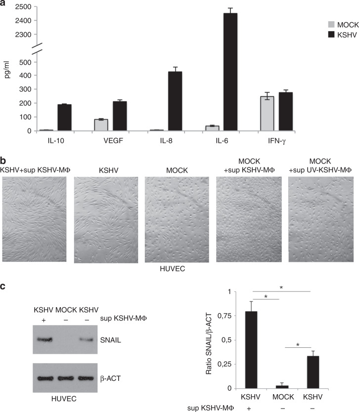
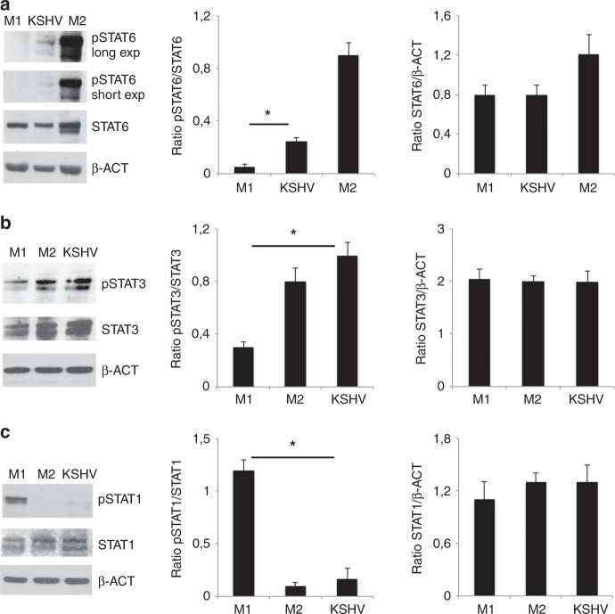
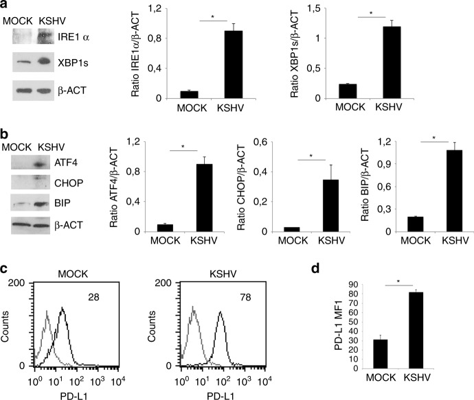
Results: We found that KSHV infection reduced macrophage survival and skewed their polarisation towards M2 like/TAM cells, based on the expression of CD163, on the activation of STAT3 and STAT6 pathways and the release of pro-tumorigenic cytokines such as IL-10, VEGF, IL-6 and IL-8. We also found that KSHV triggered Ire1 α-XBP1 axis activation in infected macrophages to increase the release of pro-tumorigenic cytokines and to up-regulate PD-L1 surface expression.
Conclusions: The findings that KSHV infection of macrophages skews their polarisation towards M2/TAM and that activate Ire1 α-XBP1 to increase the release of pro-tumorigenic cytokines and the expression of PD-L1, suggest that manipulation of UPR could be exploited to prevent or improve the treatment of KSHV-associated malignancies.
Conflict of interest statement
The authors declare no competing interests.
Figures

Similar articles
-
Kaposi's Sarcoma-Associated Herpesvirus Increases PD-L1 and Proinflammatory Cytokine Expression in Human Monocytes.mBio. 2017 Oct 10;8(5):e00917-17. doi: 10.1128/mBio.00917-17. mBio. 2017. PMID: 29018115 Free PMC article.
-
Intracellular Tat of human immunodeficiency virus type 1 activates lytic cycle replication of Kaposi's sarcoma-associated herpesvirus: role of JAK/STAT signaling.J Virol. 2007 Mar;81(5):2401-17. doi: 10.1128/JVI.02024-06. Epub 2006 Dec 6. J Virol. 2007. PMID: 17151125 Free PMC article.
-
Persistent activation of STAT3 by latent Kaposi's sarcoma-associated herpesvirus infection of endothelial cells.J Virol. 2007 Mar;81(5):2449-58. doi: 10.1128/JVI.01769-06. Epub 2006 Dec 6. J Virol. 2007. PMID: 17151100 Free PMC article.
-
Pathological Features of Kaposi's Sarcoma-Associated Herpesvirus Infection.Adv Exp Med Biol. 2018;1045:357-376. doi: 10.1007/978-981-10-7230-7_16. Adv Exp Med Biol. 2018. PMID: 29896675 Review.
-
Cellular and viral oncogenes: the key to unlocking unknowns of Kaposi's sarcoma-associated herpesvirus pathogenesis.Arch Virol. 2018 Oct;163(10):2633-2643. doi: 10.1007/s00705-018-3918-3. Epub 2018 Jun 23. Arch Virol. 2018. PMID: 29936609 Review.
Cited by
-
Periodontitis promotes tumor growth and immune evasion via PD-1/PD-L1.Cancer Immunol Immunother. 2024 Nov 13;74(1):22. doi: 10.1007/s00262-024-03865-5. Cancer Immunol Immunother. 2024. PMID: 39535607 Free PMC article.
-
LARRPM restricts lung adenocarcinoma progression and M2 macrophage polarization through epigenetically regulating LINC00240 and CSF1.Cell Mol Biol Lett. 2022 Oct 11;27(1):91. doi: 10.1186/s11658-022-00376-y. Cell Mol Biol Lett. 2022. PMID: 36221069 Free PMC article.
-
Chemoradiation induces upregulation of immunogenic cell death-related molecules together with increased expression of PD-L1 and galectin-9 in gastric cancer.Sci Rep. 2021 Jun 10;11(1):12264. doi: 10.1038/s41598-021-91603-7. Sci Rep. 2021. PMID: 34112882 Free PMC article.
-
The signature of a T-cell response to KSHV persists across space and time in individuals with epidemic and endemic KS from Uganda.bioRxiv [Preprint]. 2024 Feb 8:2024.02.06.579223. doi: 10.1101/2024.02.06.579223. bioRxiv. 2024. PMID: 38370623 Free PMC article. Preprint.
-
Viral Infection and Autophagy Dysregulation: The Case of HHV-6, EBV and KSHV.Cells. 2020 Dec 7;9(12):2624. doi: 10.3390/cells9122624. Cells. 2020. PMID: 33297368 Free PMC article. Review.
References
-
- Cirone M, Lucania G, Bergamo P, Trivedi P, Frati L, Faggioni A. Human herpesvirus 8 (HHV-8) inhibits monocyte differentiation into dendritic cells and impairs their immunostimulatory activity. Immunol. Lett. 2007;113:40–46. - PubMed
-
- Gilardini Montani MS, Falcinelli L, Santarelli R, Romeo MA, Granato M, Faggioni A, et al. Kaposi Sarcoma Herpes Virus (KSHV) infection inhibits macrophage formation and survival by counteracting Macrophage Colony-Stimulating Factor (M-CSF)-induced increase of Reactive Oxygen Species (ROS), c-Jun N-terminal kinase (JNK) phosphorylation and autophagy. Int J. Biochem Cell Biol. 2019;114:105560. - PubMed
Publication types
MeSH terms
Substances
Grants and funding
LinkOut - more resources
Full Text Sources
Medical
Research Materials
Miscellaneous
