

Machine learning techniques for biomedical image segmentation: An overview of technical aspects and introduction to state-of-art applications
- PMID: 32418337
- PMCID: PMC7338207
- DOI: 10.1002/mp.13649
Machine learning techniques for biomedical image segmentation: An overview of technical aspects and introduction to state-of-art applications
Abstract
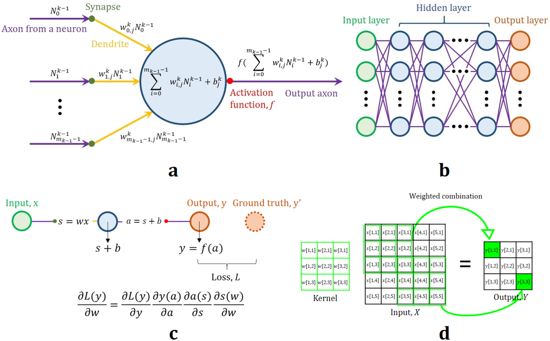
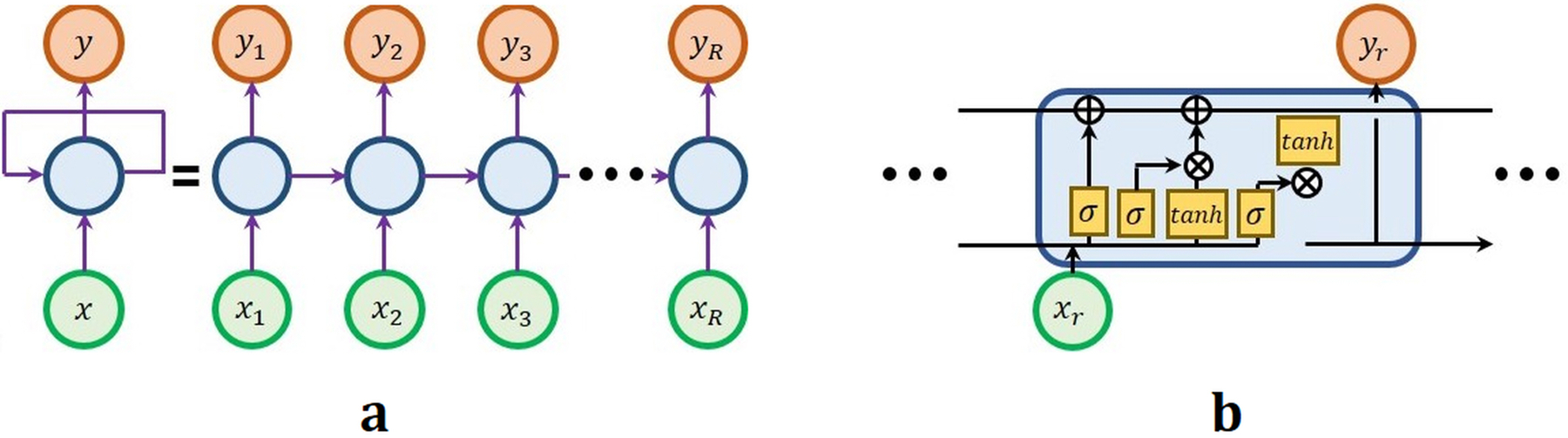
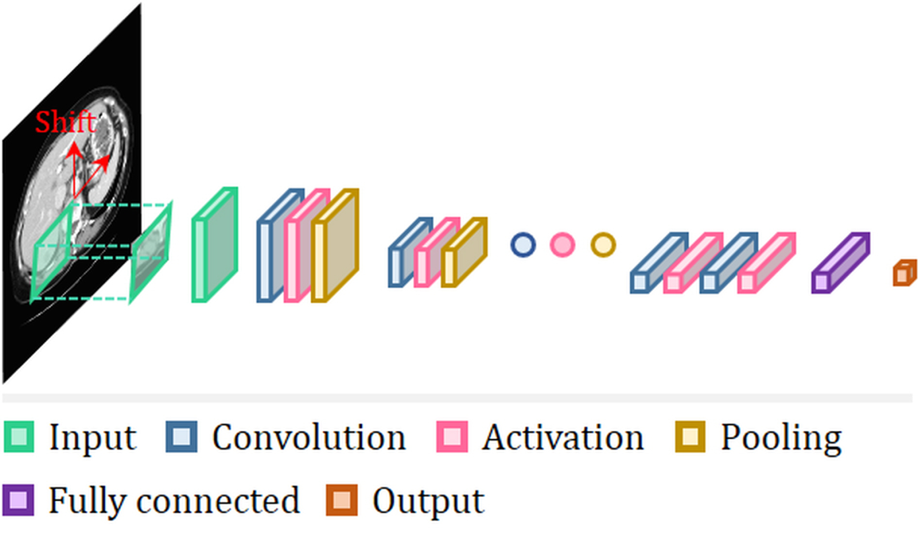
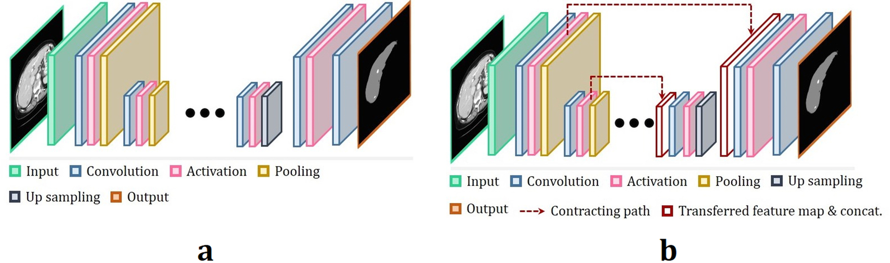
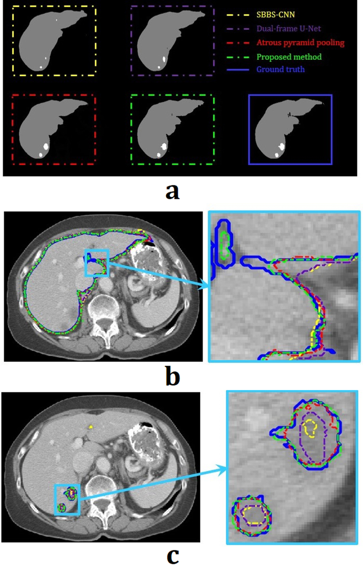
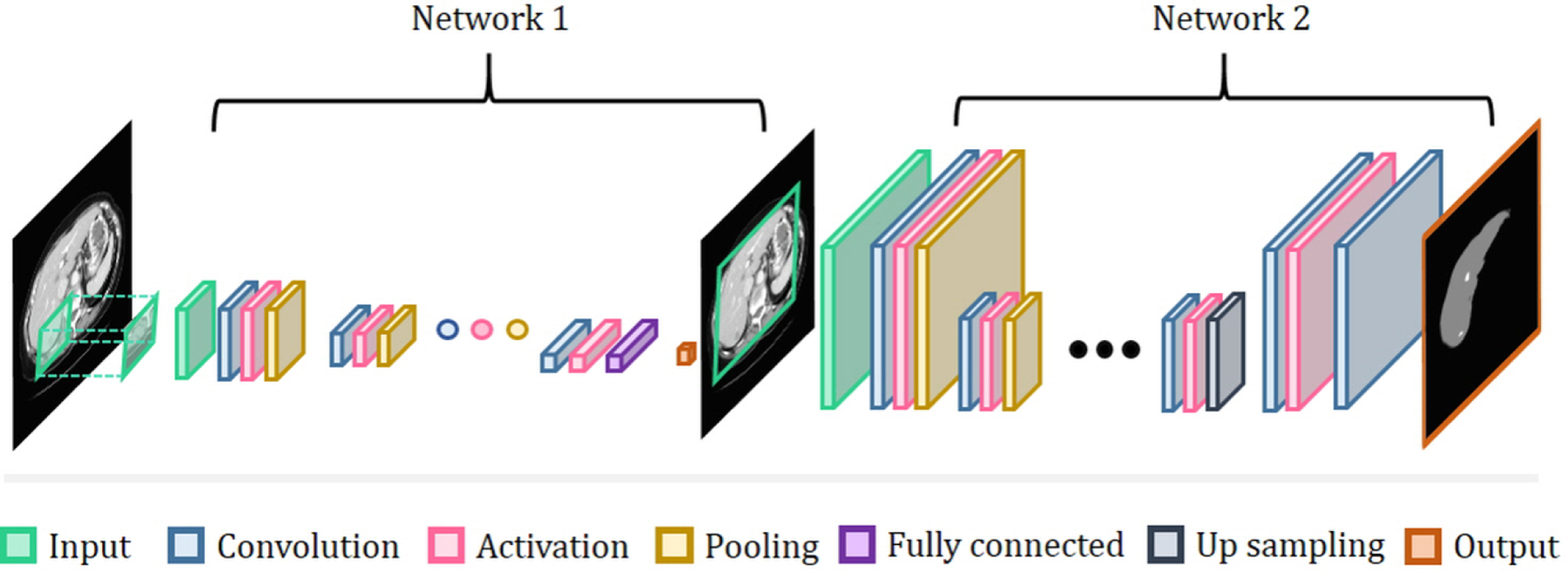
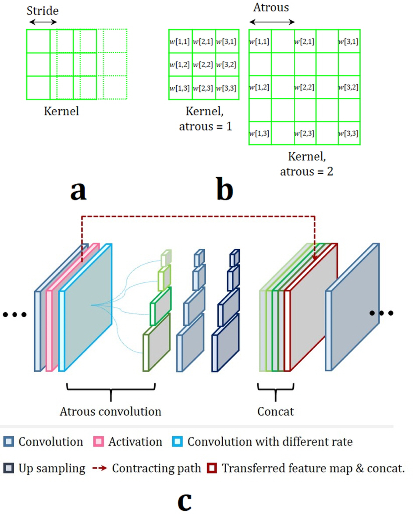
In recent years, significant progress has been made in developing more accurate and efficient machine learning algorithms for segmentation of medical and natural images. In this review article, we highlight the imperative role of machine learning algorithms in enabling efficient and accurate segmentation in the field of medical imaging. We specifically focus on several key studies pertaining to the application of machine learning methods to biomedical image segmentation. We review classical machine learning algorithms such as Markov random fields, k-means clustering, random forest, etc. Although such classical learning models are often less accurate compared to the deep-learning techniques, they are often more sample efficient and have a less complex structure. We also review different deep-learning architectures, such as the artificial neural networks (ANNs), the convolutional neural networks (CNNs), and the recurrent neural networks (RNNs), and present the segmentation results attained by those learning models that were published in the past 3 yr. We highlight the successes and limitations of each machine learning paradigm. In addition, we discuss several challenges related to the training of different machine learning models, and we present some heuristics to address those challenges.
Keywords: deep learning; machine learning; medical Image; overview; segmentation.
© 2019 American Association of Physicists in Medicine.
Conflict of interest statement
“The authors have no conflicts to disclose.”
Figures
Similar articles
-
Variability and reproducibility in deep learning for medical image segmentation.Sci Rep. 2020 Aug 13;10(1):13724. doi: 10.1038/s41598-020-69920-0. Sci Rep. 2020. PMID: 32792540 Free PMC article.
-
Machine Learning Algorithms in Neuroimaging: An Overview.Acta Neurochir Suppl. 2022;134:125-138. doi: 10.1007/978-3-030-85292-4_17. Acta Neurochir Suppl. 2022. PMID: 34862537 Review.
-
Fully Automatic Brain Tumor Segmentation using End-To-End Incremental Deep Neural Networks in MRI images.Comput Methods Programs Biomed. 2018 Nov;166:39-49. doi: 10.1016/j.cmpb.2018.09.007. Epub 2018 Sep 21. Comput Methods Programs Biomed. 2018. PMID: 30415717
-
A survey on deep learning in medical image analysis.Med Image Anal. 2017 Dec;42:60-88. doi: 10.1016/j.media.2017.07.005. Epub 2017 Jul 26. Med Image Anal. 2017. PMID: 28778026 Review.
-
Advances in Medical Image Segmentation: A Comprehensive Review of Traditional, Deep Learning and Hybrid Approaches.Bioengineering (Basel). 2024 Oct 16;11(10):1034. doi: 10.3390/bioengineering11101034. Bioengineering (Basel). 2024. PMID: 39451409 Free PMC article. Review.
Cited by
-
Automated Field of Interest Determination for Quantitative Ultrasound Analyses of Cervical Tissues: Toward Real-time Clinical Translation in Spontaneous Preterm Birth Risk Assessment.Ultrasound Med Biol. 2024 Dec;50(12):1861-1867. doi: 10.1016/j.ultrasmedbio.2024.08.011. Epub 2024 Sep 12. Ultrasound Med Biol. 2024. PMID: 39271408
-
On-board MRI image compression using video encoder for MR-guided radiotherapy.Quant Imaging Med Surg. 2023 Aug 1;13(8):5207-5217. doi: 10.21037/qims-22-1378. Epub 2023 Jun 20. Quant Imaging Med Surg. 2023. PMID: 37581063 Free PMC article.
-
Automation and artificial intelligence in radiation therapy treatment planning.J Med Radiat Sci. 2024 Jun;71(2):290-298. doi: 10.1002/jmrs.729. Epub 2023 Oct 4. J Med Radiat Sci. 2024. PMID: 37794690 Free PMC article.
-
Optical coherence tomography confirms non-malignant pigmented lesions in phacomatosis pigmentokeratotica using a support vector machine learning algorithm.Skin Res Technol. 2023 Jun;29(6):e13377. doi: 10.1111/srt.13377. Skin Res Technol. 2023. PMID: 37357662 Free PMC article.
-
Automated, high-throughput quantification of EGFP-expressing neutrophils in zebrafish by machine learning and a highly-parallelized microscope.PLoS One. 2023 Dec 7;18(12):e0295711. doi: 10.1371/journal.pone.0295711. eCollection 2023. PLoS One. 2023. PMID: 38060605 Free PMC article.
References
-
- Mao KZ, Zhao P, Tan P-H. Supervised learning-based cell image segmentation for p53 immunohistochemistry. IEEE Transactions on Biomedical Engineering. 2006;53(6):1153–1163. - PubMed
-
- Li D, Liu L, Chen J, et al. Augmenting atlas-based liver segmentation for radiotherapy treatment planning by incorporating image features proximal to the atlas contours. Physics in Medicine & Biology. 2016;62(1):272. - PubMed
-
- Noh H, Hong S, Han B. Learning Deconvolution Network for Semantic Segmentation. arXiv e-prints. 2015. https://ui.adsabs.harvard.edu/\#abs/2015arXiv150504366N. Accessed May 01, 2015.
-
- He K, Zhang X, Ren S, Sun J. Deep residual learning for image recognition. Paper presented at: Proceedings of the IEEE conference on computer vision and pattern recognition2016.
Publication types
MeSH terms
Grants and funding
LinkOut - more resources
Full Text Sources
Other Literature Sources
Medical
Miscellaneous
