. 2020 May 15;15(5):e0233177.
doi: 10.1371/journal.pone.0233177. eCollection 2020.

Atco, a yeast mitochondrial complex of Atp9 and Cox6, is an assembly intermediate of the ATP synthase

Affiliations

Atco, a yeast mitochondrial complex of Atp9 and Cox6, is an assembly intermediate of the ATP synthase

Leticia Veloso Ribeiro Franco et al. PLoS One. .

Abstract

Mitochondrial oxidative phosphorylation (oxphos) is the process by which the ATP synthase conserves the energy released during the oxidation of different nutrients as ATP. The yeast ATP synthase consists of three assembly modules, one of which is a ring consisting of 10 copies of the Atp9 subunit. We previously reported the existence in yeast mitochondria of high molecular weight complexes composed of mitochondrially encoded Atp9 and of Cox6, an imported structural subunit of cytochrome oxidase (COX). Pulse-chase experiments indicated a correlation between the loss of newly translated Atp9 complexed to Cox6 and an increase of newly formed Atp9 ring, but did not exclude the possibility of an alternate source of Atp9 for ring formation. Here we have extended studies on the functions and structure of this complex, referred to as Atco. We show that Atco is the exclusive source of Atp9 for the ATP synthase assembly. Pulse-chase experiments show that newly translated Atp9, present in Atco, is converted to a ring, which is incorporated into the ATP synthase with kinetics characteristic of a precursor-product relationship. Even though Atco does not contain the ring form of Atp9, cross-linking experiments indicate that it is oligomeric and that the inter-subunit interactions are similar to those of the bona fide ring. We propose that, by providing Atp9 for biogenesis of ATP synthase, Atco complexes free Cox6 for assembly of COX. This suggests that Atco complexes may play a role in coordinating assembly and maintaining proper stoichiometry of the two oxphos enzymes.


Conflict of interest statement

The authors have declared that no competing interests exist.

Figures

Fig 1
Fig 1. Properties of newly translated Atp9.
Mitochondrial translation products, labeled in organello with 35S-methionine/cysteine, were extracted with 2% digitonin and separated in the 1st dimension on a 5–20% gel by BN-PAGE and in the 2nd dimension on a 12% gel by SDS-PAGE. The radiolabeled bands are identified in the X-ray. The identities of some minor bands are not known. The migrations of the supercomplex, ATP synthase, Atco complexes and Atp9 ring are indicated above the 1st dimension gel strip. The monomer is expected to migrate near the dye front in the blue native gel.
Fig 2
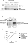
Fig 2. Pulse-chase analysis of Atco complexes.
A. Mitochondria from W303/COX6-HAC,ATP6-HApH, a strain expressing Atp6 tagged with poly histidine and Cox6 with protein C epitope was pulse-labeled with 35S-methionine/cysteine for 10 minutes and chased for 0, 30 and 60 minutes. The mitochondria were extracted with 2% digitonin and purified on Ni-NTA beads to pull down partially and fully assembled ATP synthase. The same volume aliquots of the digitonin extracts were purified on protein C antibody (PC) beads to pull down Atco complexes. The affinity purified proteins were separated by SDS-PAGE on a 12% polyacrylamide gel and by BN-PAGE on a 4–13% polyacrylamide gel. B. The proteins purified on Ni-NTA and on the protein C antibody beads were separated by BN-PAGE. C. The radiolabeled Atco complexes (1D BN gel of PC eluates), ATP synthase intermediates and Atp9 ring (1D SDS gel of PC eluate) were quantified with a phosphorimager. The Atco complexes overlap with cytochrome oxidase and Cox1 intermediates, which contribute approximately 10% of the radiolabel in that region of the 1D blue-native gel.
Fig 3
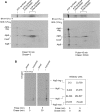
Fig 3. Pulse-chase analysis of ATP synthase.
A. Digitonin extracts of W303/COX6-HAC, ATP6-HApH mitochondria pulse-labeled with 35S-methionine/cysteine for 10 minutes and chased for 0 and 30 minutes were purified on Ni-NTA beads to pull down ATP synthase and its assembly intermediates. The purified proteins were then separated on the 1st dimension (1D) in a 4–13% polyacrylamide gel by BN-PAGE followed by separation in the 2nd (2D) dimension on a 12% gel by SDS-PAGE. B. Same as A. except that the pulse time was 5 min. Following transfer to a PVDF membrane the bands corresponding to each of the three mitochondrially encoded subunits of the fully assembled ATP synthase were quantified. In this experiment some of the ring associated with the synthase was depolymerized by SDS.
Fig 4
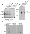
Fig 4. Effect of growth in chloramphenicol on Atco.
Mitochondria were isolated from the parental strain W303-1B and from W303/COX6-HAC and W303ΔMSS51/COX6-HAC that had been grown to early stationary phase in rich galactose and grown for an additional 2 hours in fresh medium containing 2 mg/ml chloramphenicol. Mitochondria were also isolated from W303/COX6-HAC without and with the mss51 null mutation that had not been treated with chloramphenicol. Mitochondria were labeled with 35S-methionine and cysteine for 20 min as described in the Materials and Methods section; they were extracted with digitonin at a final concentration of 2% and purified on protein C antibody beads. The digitonin extracts and purified fractions were separated by SDS-PAGE on a 12% polyacrylamide gel (A) and by BN-PAGE on a 4–13% polyacrylamide gel (B). Proteins were transferred to a PVDF membrane and exposed to X-ray film. The radiolabeled mitochondrial gene products are identified in the margins.
Fig 5
Fig 5. Analysis of Atco complexes in oxa1 and cox5 mutants.
Mitochondria were prepared from the wild type W303-1A, a strain expressing Cox6-HAC without (W303/COX6-HAC) and with a null mutation in oxa1 (W303DOXA1/COX6-HAC) or cox5a (aW303DCOX5/COX6-HAC) and an oxa1 null mutant (aW303DOXA1). The mitochondria were labeled with 35S-methionine/cysteine for 20 min, extracted with 2% digitonin and the extracts purified on protein C antibody beads. A. The digitonin extracts and the purified protein fraction in the eluates from the beads (PC eluates) were separated by SDS-PAGE on a 12% polyacrylamide gel. B. The eluates from the protein C antibody beads were separated in a single dimension on a 4–13% polyacrylamide gel by BN-PAGE. C. The eluates from the indicated strains were separated by BN-PAGE in the first dimension and the region containing Atco, by SDS-PAGE in the second dimension. The radiolabeled band of each gel is identified in the margins.
Fig 6
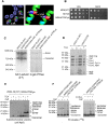
Fig 6. Cross-linking of Atp9 in Atco.
A. Ribbon structure of the wild type dimeric Atp9 (left panel) and of the V68C and S69C mutant (right panel). The inter-subunit distance of 3.3 Å between cysteines at residues 68 and 69 from two adjacent monomers is based on the structure of the Atp9 reported by Srivastava et al [24]. B. Growth of aMR6/ATP9Cys containing Atp9 with the two cysteine mutations on non-fermentable carbon sources (YEPG). The wild type parental strain MR6, the atp9 deletion mutant (RKY26) and the mutant with the cysteine modified Atp9 were grown in liquid YPD and serial dilutions spotted on YPD and YEPG media and grown for 2 days at 30°C. C. Mitochondria of the wild type (W303-1B), a strain expressing Cox6-HAC without (W303/COX-HAC) and with Atp9 with the two cysteine mutation (W303/COX6-HAC,ATP9Cys) were extracted with 2% digitonin and separated on a 4–13% polyacrylamide gel by BN-PAGE (left panel). Proteins were transferred to a PVDF membrane and reacted with a primary rabbit antibody against the β-subunit of F1. The digitonin extract was also separated on a clear native 4–13% polyacrylamide gel by CN-PAGE (right panel). The gel was incubated in the presence of 4 mM ATP and 0.05% lead acetate to stain for ATPase activity [25]. D. Mitochondria from the wild type W303-1A and a cox6 null mutant (W303ΔCOX6) were labeled with 35S-methionine/cysteine for 20 min and extracted with 2% digitonin. The extracts were analyzed in a 12% polyacrylamide gel by SDS-PAGE. One half of each sample was precipitated with 5% TCA before addition of the SDS sample buffer to depolymerize the Atp9 ring. The migration of the ATP synthase monomer and dimer are indicated in the margin. E. Mitochondria from the wild type (W303-1B) and from the strain with the V68C and S69C mutations were converted to submitochondrial particles by sonication. The submitochondrial particles were sedimented at 70,000 x gave for 10 min, suspended in sample buffer [26] without β-mercaptoethanol (β-m.e.) and separated by SDS-PAGE on a 15% polyacrylamide gel without and with a prior treatment with 1.5 mM CuP for 1 h. One half of each sample was precipitated with 5% TCA before addition of the SDS sample buffer to depolymerize the Atp9 ring. F. (Left panel) Mitochondria of an mss51 null mutant strain expressing Cox6-HAC (ΔMSS51/COX6-HAC) and wild type Atp9, and from an mss51, pet494 double mutant expressing Cox6-HAC and Atp9 with the V68C and S69C mutations (ΔMSS51,PET494/COX6-HAC/ATP9Cys) were labeled with 35S-methionine/cysteine for 20 min, extracted with 2% digitonin and purified in protein C antibody beads. The band marked with an arrow that is missing in the pet494 null mutant is Cox3 that has a tendency to non-specifically adsorb to the protein C beads. (Right panel). Mitochondria of an mss51 and pet494 double mutant expressing Cox6-HAC and Atp9 with the two cysteine mutations (ΔMSS51ΔPET494/COX6-HAC/ATP9Cys) were labeled and Atco purified protein C antibody beads as above. The purified fraction was analyzed on a 15% polyacrylamide gel by SDS-PAGE. Equal size sample were dissolved in sample buffer with and without 1% β-mercaptoethanol to reduce the disulfide bonds. The identity of the two bands marked with asterisks that remain undiminished after treatment with β-mercaptoethanol have not been identified (left panel).
Fig 7
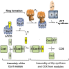
Fig 7. Model of the role of Atco complexes in coordinating assembly of cytochrome oxidase and ATP synthase.
Atco is shown to co-regulate assembly of ATP synthase and cytochrome oxidase by supplying Cox6 and mitochondrially translated Atp9 to the respective enzymes.

Similar articles

Cited by

References

    1. Schagger H, Pfeiffer K. Supercomplexes in the respiratory chains of yeast and mammalian mitochondria. EMBO J. 2000;19: 1777–1783. 10.1093/emboj/19.8.1777 - DOI - PMC - PubMed
    1. Sharma LK, Jianxin Lu J, Yidong Bai Y. Mitochondrial Respiratory Complex I, Structure, Function and Implication in Human Diseases. Curr Med Chem. 2009;16: 1266–1277. 10.2174/092986709787846578 - DOI - PMC - PubMed
    1. Luttik MA, Overkamp KM, Kotter P, De Vriess S, Van Dijken JP, Pronk JT. The Saccharomyces cerevisiae NDE1 and NDE2 genes encode separate mitochondrial NADH dehydrogenases catalyzing the oxidation of cytosolic NADH. J Biol Chem. 1998; 273: 24529–24534. 10.1074/jbc.273.38.24529 - DOI - PubMed
    1. Heinemeyer J, Braun HP, Boekema EJ, Kouril R. A structural model of the cytochrome C reductase/oxidase supercomplex from yeast mitochondria. J. Biol. Chem. 2007; 282: 12240–12248 10.1074/jbc.M610545200 - DOI - PubMed
    1. Rathore S, Berndtsson J, Marin-Buera L, Conrad J, Carroni M, Brzezinski P, et al. Cryo-EM structure of the yeast respiratory supercomplex. Nat. Struct. Mol Biol 2019;26: 50–57. 10.1038/s41594-018-0169-7 - DOI - PubMed

Publication types

MeSH terms

LinkOut - more resources