

Tyrosol as a Neuroprotector: Strong Effects of a "Weak" Antioxidant
- PMID: 32379590
- PMCID: PMC8206466
- DOI: 10.2174/1570159X18666200507082311
Tyrosol as a Neuroprotector: Strong Effects of a "Weak" Antioxidant
Abstract
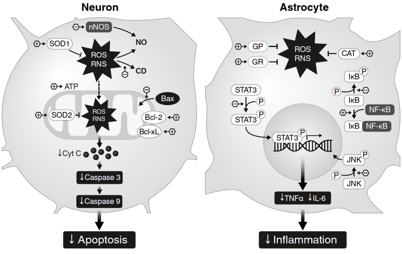
The use of neuroprotective agents for stroke is pathogenetically justified, but the translation of the results of preclinical studies of neuroprotectors into clinical practice has been a noticeable failure. One of the leading reasons for these failures is the one-target mechanism of their activity. p-Tyrosol (Tyr), a biophenol, is present in a variety of natural sources, mainly in foods, such as olive oil and wine. Tyr has a wide spectrum of biological activity: antioxidant, stress-protective, anti-inflammatory, anticancer, cardioprotective, neuroprotective and many others. This review analyzes data on the neuroprotective, antioxidant, anti-inflammatory, anti-apoptotic and other kinds of Tyr activity as well as data on the pharmacokinetics of the substance. The data presented in the review substantiate the acceptability of tyr as the basis for the development of a new neuroprotective drug with multitarget activity for the treatment of ischemic stroke. Tyr is a promising molecule for the development of an effective neuroprotective agent for use in ischemic stroke.
Keywords: Ischemic stroke; anti-apoptotic activity; anti-inflammatory activity; antioxidant activity; neuroprotective activity; p-tyrosol; pharmacokinetics.
Copyright© Bentham Science Publishers; For any queries, please email at epub@benthamscience.net.
Figures
Similar articles
-
Neuroprotective effect of tyrosol on transient focal cerebral ischemia in rats.Neurosci Lett. 2007 Mar 13;414(3):218-21. doi: 10.1016/j.neulet.2006.08.094. Epub 2006 Dec 15. Neurosci Lett. 2007. PMID: 17316989
-
Neuroprotective effects of Salidroside and its analogue tyrosol galactoside against focal cerebral ischemia in vivo and H2O2-induced neurotoxicity in vitro.Neurotox Res. 2012 May;21(4):358-67. doi: 10.1007/s12640-011-9290-7. Epub 2011 Nov 18. Neurotox Res. 2012. PMID: 22095090
-
Anti-inflammatory effect of white wine in CKD patients and healthy volunteers.Blood Purif. 2015;39(1-3):218-223. doi: 10.1159/000371570. Epub 2015 Mar 31. Blood Purif. 2015. PMID: 25833063 Clinical Trial.
-
Bioavailability of tyrosol, an antioxidant phenolic compound present in wine and olive oil, in humans.Drugs Exp Clin Res. 2003;29(5-6):203-6. Drugs Exp Clin Res. 2003. PMID: 15134375 Review.
-
Hydroxytyrosol, Tyrosol and Derivatives and Their Potential Effects on Human Health.Molecules. 2019 May 24;24(10):2001. doi: 10.3390/molecules24102001. Molecules. 2019. PMID: 31137753 Free PMC article. Review.
Cited by
-
Association between alcohol consumption and all-cause mortality, cardiovascular disease, and chronic kidney disease: A prospective cohort study.Medicine (Baltimore). 2024 Jul 5;103(27):e38857. doi: 10.1097/MD.0000000000038857. Medicine (Baltimore). 2024. PMID: 38968463 Free PMC article.
-
Gut microbiota-based pharmacokinetic-pharmacodynamic study and molecular mechanism of specnuezhenide in the treatment of colorectal cancer targeting carboxylesterase.J Pharm Anal. 2023 Sep;13(9):1024-1040. doi: 10.1016/j.jpha.2023.06.012. Epub 2023 Jun 28. J Pharm Anal. 2023. PMID: 37842660 Free PMC article.
-
Tyrosol blocks E. coli anaerobic biofilm formation via YbfA and FNR to increase antibiotic susceptibility.Nat Commun. 2024 Jul 6;15(1):5683. doi: 10.1038/s41467-024-50116-3. Nat Commun. 2024. PMID: 38971825 Free PMC article.
-
Anti-Inflammatory Effects Induced by a Polyphenolic Granular Complex from Olive (Olea europaea, Mainly Cultivar coratina): Results from In Vivo and Ex Vivo Studies in a Model of Inflammation and MIA-Induced Osteoarthritis.Nutrients. 2022 Apr 2;14(7):1487. doi: 10.3390/nu14071487. Nutrients. 2022. PMID: 35406100 Free PMC article.
-
Tyrosol improves ovalbumin (OVA)-induced asthma in rat model through prevention of airway inflammation.Naunyn Schmiedebergs Arch Pharmacol. 2021 Oct;394(10):2061-2075. doi: 10.1007/s00210-021-02117-y. Epub 2021 Jul 21. Naunyn Schmiedebergs Arch Pharmacol. 2021. PMID: 34287677
References
-
- Hachinski V., Donnan G.A., Gorelick P.B., Hacke W., Cramer S.C., Kaste M., Fisher M., Brainin M., Buchan A.M., Lo E.H., Skolnick B.E., Furie K.L., Hankey G.J., Kivipelto M., Morris J., Rothwell P.M., Sacco R.L., Smith S.C., Jr, Wang Y., Bryer A., Ford G.A., Iadecola C., Martins S.C., Saver J., Skvortsova V., Bayley M., Bednar M.M., Duncan P., Enney L., Finklestein S., Jones T.A., Kalra L., Kleim J., Nitkin R., Teasell R., Weiller C., Desai B., Goldberg M.P., Heiss W.D., Saarelma O., Schwamm L.H., Shinohara Y., Trivedi B., Wahlgren N., Wong L.K., Hakim A., Norrving B., Prudhomme S., Bornstein N.M., Davis S.M., Goldstein L.B., Leys D. Tuomilehto, J. Stroke: working toward a prioritized world agenda. Stroke. 2010;41(6):1084–1099. doi: 10.1161/STROKEAHA.110.586156. - DOI - PMC - PubMed
-
- Sarwal A., Hussain M.S., Shuaib A. In: Neuroprotection in stroke. Translational Stroke Research, Springer Series in Translational Stroke Research; Lapchak, P. Zhang J., editor. New York: Springer; 2012. pp. 79–90.
Publication types
MeSH terms
Substances
LinkOut - more resources
Full Text Sources
