. 2020 May 28;181(5):1004-1015.e15.
doi: 10.1016/j.cell.2020.04.031. Epub 2020 May 5.

Structural Basis for Potent Neutralization of Betacoronaviruses by Single-Domain Camelid Antibodies

Affiliations

Structural Basis for Potent Neutralization of Betacoronaviruses by Single-Domain Camelid Antibodies

Daniel Wrapp et al. Cell. .

Erratum in

Abstract

Coronaviruses make use of a large envelope protein called spike (S) to engage host cell receptors and catalyze membrane fusion. Because of the vital role that these S proteins play, they represent a vulnerable target for the development of therapeutics. Here, we describe the isolation of single-domain antibodies (VHHs) from a llama immunized with prefusion-stabilized coronavirus spikes. These VHHs neutralize MERS-CoV or SARS-CoV-1 S pseudotyped viruses, respectively. Crystal structures of these VHHs bound to their respective viral targets reveal two distinct epitopes, but both VHHs interfere with receptor binding. We also show cross-reactivity between the SARS-CoV-1 S-directed VHH and SARS-CoV-2 S and demonstrate that this cross-reactive VHH neutralizes SARS-CoV-2 S pseudotyped viruses as a bivalent human IgG Fc-fusion. These data provide a molecular basis for the neutralization of pathogenic betacoronaviruses by VHHs and suggest that these molecules may serve as useful therapeutics during coronavirus outbreaks.

Keywords: COVID-19; MERS; SARS; SARS-CoV-2; nanobody.


Conflict of interest statement

Declaration of Interests K.S.C., N.W., B.S.G., and J.S.M. are inventors on US patent application no. 62/412,703, entitled “Prefusion Coronavirus Spike Proteins and Their Use.” D.W., K.S.C., N.W., B.S.G., and J.S.M. are inventors on US patent application no. 62/972,886, entitled “2019-nCoV Vaccine.” D.W., D.D.V., B.S.G., B.S., X.S., and J.S.M. are inventors on US patent application no. 62/988,610, entitled “Coronavirus Binders.” D.W., L.v.S., N.C., B.S., X.S., and J.S.M. are inventors on US patent application no. 62/991,408, entitled “SARS-CoV-2 Virus Binders.”

Figures

None
Graphical abstract
Figure S1
Figure S1
CoV VHH Immunization and Panning, Related to Figure 1 (A) Schematic depicting the immunization strategy that was used to isolate both SARS-CoV-1 S and MERS-CoV S-directed VHHs from a single llama. The prefusion stabilized SARS-CoV-1 spike is shown in pink and the prefusion stabilized MERS-CoV spike is shown in tan. (B) Phylogenetic tree of the isolated MERS-CoV and SARS-CoV S-directed VHHs, based on the neighbor joining method. (C) Reactivity of MERS-CoV and SARS-CoV S-directed VHHs with the prefusion stabilized MERS-CoV S and SARS-CoV-1 S protein, respectively. A VHH against an irrelevant antigen (F-VHH) was included as a control.
Figure S2
Figure S2
Sequence Alignment of Neutralizing SARS-CoV and MERS-CoV S-Directed VHHs, Related to Figure 1 Invariant residues are shown as black dots. The CDRs are shown in boxes and Kabat numbering is shown above.
Figure 1
Figure 1
Epitope Determination and Biophysical Characterization of MERS VHH-55 and SARS VHH-72 (A) Reactivity of MERS-CoV and SARS-CoV RBD-directed VHHs against the MERS-CoV and SARS-CoV-1 RBD, respectively. A VHH against an irrelevant antigen (F-VHH) was included as a control. Datapoints represent the mean of three replicates and error bars represent the standard errors of the mean. (B) SPR sensorgrams showing binding between the MERS-CoV RBD and MERS VHH-55 (left) and SARS-CoV-1 RBD and SARS VHH-72 (right). Binding curves are colored black, and fit of the data to a 1:1 binding model is colored red.
Figure S3
Figure S3
Lack of Binding of MERS-CoV and SARS-CoV-Directed VHHs to Non-RBD Epitopes, Related to Figure 1 ELISA data showing binding of the MERS-CoV specific VHHs to the MERS-CoV S1 protein and absence of binding of the MERS-CoV and SARS-CoV specific VHHs against the MERS-CoV NTD and SARS-CoV-1 NTD, respectively. A VHH against an irrelevant antigen (F-VHH) was included as a control.
Figure 2
Figure 2
The Crystal Structure of MERS VHH-55 Bound to the MERS-CoV RBD (A) MERS VHH-55 is shown as blue ribbons and the MERS-CoV RBD is shown as a tan-colored molecular surface. The DPP4 binding interface on the MERS-CoV RBD is colored red. (B) The structure of DPP4 bound to the MERS-CoV RBD (PDB ID: 4L72) is aligned to the crystal structure of MERS VHH-55 bound to the MERS-CoV RBD. A single monomer of DPP4 is shown as a red, transparent molecular surface. (C) A zoomed-in view of the panel from (A), with the MERS-CoV RBD now displayed as tan-colored ribbons. Residues that form interactions are shown as sticks, with nitrogen atoms colored dark blue and oxygen atoms colored red. Hydrogen-bonds and salt bridges between MERS VHH-55 and the MERS-CoV RBD are shown as black dots. (D) The same view from (C) has been turned by approximately 90° to show additional contacts. Residues that form interactions are shown as sticks, with nitrogen atoms colored dark blue and oxygen atoms colored red. Hydrogen bonds and salt bridges between MERS VHH-55 and the MERS-CoV RBD are shown as black dots.
Figure S4
Figure S4
MERS VHH-55 Binds to a Relatively Conserved Epitope on the MERS-CoV RBD, Related to Figure 2 (A) The crystal structure of MERS VHH-55 bound to the MERS-CoV RBD is shown with MERS VHH-55 in white ribbons and the MERS-CoV RBD as a multicolored molecular surface. More variable residues are shown in warm colors and more conserved residues are shown in cool colors according to the spectrum (bottom). Sequence alignments and variability mapping was performed using ConSurf. (B) The crystal structure of MERS VHH-55 bound to the MERS-CoV RBD is shown as ribbons with MERS VHH-55 colored blue and the MERS-CoV RBD colored tan. Phe506 from the MERS-CoV RBD and Trp99 from MERS VHH-55, which are thought to form hydrophobic interactions with one another are shown as sticks surrounded by a transparent molecular surface. (C) SPR sensorgram measuring the binding of MERS VHH-55 to the naturally occurring MERS-CoV RBD F506L variant. Binding curves are colored black and the fit of the data to a 1:1 binding model is colored red.
Figure 3
Figure 3
The Crystal Structure of SARS VHH-72 Bound to the SARS-CoV-1 RBD (A) SARS VHH-72 is shown as dark blue ribbons and the SARS-CoV-1 RBD is shown as a pink-colored molecular surface. The ACE2 binding interface on the SARS-CoV-1 RBD is colored red. (B) The structure of ACE2 bound to the SARS-CoV-1 RBD (PDB ID: 2AJF) is aligned to the crystal structure of SARS VHH-72 bound to the SARS-CoV-1 RBD. ACE2 is shown as a red, transparent molecular surface. (C) A simulated N-linked glycan containing an energy-minimized trimannosyl core (derived from PDB ID: 1HD4) is modeled as red sticks, coming from Asn322 in ACE2. ACE2 is shown as a red molecular surface, the SARS-CoV-1 RBD is shown as pink ribbons, and SARS VHH-72 is shown as a dark blue, transparent molecular surface. (D) A zoomed-in view of the panel from (A) is shown, with the SARS-CoV-1 RBD now displayed as pink-colored ribbons. Residues that form interactions are shown as sticks, with nitrogen atoms colored dark blue and oxygen atoms colored red. Hydrogen bonds and salt bridges between SARS VHH-72 and the SARS-CoV-1 RBD are shown as black dots. (E) The same view from (D) has been turned by 60° to show additional contacts. Residues that form interactions are shown as sticks, with nitrogen atoms colored dark blue and oxygen atoms colored red. Interactions between SARS VHH-72 and the SARS-CoV-1 RBD are shown as black dots.
Figure S5
Figure S5
SARS VHH-72 Binds to a Broadly Conserved Epitope on the SARS-CoV-1 RBD, Related to Figure 3 (A) The crystal structure of SARS VHH-72 bound to the SARS-CoV-1 RBD is shown, with colors corresponding to those of Figure S4A. (B) The crystal structure of SARS VHH-72 bound to the SARS-CoV-1 RBD is shown with SARS VHH-72 as dark blue ribbons and the RBD as a pink molecular surface. Amino acids that vary between SARS-CoV-1 and WIV1-CoV are colored teal. (C) SPR sensorgram measuring the binding of SARS VHH-72 to the WIV1-CoV RBD. Binding curves are colored black and the fit of the data to a 1:1 binding model is colored red.
Figure 4
Figure 4
SARS VHH-72 Cross-Reacts with SARS-CoV-2 (A) An SPR sensorgram measuring the binding of SARS VHH-72 to the SARS-CoV-2 RBD-SD1. Binding curves are colored black, and fit of the data to a 1:1 binding model is colored red. (B) The crystal structure of SARS VHH-72 bound to the SARS-CoV-1 RBD is shown with SARS VHH-72 as dark blue ribbons and the RBD as a pink molecular surface. Amino acids that vary between SARS-CoV-1 and SARS-CoV-2 are colored green.
Figure 5
Figure 5
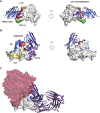
Neutralizing Mechanisms of MERS VHH-55 and SARS VHH-72 (A) The MERS-CoV spike (PDB ID: 5W9H) is shown as a transparent molecular surface, with each monomer colored either white, gray, or tan. Each monomer is bound by MERS VHH-55, shown as blue ribbons. The clash between MERS VHH-55 bound to the white monomer and the neighboring tan RBD is highlighted by the red ellipse. (B) The SARS-CoV-1 spike (PDB ID: 5X58) is shown as a transparent molecular surface, with each protomer colored either white, gray, or pink. Every monomer is bound by a copy of SARS VHH-72, shown as dark blue ribbons. The clashes between copies of SARS VHH-72 and the two neighboring spike monomers are highlighted by the red circle. (C) The SARS-CoV-2 spike (PDB ID: 6VXX) is shown as a transparent molecular surface, with each protomer colored either white, gray, or green. Every monomer is bound by a copy of SARS VHH-72, shown as dark blue ribbons. The clashes between copies of SARS VHH-72 and the two neighboring spike monomers are highlighted by the red circle. The SARS-CoV-2 trimer appears smaller than SARS-CoV-1 S because of the absence of flexible NTD-distal loops, which could not be built during cryo-EM analysis. (D) CoV VHHs prevent MERS-CoV RBD, SARS-CoV-1 RBD, and SARS-CoV-2 RBD-SD1 from interacting with their receptors. The results of the BLI-based receptor-blocking experiment are shown. The legend lists the immobilized RBDs and the VHHs or receptors that correspond to each curve.
Figure 6
Figure 6
SARS VHH-72 Bivalency Permits SARS-CoV-2 Pseudovirus Neutralization (A and B) SARS-CoV-1 S (A) and SARS-CoV-2 S (B) VSV pseudoviruses were used to evaluate the neutralization capacity of SARS VHH-72 and SARS VHH-6. MERS VHH-55 and PBS were included as negative controls. Luciferase activity is reported (n = 3 ±SEM) in counts per second (c.p.s.). NI, cells were not infected. (C and D) Binding of monovalent and bivalent VHHs was tested by ELISA against SARS-CoV-1 S (C) and SARS-CoV-2 RBD-SD1 (D). VHH-72-Fc refers to SARS VHH-72 fused to a human IgG1 Fc domain by a GS(GGGGS)2 linker. VHH-72-Fc (S) is the same Fc fusion with a GS, rather than a GS(GGGGS)2, linker. GBP is an irrelevant GFP-binding protein. VHH-72-VHH-72 refers to the tail-to-head construct with two SARS VHH-72 proteins connected by a (GGGGS)3 linker. VHH-23-VHH-23 refers to the two irrelevant VHHs linked via the same (GGGGS)3 linker. (E and F) SARS-CoV-1 S (E) and SARS-CoV-2 S (F) pseudoviruses were used to evaluate the neutralization capacity of bivalent VHH-72-Fc. GBP and PBS were included as negative controls. NI, cells were not infected.
Figure S6
Figure S6
Engineering a Functional Bivalent VHH Construct, Related to Figure 6 (A) Flow cytometry measuring the binding of the bivalent SARS VHH-72 tail-to-head fusion (VHH-72-VHH-72) to SARS-CoV-1 or SARS-CoV-2 S expressed on the cell surface. VHH-23-VHH-23, a bivalent tail-to-head fusion of an irrelevant nanobody, was included as a negative control. (B) Binding of SARS-CoV-2 RBD-SD1 to Vero E6 cells is prevented by VHH-72-VHH-72 in a dose-dependent fashion. Binding of SARS-CoV-2 RBD-SD1 to Vero E6 cells was detected by flow cytometry in the presence of the indicated bivalent VHHs (n = 2 except VHH-72-VHH-72 and VHH-23-VHH-23 at 5 μg/mL, n = 5). (C) Binding of SARS-CoV-2 RBD-SD1 to Vero E6 cells is prevented by bivalent VHH-72-Fc fusion proteins in a dose-dependent fashion. Binding of SARS-CoV-2 RBD-SD1-Fc to Vero E6 cells was detected by flow cytometry in the presence of the indicated constructs and amounts (n = 2 except no RBD, n = 4). (D) Cell surface binding of SARS VHH-72 and SARS VHH-6 to SARS-CoV-1 S. 293S cells were transfected with a GFP expression plasmid together with a SARS-CoV-1 S expression plasmid. Binding of the indicated protein at the indicated concentration is expressed as the median fluorescent intensity (MFI), measured to detect the His-tagged MERS VHH-55 or SARS VHH-6 or SARS VHH-72 or the SARS VHH-72-Fc fusions, of the GFP positive cells divided by the MFI of the GFP negative cells. (E) Cell surface binding of SARS VHH-72 to SARS-CoV-2. MFI was calculated using the same equation as Figure S6D.
Figure 7
Figure 7
VHH-72-Fc Neutralizes SARS-CoV-2 S Pseudoviruses (A) BLI sensorgram measuring apparent binding affinity of VHH-72-Fc to immobilized SARS-CoV-2 RBD-Fc. Binding curves are colored black, buffer-only blanks are colored gray, and the fit of the data to a 1:1 binding curve is colored red. (B) Time course analysis of VHH-72-Fc expression in ExpiCHO cells. Cell culture supernatants of transiently transfected ExpiCHO cells were removed on days 3–7 after transfection (or until cell viability dropped below 75%), as indicated. Two control mAbs were included for comparison, along with the indicated amounts of purified GBP-Fc as a loading control. (C) SARS-CoV-2 S pseudotyped VSV neutralization assay. Monolayers of Vero E6 cells were infected with pseudoviruses that had been pre-incubated with the mixtures indicated by the legend. The VHH-72-Fc used in this assay was purified after expression in ExpiCHO cells (n = 4). VHH-23-Fc is an irrelevant control VHH-Fc (n = 3). NI, cells were not infected. Luciferase activity is reported in counts per second (c.p.s.) ± SEM.
Figure S7
Figure S7
Comparison of the CoV VHH Epitopes with Known RBD-Directed Antibodies, Related to Figures 2 and 3 (A) The structure of MERS VHH-55 bound to the MERS-CoV RBD is shown with MERS VHH-55 as blue ribbons and the MERS-CoV RBD as a white molecular surface. Epitopes from previously reported crystal structures of the MERS-CoV RBD bound by RBD-directed antibodies are shown as colored patches on the MERS-CoV RBD surface. The LCA60 epitope is shown in yellow, the MERS S4 epitope is shown in green, the overlapping C2/MCA1/m336 epitopes are shown in red and the overlapping JC57-14/D12/4C2/MERS-27 epitopes are shown in purple. (B) The structure of SARS VHH-72 bound to the SARS-CoV-1 RBD is shown with SARS VHH-72 as dark blue ribbons and the SARS-CoV-1 RBD as a white molecular surface. Epitopes from previously reported crystal structures of the SARS-CoV-1 RBD bound by RBD-directed antibodies are shown as colored patches on the SARS-CoV-1 RBD surface. The 80R epitope is shown in blue, the S230 epitope is shown in yellow, the CR3022 epitope is shown in purple and the overlapping m396/F26G19 epitopes are shown in red. (C) The SARS-CoV-1 RBD is shown as a white molecular surface, ACE2 is shown as a transparent red molecular surface, SARS VHH-72 is shown as dark blue ribbons and CR3022 Fab is shown as purple ribbons.

Comment in

Similar articles

  • Nanobodies from camelid mice and llamas neutralize SARS-CoV-2 variants.
    Xu J, Xu K, Jung S, Conte A, Lieberman J, Muecksch F, Lorenzi JCC, Park S, Schmidt F, Wang Z, Huang Y, Luo Y, Nair MS, Wang P, Schulz JE, Tessarollo L, Bylund T, Chuang GY, Olia AS, Stephens T, Teng IT, Tsybovsky Y, Zhou T, Munster V, Ho DD, Hatziioannou T, Bieniasz PD, Nussenzweig MC, Kwong PD, Casellas R. Xu J, et al. Nature. 2021 Jul;595(7866):278-282. doi: 10.1038/s41586-021-03676-z. Epub 2021 Jun 7. Nature. 2021. PMID: 34098567 Free PMC article.
  • An alpaca nanobody neutralizes SARS-CoV-2 by blocking receptor interaction.
    Hanke L, Vidakovics Perez L, Sheward DJ, Das H, Schulte T, Moliner-Morro A, Corcoran M, Achour A, Karlsson Hedestam GB, Hällberg BM, Murrell B, McInerney GM. Hanke L, et al. Nat Commun. 2020 Sep 4;11(1):4420. doi: 10.1038/s41467-020-18174-5. Nat Commun. 2020. PMID: 32887876 Free PMC article.
  • Cross-neutralization of SARS-CoV-2 by a human monoclonal SARS-CoV antibody.
    Pinto D, Park YJ, Beltramello M, Walls AC, Tortorici MA, Bianchi S, Jaconi S, Culap K, Zatta F, De Marco A, Peter A, Guarino B, Spreafico R, Cameroni E, Case JB, Chen RE, Havenar-Daughton C, Snell G, Telenti A, Virgin HW, Lanzavecchia A, Diamond MS, Fink K, Veesler D, Corti D. Pinto D, et al. Nature. 2020 Jul;583(7815):290-295. doi: 10.1038/s41586-020-2349-y. Epub 2020 May 18. Nature. 2020. PMID: 32422645
  • Therapeutic nanobodies against SARS-CoV-2 and other pathogenic human coronaviruses.
    Yang Y, Li F, Du L. Yang Y, et al. J Nanobiotechnology. 2024 May 31;22(1):304. doi: 10.1186/s12951-024-02573-7. J Nanobiotechnology. 2024. PMID: 38822339 Free PMC article. Review.
  • Targeting SARS-CoV2 Spike Protein Receptor Binding Domain by Therapeutic Antibodies.
    Hussain A, Hasan A, Nejadi Babadaei MM, Bloukh SH, Chowdhury MEH, Sharifi M, Haghighat S, Falahati M. Hussain A, et al. Biomed Pharmacother. 2020 Oct;130:110559. doi: 10.1016/j.biopha.2020.110559. Epub 2020 Aug 1. Biomed Pharmacother. 2020. PMID: 32768882 Free PMC article. Review.

Cited by

References

    1. Adams P.D., Grosse-Kunstleve R.W., Hung L.W., Ioerger T.R., McCoy A.J., Moriarty N.W., Read R.J., Sacchettini J.C., Sauter N.K., Terwilliger T.C. PHENIX: building new software for automated crystallographic structure determination. Acta Crystallogr. D Biol. Crystallogr. 2002;58:1948–1954. - PubMed
    1. Afonine P.V., Poon B.K., Read R.J., Sobolev O.V., Terwilliger T.C., Urzhumtsev A., Adams P.D. Real-space refinement in PHENIX for cryo-EM and crystallography. Acta Crystallogr D. Struct. Biol. 2018;74:531–544. - PMC - PubMed
    1. Battye T.G., Kontogiannis L., Johnson O., Powell H.R., Leslie A.G. iMOSFLM: a new graphical interface for diffraction-image processing with MOSFLM. Acta Crystallogr. D Biol. Crystallogr. 2011;67:271–281. - PMC - PubMed
    1. Berger Rentsch M., Zimmer G. A vesicular stomatitis virus replicon-based bioassay for the rapid and sensitive determination of multi-species type I interferon. PLoS ONE. 2011;6:e25858. - PMC - PubMed
    1. Bosch B.J., van der Zee R., de Haan C.A., Rottier P.J. The coronavirus spike protein is a class I virus fusion protein: structural and functional characterization of the fusion core complex. J. Virol. 2003;77:8801–8811. - PMC - PubMed

Publication types

MeSH terms