. 2020 Aug;14(8):1779-1799.
doi: 10.1002/1878-0261.12701. Epub 2020 Jun 28.

Estrogen receptor α promotes lung cancer cell invasion via increase of and cross-talk with infiltrated macrophages through the CCL2/CCR2/MMP9 and CXCL12/CXCR4 signaling pathways

Affiliations

Estrogen receptor α promotes lung cancer cell invasion via increase of and cross-talk with infiltrated macrophages through the CCL2/CCR2/MMP9 and CXCL12/CXCR4 signaling pathways

Miao He et al. Mol Oncol. 2020 Aug.

Abstract

Data analysis of clinical samples suggests that higher estrogen receptor α (ERα) expression could be associated with worse overall survival in some patients with non-small-cell lung cancer (NSCLC). Immunofluorescence results further showed that higher ERα expression was linked to larger numbers of infiltrated macrophages in NSCLC tissues. However, the detailed mechanisms underlying this phenomenon remain unclear. Results from in vitro studies with multiple cell lines revealed that, in NSCLC cells, ERα can activate the CCL2/CCR2 axis to promote macrophage infiltration, M2 polarization, and MMP9 production, which can then increase NSCLC cell invasion. Mechanistic studies using chromatin immunoprecipitation and promoter luciferase assays demonstrated that ERα could bind to estrogen response elements (EREs) on the CCL2 promoter to increase CCL2 expression. Furthermore, ERα-increased macrophage infiltration can induce a positive feedback mechanism to increase lung cancer cell ERα expression via the up-regulation of the CXCL12/CXCR4 pathway. Targeting these newly identified pathways, NSCLC ERα-increased macrophage infiltration or the macrophage-to-NSCLC CXCL12/CXCR4/ERα signal, with anti-estrogens or CCR2/CXCR4 antagonists, may help in the development of new alternative therapies to better treat NSCLC.

Keywords: estrogen receptor α; macrophage; non-small-cell lung cancer.


Conflict of interest statement

The authors declare no conflict of interest.

Figures
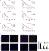
Fig. 1
Fig. 1
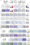
Estrogen receptor α is correlated with worse prognosis and macrophage infiltration in early‐stage NSCLC patients. (A,B) Survival curve based on TCGA database indicated that high levels of ERα mRNA are associated with shorter overall survival in both LUAD and LUSC. (C–F) Differential analysis based on gender showed that ERα mRNA is associated with poorer overall survival both in male (C, E) and in female (D, F) NSCLC patients. (G) Immunofluorescence assay comparing ERα, CD68, and CD163 in human ERα‐positive and ERα‐negative NSCLC samples. Nuclei (blue), ERα (red), CD68 (green), and CD163 (green) were shown. 200× magnification was shown for the left image, and 400× magnification was shown for the right image. Log‐rank (Mantel–Cox) test was applied to compare survival data in Fig. 1A–F, and Student’s t‐test was used to compare the means between groups in Fig. 1G. Experiments were done at least in 3 replicates. Results were presented as mean ± SD, ***P < 0.001.
Fig. 2
Fig. 2
E2/ERα signals in lung cancer cells could promote the macrophage recruitment to induce cancer invasion. (A) The carton (left) illustrates macrophage migration systems. PMA‐activated THP‐1 or primary B6 Mφ cells were seeded in the upper chambers, and lung cancer cells were seeded in the lower chambers of the 24‐well transwell migration systems. Migrated THP‐1 cells/B6 Mφ were checked after 48 h incubation. (B) E2 pretreated ERα‐positive cells can promote the macrophage recruitment. H1299 (vector or shERα #1, #2), A549 (vector or oeERα), or LLC1 (vector, or shERα #1, or shERα #2) cultured in charcoal‐stripped FBS media were treated with control and 10 nm E2, with/without 1 μm MPP, with/without 10 μm fulvestrant for 48 h, and then seeded in the lower chamber of transwell migration systems for checking the ability to recruit THP‐1 cells or B6 Mφ, respectively. (C) CMs were collected from coculture of lung cancer cells with/without THP‐1 or B6 Mφ cells (4 : 1) for 48 h. The CMs were then added to the bottom well of 24‐well plates. Lung cancer cells were seeded into inserted transwells precoated with Matrigel. After 24 h incubation, invaded lung cancer cells were counted and compared. (D) CMs were collected from coculture of A549 (vector or oeERα) or H1299 (vector, or shERα #1, or shERα #2) cells with PMA‐activated THP‐1 cells. The CMs were then added in the lower chambers of transwells to test the effect on lung cancer cell (A549 and H1299) invasion. (E) CMs were collected from coculture of LLC1 (vector or oeERα) or LLC1 (vector, or shERα #1, or shERα #2) cells with primary B6 Mφ cells. The CMs were then added in the lower chambers of transwells to test the effect on lung cancer cell (LLC1) invasion. 100 × magnification of images was shown for the migration and invasion assay. Student’s t‐test was used to analyze data in Fig. 2A–E. Experiments were done at least in 3 replicates. Results were presented as mean ± SD, **P < 0.01, ***P < 0.001. ns, not significant.
Fig. 3
Fig. 3
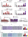
Estrogen receptor α in lung cancer cells can promote the M2 polarization and macrophage MMP9 production to facilitate cancer cell invasion. (A) THP‐1 cells were collected after coculture with A549 (upper left, vector or oeERα) and H1299 (upper right, vector or shERα) human lung cancer cells, and B6 Mφ cells were collected after coculture with LLC1 (lower left, vector or oeERα) and LLC1 (lower right, vector or shERα) mouse lung cancer cells, in order to test the change of macrophage polarization markers. (B) A group of proteases reported to promote cancer cell invasion were tested on THP‐1 cells for mRNA expression change after they were cocultured with A549 (left, vector or oeERα) and H1299 (middle, vector or shERα) by qPCR. Western blot was done to confirm the protein expression change (right). (C,D) CMs were collected from THP‐1 cells (vector or shMMP9) cocultured with (C) A549 (vector or oeERα) and (D) H1299 (vector or oeERα) to test the effect on lung cancer cell invasion. 100× magnification was shown for the images in the invasion assay. Student’s t‐test was used to compare the means between groups in A–D. Experiments were done in at least 3 replicates. Results were presented as mean ± SD, *P < 0.05, **P < 0.01, ***P < 0.001.
Fig. 4
Fig. 4
Estrogen receptor α in lung cancer cells could increase CCL2 expression to promote infiltration and MMP9 production of macrophages. (A) A group of chemokines and cytokines related to macrophage recruitment and polarization were tested by qPCR through overexpression and knockdown of ERα in A549 and H1299, respectively (left and middle). Western blots confirm that CCL2 expression is correlated with ERα expression (right). (B) Dose‐dependent increase of CCL2 mRNA levels in H1299 cells with serial doses of E2 treatments for 24h. (C) CCL2 mRNA levels were checked in H1299 cells treated with vehicle or E2 (10 nm), and with/without MPP (1 μm), for 24 h. (D) Western blot assays confirmed the efficiency of knockdown and overexpression of CCL2 in A549 (vector or oeERα) and H1299 (vector, or shERα#1, or shERα#2), respectively. (E,F) CCL2 was knocked down or overexpressed in A549 (vector or oeERα, E) or H1299 (vector, or shERα#1, or shERα#2, F) cells to test the effect on PMA‐treated THP‐1 infiltration. (G) A549 (vector or oeERα) cells were cultured in the lower wells and incubated with 25 nm CCR2 antagonist or DMSO. After 24 h, PMA‐treated THP‐1 cells were added to the upper wells for 48 h. Both the top and bottom wells contained 25 nm CCR2 antagonist. The migrated THP‐1 cells were counted and compared. (H,I) THP‐1 cells were cocultured with A549 (vector or oeERα) +/− CCR2 antagonist for 48 h to test the change of mRNA level change of macrophage polarization markers (H), and MMP9 (I). (J) Western blot assays to detect the THP‐1 MMP9 production after cocultured with A549 (vector or oeERα) with or without CCL2‐shRNA or with H1299 (vector or shERα #1, #2) with or without CCL2‐cDNA. (K,L) CMs were collected from THP‐1 cells cocultured with A549 (vector or oeERα) cells +/− CCL2‐shRNA (K), or +/− CCR2 antagonist (L). After 48 h coculture or treatment(s), CMs were collected to test their effects on A549 cell invasion. The images with 100 × magnification were shown for the migration and invasion assay. Student’s t‐test was used to analyze data in A–C, E–I, K–L. Experiments were done in at least 3 replicates. Results were presented as mean ± SD, *P < 0.05. **P < 0.01. ***P < 0.001.
Fig. 5
Fig. 5
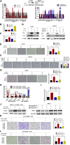
Estrogen receptor α transcriptionally regulates CCL2 production in lung cancer cells. (A) Illustration of the potential ERE sites on the CCL2 promoter region. (B) ChIP assays were performed to show ERα binding on the proposed ERE3 on CCL2 promoter region. (C) Luciferase reporter assays were used to determine whether ERα transcriptionally regulates the CCL2 promoter. (D,E) Wild‐type or mutant CCL2 promoter (ERE3)‐luciferase reporter was transfected into A549 with vector or oeERα (D), or into H1299 with vector or shERα (E). CCL2 promoter reporter activity was analyzed using the Dual‐Luciferase Assay. Experiments were done at least in 3 replicates. Results were presented as mean ± SD, and P values were calculated by Student’s t‐test. *P < 0.05, **P < 0.01, ***P < 0.001. ns, not significant.
Fig. 6
Fig. 6
Infiltrated macrophages induce the expressions of ERα and its downstream gene CCL2 in lung cancer cells. (A,B) A549 and H1299 cells were cultured in control media or THP‐1 conditioned media for 48 h, and LLC1 cells were cultured in control media or primary B6 Mφ‐conditioned media for 48 h to test the ERα expression by western blots (A), as well as CCL2 mRNA by qPCR (B). (C,D) We collected lung cancer cells from coculture of increasing ratio of THP‐1 cells for 48 h and tested the expression of ERα in lung cancer cells by western blots (C) and CCL2 mRNA expression by qPCR (D). Student’s t‐test was used to analyze data in (B) and (D). Experiments were done at least in 3 replicates. Results were presented as mean ± SD, *P < 0.05, **P < 0.01, ***P < 0.001.
Fig. 7
Fig. 7
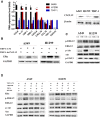
Infiltrated macrophages induce ERα expression in lung cancer cells through CXCL12. (A) We tested some cytokines that have been reported to increase ERα expression between lung cancer cells and macrophages (left). Western blot was done to confirm the protein expression change (right). (B) We collected THP‐1 (vehicle or shCXCL12)‐CM to culture lung cancer cells for 48 h and tested the change of effect on ERα expression. (C) Downstream pathway of CXCL12 was tested in A549 and H1299 cells after coculture in THP‐1‐CM for 48 h. (D) Specific antagonist of CXCR4 (AMD3100, 1µM), ERK phosphorylation (U0126, 10 μm), and Akt phosphorylation (MK2206, 1 µm) was added during culture of lung cancer cells, and the effect of ERα expression, as well as ERK phosphorylation and Akt phosphorylation, was tested by western blot. Experiments were done at least in 3 replicates. Student’s t‐test was used to analyze data in 7A. Results were presented as mean ± SD, *P < 0.05, **P < 0.01, ***P < 0.001.
Fig. 8
Fig. 8
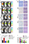
Estrogen receptor α promotes lung cancer progression using the in vivo mouse model. (A) Representative IVIS imaging for mice with A549‐Luc cells, with or without oeERα, after the respective treatments. (B) After 4 weeks of treatments, the mice were sacrificed and tumor weights were measured. (C,D) The IHC staining (C), and quantification (D) for detection of ERα and F4/80 expressions in different groups. 200× magnification was shown for the upper image, and 400× magnification was shown for the lower image for the IHC results. Student’s t‐test was used to analyze data in B,D. Experiments were done at least in 3 replicates. Results were presented as mean ± SD, ***P < 0.001.
Fig. 9
Fig. 9
Schematic illustration of the cross‐talk between lung cancer cells and macrophages showing that (i) a higher ERα activity in lung cancer cells can promote the infiltration of macrophage via an increased CCL2 secretion, which can secrete MMP9 to promote lung cancer cell invasion; (ii) the infiltrated macrophages can secrete CXCL12 to bind to lung cancer cell surface gene CXCR4 to increase ERα expression.

Similar articles

Cited by

References

    1. Albain KS, Crowley JJ, LeBlanc M and Livingston RB (1991) Survival determinants in extensive‐stage non‐small‐cell lung cancer: the Southwest Oncology Group experience. J Clin Oncol 9, 1618–1626. - PubMed
    1. Ao J‐Y, Zhu X‐D, Chai Z‐T, Cai H, Zhang Y‐Y, Zhang K‐Z, Kong L‐Q, Zhang N, Ye B‐G, Ma D‐N et al (2017) Colony‐stimulating factor 1 receptor blockade inhibits tumor growth by altering the polarization of tumor‐associated Macrophages in Hepatocellular Carcinoma. Mol Cancer Ther 16, 1544–1554. - PubMed
    1. Arendt LM, McCready J, Keller PJ, Baker DD, Naber SP, Seewaldt V and Kuperwasser C (2013) Obesity promotes breast cancer by CCL2‐mediated macrophage recruitment and angiogenesis. Cancer Res 73, 6080–6093. - PMC - PubMed
    1. Bröker LE, Huisman C, Span SW, Rodriguez JA, Kruyt FAE and Giaccone G (2004) Cathepsin B Mediates caspase‐independent cell death induced by microtubule stabilizing agents in non‐small cell lung cancer cells. Can Res 64, 27–30. - PubMed
    1. Brueckl WM, Al‐Batran SE, Ficker JH, Claas S, Atmaca A, Hartmann A, Rieker RJ and Wirtz RM (2013) Prognostic and predictive value of estrogen receptor 1 expression in completely resected non‐small cell lung cancer. Int J Cancer 133, 1825–1831. - PubMed

MeSH terms