

LncRNA HOTTIP facilitates the stemness of breast cancer via regulation of miR-148a-3p/WNT1 pathway
- PMID: 32307830
- PMCID: PMC7294123
- DOI: 10.1111/jcmm.15261
LncRNA HOTTIP facilitates the stemness of breast cancer via regulation of miR-148a-3p/WNT1 pathway
Abstract
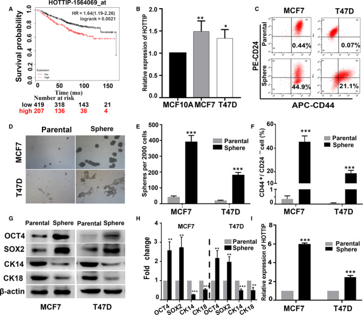
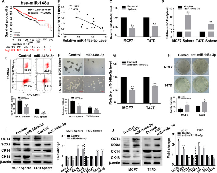
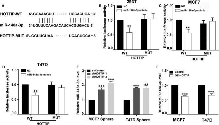
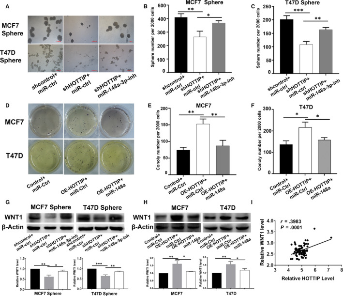
Emerging evidence suggests that dysregulation of long non-coding RNA (lncRNA) plays a key role in tumorigenesis. The lncRNA, HOXA transcript at the distal tip (HOTTIP), has been reported to be up-regulated in multiple cancers, including breast cancer, and is involved in various biological processes, including the maintenance of stemness. However, the biological function and underlying modulatory mechanism of HOTTIP in breast cancer stem cells (BCSCs) remains unknown. In this study, we found that HOTTIP was markedly up-regulated in BCSCs and had a positive correlation with breast cancer progression. Functional studies revealed that overexpression of HOTTIP markedly promoted cell clonogenicity, increased the expression of the stem cell markers, OCT4 and SOX2, and decreased the expression of the differentiation markers, CK14 and CK18, in breast cancer cells. Knockdown of HOTTIP inhibited the CSC-like properties of BCSCs. Consistently, depletion of HOTTIP suppressed tumour growth in a humanized model of breast cancer. Mechanistic studies demonstrated that HOTTIP directly binds to miR-148a-3p and inhibits the mediation of WNT1, which leads to inactivation of the Wnt/β-catenin signalling pathway. Our study is the first to report that HOTTIP regulates the CSC-like properties of BCSCs by as a molecular sponge for miR-148a-3p to increase WNT1 expression, offering a new target for breast cancer therapy.
Keywords: HOXA transcript at the distal tip (HOTTIP); WNT1; breast cancer; miR-148a-3p; stemness.
© 2020 The Authors. Journal of Cellular and Molecular Medicine published by Foundation for Cellular and Molecular Medicine and John Wiley & Sons Ltd.
Conflict of interest statement
All authors declare that they have no competing interests.
Figures


Similar articles
-
MicroRNA-148a-3p suppresses epithelial-to-mesenchymal transition and stemness properties via Wnt1-mediated Wnt/β-catenin pathway in pancreatic cancer.J Cell Mol Med. 2020 Nov;24(22):13020-13035. doi: 10.1111/jcmm.15900. Epub 2020 Oct 7. J Cell Mol Med. 2020. PMID: 33026174 Free PMC article.
-
LncCCAT1 Promotes Breast Cancer Stem Cell Function through Activating WNT/β-catenin Signaling.Theranostics. 2019 Oct 1;9(24):7384-7402. doi: 10.7150/thno.37892. eCollection 2019. Theranostics. 2019. PMID: 31695775 Free PMC article.
-
Long non-coding RNA LUCAT1/miR-5582-3p/TCF7L2 axis regulates breast cancer stemness via Wnt/β-catenin pathway.J Exp Clin Cancer Res. 2019 Jul 12;38(1):305. doi: 10.1186/s13046-019-1315-8. J Exp Clin Cancer Res. 2019. PMID: 31300015 Free PMC article.
-
Tumour-regulatory role of long non-coding RNA HOXA-AS3.Prog Biophys Mol Biol. 2024 Jul;189:13-25. doi: 10.1016/j.pbiomolbio.2024.04.003. Epub 2024 Apr 7. Prog Biophys Mol Biol. 2024. PMID: 38593905 Review.
-
LncRNAs and microRNAs as Essential Regulators of Stemness in Breast Cancer Stem Cells.Biomolecules. 2021 Mar 3;11(3):380. doi: 10.3390/biom11030380. Biomolecules. 2021. PMID: 33802575 Free PMC article. Review.
Cited by
-
The expression and biological role of complement C1s in esophageal squamous cell carcinoma.Open Life Sci. 2024 Jul 24;19(1):20220915. doi: 10.1515/biol-2022-0915. eCollection 2024. Open Life Sci. 2024. PMID: 39071493 Free PMC article.
-
Long non-coding RNA HOX transcript antisense intergenic RNA depletion protects against alcoholic hepatitis through the microRNA-148a-3p/sphingosine 1-phosphate receptor 1 axis.Cell Tissue Res. 2023 Dec;394(3):471-485. doi: 10.1007/s00441-023-03835-w. Epub 2023 Oct 18. Cell Tissue Res. 2023. PMID: 37851113
-
Knockdown of lncRNA HOTTIP Inhibits Retinoblastoma Progression by Modulating the miR-101-3p/STC1 Axis.Technol Cancer Res Treat. 2021 Jan-Dec;20:1533033821997831. doi: 10.1177/1533033821997831. Technol Cancer Res Treat. 2021. PMID: 33784880 Free PMC article.
-
MicroRNA-148a-3p suppresses epithelial-to-mesenchymal transition and stemness properties via Wnt1-mediated Wnt/β-catenin pathway in pancreatic cancer.J Cell Mol Med. 2020 Nov;24(22):13020-13035. doi: 10.1111/jcmm.15900. Epub 2020 Oct 7. J Cell Mol Med. 2020. PMID: 33026174 Free PMC article.
-
Sauchinone inhibits breast cancer cell proliferation through regulating microRNA-148a-3p/HER-2 axis.Thorac Cancer. 2023 May;14(13):1135-1144. doi: 10.1111/1759-7714.14834. Epub 2023 Mar 23. Thorac Cancer. 2023. PMID: 36959089 Free PMC article.
References
-
- Siegel RL, Miller KD, Jemal A. Cancer statistics, 2019. CA Cancer J Clin. 2019;69:7‐34. - PubMed
-
- O'Brien CA, Kreso A, Jamieson CH. Cancer stem cells and self‐renewal. Clin Cancer Res. 2010;16:3113‐3120. - PubMed
-
- Kreso A, Dick JE. Evolution of the cancer stem cell model. Cell Stem Cell. 2014;14:275‐291. - PubMed
Publication types
MeSH terms
Substances
LinkOut - more resources
Full Text Sources
Medical
