. 2020 Jun:109:182-194.
doi: 10.1016/j.actbio.2020.04.017. Epub 2020 Apr 16.

Functionally engineered extracellular vesicles improve bone regeneration

Affiliations

Functionally engineered extracellular vesicles improve bone regeneration

Chun-Chieh Huang et al. Acta Biomater. 2020 Jun.

Abstract

Lineage specific differentiation of host mesenchymal stem cells (MSCs) is a necessary step for bone repair/regeneration. Clinically, growth factors such as bone morphogenetic protein 2 (BMP2) are used to enhance/hasten this process to heal critical sized defects. However, the clinical application of such growth factors is fraught with dosage challenges as well as immunological and ectopic complications. The identification of extracellular vesicles (EVs) as active components of the MSC secretome suggest alternative approaches to enhancing bone regeneration. Based on our earlier studies on the properties of EVs from lineage specified MSCs, this study sought to engineer EVs to enhance osteogenic differentiation. To generate MSC EVs with enhanced osteoinductive abilities, genetically modified human bone marrow derived MSCs (HMSCs) were generated by constitutively expressing BMP2. We hypothesized that these cells would generate functionally engineered EVs (FEEs) with enhanced osteoinductive properties. Our results show that these FEEs maintained the general physical and biochemical characteristics of naïve HMSC EVs in the form of size distribution, EV marker expression and endocytic properties but show increased bone regenerative potential compared to MSC EVs in a rat calvarial defect model in vivo. Mechanistic studies revealed that although BMP2 was constitutively expressed in the parental cells, the corresponding EVs (FEEs) do not contain BMP2 protein as an EV constituent. Further investigations revealed that the FEEs potentiate the BMP2 signaling cascade possibly due to an altered miRNA composition. Collectively, these studies indicate that EVs' functionality may be engineered by genetic modification of the parental MSCs to induce osteoinduction and bone regeneration. SIGNIFICANCE STATEMENT: With mounting evidence for the potential of MSC EVs in treatment of diseases and regeneration of tissues, it is imperative to evaluate if they can be modified for application specificity. The results presented here indicate the possibility for generating Functionally Engineered EVs (FEEs) from MSC sources. As a proof of concept approach, we have shown that EVs derived from genetically modified MSCs (BMP2 overexpression) can be effective as biomimetic substitutes for growth factors for enhanced tissue-specific regeneration (bone regeneration) in vivo. Mechanistic studies highlight the role of EV miRNAs in inducing pathway-specific changes. We believe that this study will be useful to researchers evaluating EVs for regenerative medicine applications.

Keywords: BMP2; Bone regeneration; Exosomes; Extracellular vesicles; Mesenchymal stem cells.


Conflict of interest statement

Declaration of Competing Interest The authors declare that they have no known competing financial interests or personal relationships that could have appeared to influence the work reported in this paper.

Figures

Figure 1:
Figure 1:. Generation, isolation and characterization of BMP2 FEEs:
A) Graph representing the fold change in the expression levels of BMP2 gene in vector control and BMP2 OE HMSCs with respect to untreated controls. Data represent mean foldchange +/− SD of three independent cultures. B) Representative images of alizarin red stained culture dishes of control, vector control and BMP2 OE HMSCs after 7 days of culture in osteogenic differentiation media. Scale bar represents 1.75cm. Note the increase in calcium deposits in the BMP2 OE HMSC group. C) Representative TEM image of BMP2 FEEs immunolabeled for CD63 (20nm gold dots). The insert represents the boxed area. The arrows in the insert represent the EV membrane. D) Representative NTA plot of BMP2 FEEs indicating exosomal size distribution. E) Immunoblots of Cell lysates, EV depleted conditioned medium and EV lysates from control and BMP2 OE HMSCs for the presence of CD63 and CD9 exosomal marker proteins as well as for the intracellular protein control tubulin. Note the absence of the EV markers and tubulin in the EV depleted conditioned medium and the absence of tubulin in the EV lysates.
Figure 2:
Figure 2:. Endocytosis of BMP2 FEEs by HMSCs:
A) Graphical representation of dose-dependent and saturable endocytosis of fluorescently labeled BMP2 FEEs by naïve HMSCs. Data points represent mean fluorescence +/− SD (n=6). All data points were statistically significant as measured by Tukey’s ad-hoc test post ANOVA with respect to the untreated control and with respect to adjacent data points except as indicated between the last two points. B) Representative confocal micrograph showing orthogonal view of a z-stack of confocal images of BMP2 FEE endocytosis by naïve HMSCs at 37°C. The arrows point to localization of the fluorescently labeled FEEs within the cells. Scale bar represents 20μm. C) Graph showing the reduction in BMP2 FEE endocytosis at 4°C compared to 37°C (data represent mean percentage fluorescence +/−SD, n=6). * represents statistical significance with respect to control as measured by student’s t-test. D) Confocal micrograph showing colocalization of endocytosed BMP2 FEEs (green) with caveolin1 (red). E) Confocal micrograph showing the absence of co-localization between endocytosed BMP2 FEEs (green) and clathrin (red). In images D and E, scale bar represents 20μm. F) Graph showing the reduction in HMSC endocytosis after disruption of target cell membrane cholesterol with increasing doses of MBCD. Data is presented as mean percentage fluorescence with respect to control +/− SD (n=6). * represents statistical significance with respect to control as measured by Tukey’s ad-hoc test post ANOVA.
Figure 3:
Figure 3:. BMP2 FEEs potentiate the BMP2 signaling cascade:
A) Fold change in osteogenic gene expression (w.r.t untreated control) after HMSCs were treated with BMP2 EVs (1x10⁸ EVs per 250,000 cells) for 72 hrs. * Represents statistical significance w.r.t untreated control group (n=4, student’s t-test). B) Representative western blot (n=3) showing phosphorylated SMAD 1/5/8 (red lanes to the left) and tubulin (green to the right) after treatment of HMSCs with rhBMP2, Control EVs and BMP2 EVs. Note the increase in the band intensity for phosphorylated SMAD 1/5/8 after treatment with positive control BMP2 and with BMP2 EVs. For quantitation, an in-cell western was performed in 96 well plates (n=5). The image below the blots shows representative images of 96 well plates. C) Graphical quantitation of the intensity in each well for the in-cell western assay. *represents statistical significance w.r.t control as measured by Tukey’s ad-hoc pairwise test post ANOVA. Note that in line with the western blot data, the quantitative results show the phosphorylation of SMAD1/5/8 in the presence of BMP2 and BMP2 EVs. D) Graphical representation of SMAD 1/5 specific luciferase reporter assay that shows percentage increase in luciferase activity of the SMAD 1/5 specific reporter with the different treatments. Note the increase in activity after treatment with rhBMP2, BMP2 EVs and the combination of BMP2 and BMP2 EVs. * represents statistical significance w.r.t untreated control and # represents statistical significance w.r.t the rhBMP2 treated group (n=4 for all groups) as measured by Tukey’s ad-hoc pairwise test post ANOVA. E) Dual immunoblot for BMP2 (red) and CD63 (green) showing the presence of BMP2 in the EV-depleted conditioned medium from the BMP2 OE cells but not in the EV protein isolates of the control cell conditioned medium. CD63 was observed in the EV protein isolates only.
Figure 4:
Figure 4:. Role of BMP2 FEE miRNAs:
A) Table listing the mean fold change (n=4) in the expression levels of miRNA that bind to the 3’UTR of SMAD7 and SMURF1. miRNA 3960 is a pro-osteogenic miRNA that remained unchanged and is used as a control to show pathway specific increase in EV miRNA composition. P value was calculated using student’s t-test. B) Immunoblot of SMURF1 in untreated control HMSC cell lysate and BMP2 EV treated cell lysate (top) and the corresponding tubulin blot (loading control, bottom). Note the reduction in the expression of SMURF 1 post treatment with BMP2 EVs. C) Immunoblot confirming the knockdown of AGO2 protein expression in HMSCs (top) and the corresponding tubulin loading control (bottom). D) Representative in-cell western images of wells representing PSMAD 1/5/8 (red) and tubulin (green) expression in control (WT HMSC) and AGO2 knockdown (AGO2 KD) HMSCs treated with rhBMP2 or BMP2 EVs. Note the reduction in the level of PSMAD 1/5/8 in the BMP2 EV treated AGO2 KD group compared to the WT HMSC group. The graph represents the quantitation of the in-cell western results (n=5). *represents statistical significance w.r.t control and # represents statistical significance with respect to untreated AGO2 KD group as measured by Tukey’s ad-hoc pairwise test post ANOVA.
Figure 5:
Figure 5:. BMP2 EV mediated bone regeneration:
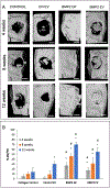
A) Representative μCT images showing regeneration of bone in 5mm calvarial defects that were treated with plain collagen sponge (Control), collagen sponge containing control EVs (Ctrl. EV, 5x10⁸ EVs per defect), collagen sponge containing BMP2 (BMP2 GF 5μg/defect) and collagen sponge containing BMP2 EV (5x10⁸ EVs per defect) at 4, 8- and 12-weeks post wounding. The arrow in the 12-week BMP2 GF group shows ectopic bone formation. Scale bar represents 2.5mm. B) Volumetric quantitation of the μCT data expressed as percentage bone volume regenerated with mineralized tissue (n=6 defects per group per time point). * represents statistical significance with respect to the collagen control group (no EV) and # represents statistical significance with respect to the control EV group. The significance was calculated using Tukey’s ad-hoc test following one-way ANOVA.
Figure 6:
Figure 6:. Histological evaluation of calvarial defects:
Images are representative light microscopy images of H&E stained demineralized calvarial samples of defects treated with plain collagen sponge (Control), collagen sponge containing control EVs (Ctrl. EV), collagen sponge containing BMP2 (BMP2 GF) and collagen sponge containing BMP2 EV after 4, 8- and 12-weeks post wounding. The black arrows in the images point to regenerated bone tissue. The yellow arrows in the BMP2 GF group point to fat deposits within the regenerated bone. Scale bar represents 200μm in all images.
Figure 7:
Figure 7:. IHC of calvarial defects:
Images represent the expression levels of osteoinductive marker proteins BMP2, BSP, DMP1 and OCN in the calvarial sections from the different groups after 4 weeks. Note the increase in the expression levels of all markers in the rhBMP2 treated (BMP2 GF) and BMP2 EV treated groups. The arrows in the BMP2 EV group shows protein expression in the cells lining the newly formed bone. Scale bar represents 50μm in all images

Similar articles

Cited by

References

    1. Gonnelli S, Caffarelli C, Nuti R, Obesity and fracture risk, Clin Cases Miner Bone Metab 11(1) (2014) 9–14. - PMC - PubMed
    1. Khazai NB, Beck GR Jr., Umpierrez GE, Diabetes and fractures: an overshadowed association, Curr Opin Endocrinol Diabetes Obes 16(6) (2009) 435–45. - PMC - PubMed
    1. Flierl MA, Smith WR, Mauffrey C, Irgit K, Williams AE, Ross E, Peacher G, Hak DJ, Stahel PF, Outcomes and complication rates of different bone grafting modalities in long bone fracture nonunions: a retrospective cohort study in 182 patients, J Orthop Surg Res 8 (2013) 33. - PMC - PubMed
    1. Smith DC, Extremity Injury and War: A Historical Reflection, Clin Orthop Relat Res 473(9) (2015) 2771–6. - PMC - PubMed
    1. Marin C, Luyten FP, Van der Schueren B, Kerckhofs G, Vandamme K, The Impact of Type 2 Diabetes on Bone Fracture Healing, Front Endocrinol (Lausanne) 9 (2018) 6. - PMC - PubMed

Publication types

MeSH terms

Substances