. 2020 Jun 16;94(13):e00123-20.
doi: 10.1128/JVI.00123-20. Print 2020 Jun 16.

Varicella-Zoster Virus (VZV) Small Noncoding RNAs Antisense to the VZV Latency-Encoded Transcript VLT Enhance Viral Replication

Affiliations

Varicella-Zoster Virus (VZV) Small Noncoding RNAs Antisense to the VZV Latency-Encoded Transcript VLT Enhance Viral Replication

Punam Bisht et al. J Virol. .

Abstract

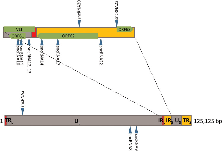
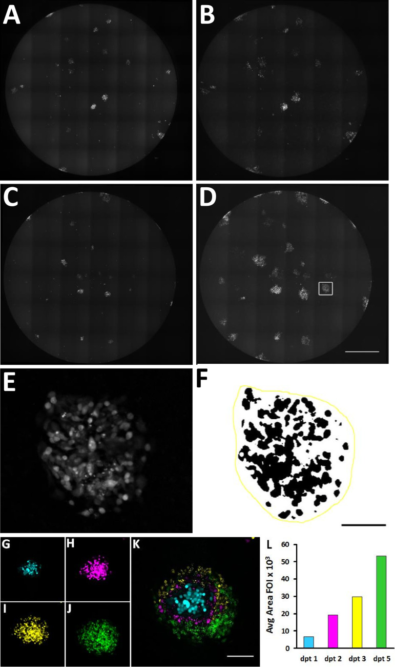
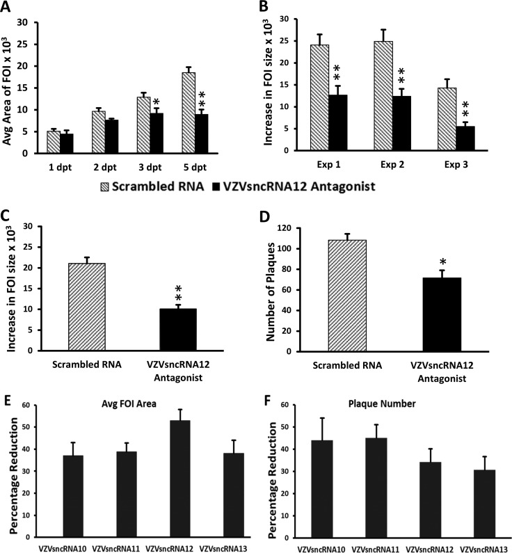
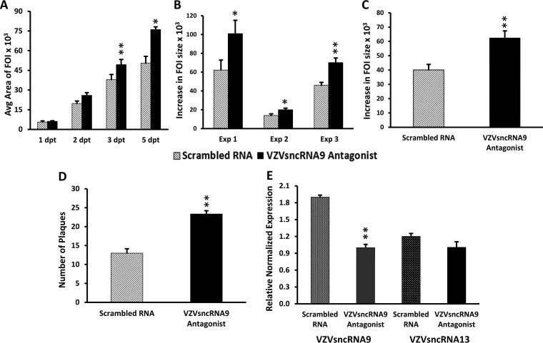
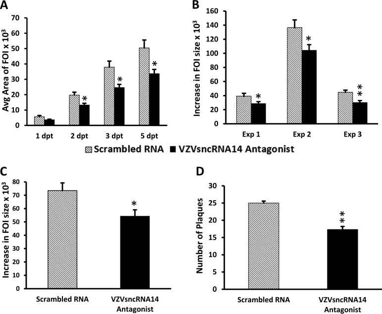
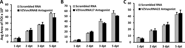
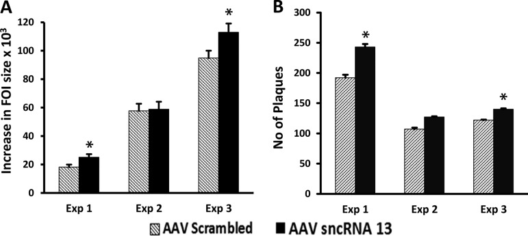
Small noncoding RNAs (sncRNA), including microRNA (miR), are expressed by many viruses to provide an additional layer of gene expression regulation. Our work has shown that varicella-zoster virus (VZV; also called human herpesvirus 3 [HHV3]), the human alphaherpesvirus causing varicella and herpes zoster, expresses 24 virally encoded sncRNA (VZVsncRNA) in infected cells. Here, we demonstrate that several VZVsncRNA can modulate VZV growth, including four VZVsncRNA (VZVsncRNA10, -11, -12, and -13) that are antisense to VLT, a transcript made in lytic infections and associated with VZV latency. The influence on productive VZV growth and spread was assessed in epithelial cells transfected with locked nucleotide analog antagonists (LNAA). LNAA to the four VZVsncRNA antisense to VLT significantly reduced viral spread and progeny titers of infectious virus, suggesting that these sncRNA promoted lytic infection. The LNAA to VZVsncRNA12, encoded in the leader to ORF61, also significantly increased the levels of VLT transcripts. Conversely, overexpression of VZVsncRNA13 using adeno-associated virus consistently increased VZV spread and progeny titers. These results suggest that sncRNA antisense to VZV may regulate VZV growth, possibly by affecting VLT expression. Transfection of LNAA to VZVsncRNA14 and VZVsncRNA9 decreased and increased VZV growth, respectively, while LNAA to three other VZVsncRNA had no significant effects on replication. These data strongly support the conclusion that VZV replication is modulated by multiple virally encoded sncRNA, revealing an additional layer of complexity of VZV regulation of lytic infections. This may inform the development of novel anti-sncRNA-based therapies for treatment of VZV diseases.IMPORTANCE Varicella-zoster virus (VZV) causes herpes zoster, a major health issue in the aging and immunocompromised populations. Small noncoding RNAs (sncRNA) are recognized as important actors in modulating gene expression. This study extends our previous work and shows that four VZVsncRNA clustering in and near ORF61 and antisense to the latency-associated transcript of VZV can positively influence productive VZV infection. The ability of multiple exogenous small oligonucleotides targeting VZVsncRNA to inhibit VZV replication strengthens the possibility that they may inform development of novel treatments for painful herpes zoster.

Keywords: herpes zoster; noncoding RNA; varicella-zoster virus.


Figures

FIG 1
FIG 1
Location in the viral genome of VZVsncRNA studied in this report. The lower diagram represents the entire VZV genome, while the top diagram is an expansion of the region containing most of the VZVsncRNA whose function was assayed for in the present study. Arrowheads facing upward indicate the VZVsncRNA coded by the bottom strand and deriving from right-to-left primary transcripts, while the arrowheads facing downward indicate the VZVsncRNA coded by the top strand of the viral genome in its standard configuration and deriving from RNAs transcribed left to right. The positions of relevant VZV ORFs and the VLT transcript coding region are indicated in green in the upper diagram.
FIG 2
FIG 2
Method used for measuring growth of VZV infectious foci (FOI) in ARPE-19 cells. Ten to thirty ARPE-19 cells infected with VZV66RFP were added to 90% confluent wells of 96-well plates. At 24 h postinfection, cells were transfected either with an LNA antagonist to a scrambled RNA (A and B) or with VZVsncRNA9 (C and D). The RFP fluorescence of entire wells was photographed with an automated microscope, and the micrographs were assembled into a single image. (E) A single fluorescent VZV FOI after image processing. (F) FOI in panel E after application of a threshold for fluorescence (and inversion of the pixel luminance values): the black pixels represent the fluorescence indicating the presence of the virus. The yellow line demarcates the area within which black pixels were counted by the computer in order to generate a value for infection in an FOI. See Materials and Methods for more details. (G to L) Growth of a single focus of infection and its quantification. (G to J) Single FOI at 1 (blue), 2 (magenta), 3 (yellow), and 5 (green) days after transfection. (K) Images were pseudocolored and overlaid in order to visualize the FOI growth. (L) Graph of the quantification of this FOI on these days. Bars, 1 mm (A to D), 250 μm (E and F), and 100 μm (G to K).
FIG 3
FIG 3
Transfection of LNA antagonists antisense to VLT reduces growth of VZV infectious foci and infectious virus and increase VLT expression. An LNA antagonist to VZVsncRNA12 was transfected into wells of ARPE-19 cells 1 day after seeding of 10 to 30 VZV66RFP-infected ARPE-19 cells onto a 90% confluent culture of naive ARPE-19 cells (black bars). An LNA RNA oligonucleotide with a scrambled sequence was transfected as a control (gray bars). (A) Graph of the average size of FOI 1 to 5 dpt in a single experiment. Significant differences are indicated with asterisks (*, P < 0.05; **, P < 0.005). Errors bars represent the standard errors of the means (SEM) of the measurements from the three wells assayed within the same experiment. (B) Average increase in size of individual FOI between 1 to 5 dpt in arbitrary (pixel) units in three independent experiments. **, P < 0.005. (C) The transfection of an LNA antagonist to VZVsncRNA12 decreased the growth of the FOI by an average of 52% for the three experiments. **, P < 0.005. (D) Results from plaque assays of cells harvested from the LNA antagonist and scrambled RNA-transfected wells at 7 dpt. The antagonist reduced the plaque count by 34% compared to scrambled RNA (average from 3 independent experiments). The asterisk indicates statistical significance between control and LNAA-transfected wells (*, P < 0.05). (E and F) Effects of LNAA on all 4 VZVsncRNA antisense to the putative VZV latency-encoded transcript VLT on viral growth. LNA antagonists to VZVsncRNA10, -11, -12, and -13 and scrambled LNA RNA were transfected into VZV-infected ARPE-19 cells and analyzed as described in Materials and Methods and the legend to Fig. 2. All four antagonists reduced the growth of FOI between 1 and 5 dpt, by averages of 37%, 39%, 52%, and 38%, respectively (n = 3 for each antagonist). (F) Plaque-forming assays of cells harvested from the antagonist and scrambled-RNA wells. The average number of plaques obtained was reduced by 44%, 45%, 34%, and 30% for the LNA antagonists to VZVsncRNA10, -11, -12, and -13, respectively. All of the reductions in FOI growth and plaques were significant (P < 0.05).
FIG 4
FIG 4
Administration of an LNAA to VZVsncRNA9 increases VZV FOI growth and number of infectious plaques. (A) The growth of FOI 1 to 5 days after transfection of an LNAA to VZVsncRNA9 (black bars) compared to control scrambled LNA RNA (gray bars) in a typical experiment is shown in arbitrary (pixel) units. A difference was observed at 2 dpt and became significant by 3 dpt. (B) Average increase in size of individual FOI between 1 and 5 dpt for each of the three experiments performed. (C) The transfection of an antagonist to VZVsncRNA9 increased the growth of FOI by an average of 55% for the three experiments. (D) Progeny virus yields quantified by plaque assays of cells harvested from the LNA antagonist and scrambled LNA RNA-transfected wells 7 dpt. The antagonist increased the plaque count compared to scrambled RNA by an average of 79% (n = 3). Asterisks indicate statistical significance between control and LNAA-transfected wells (*, P < 0.05; **, P < 0.005). (E) Transfection of an LNA antagonist to one VZVsncRNA reduced the levels of its target but not those of another VZVsncRNA. VZV-infected ARPE-19 cells were transfected with an LNA antagonist to VZVsncRNA9 or a scrambled LNA oligonucleotide control. At 7 dpt, cells were harvested, RNA was extracted, and stem-loop qRT-PCR was performed on small (<200-nt) RNAs for VZVsncRNA9 (targeted). VZVsncRNA13 was a nontargeted control. The antagonist reduced the level of the targeted VZVsncRNA9 by about 50% in two independent experiments (P < 0.05; 3 wells in each experiment). In contrast, the level of nontargeted VZVsncRNA13 from the same RNA preparation was not significantly different from that of the scrambled control.
FIG 5
FIG 5
Inhibition of VZVsncRNA14 reduced growth of VZV infectious foci and infectious virus production. An LNA antagonist to VZVsncRNA14 or control LNA RNA was transfected 1 day after seeding of 10 to 30 VZV66RFP-infected ARPE-19 cells onto monolayers of naive ARPE-19 and analyzed as described in Materials and Methods and the legend to Fig. 3. (A) Increase in FOI size in a single experiment, in which a significant difference, determined by comparing averages of FOI growth from three wells with each oligonucleotide, appeared by 2 dpt. (B) Average change in size of individual FOI in 3 independent experiments. (C) A modest but significant 19% average decrease in FOI growth was measured in the three experiments whose results are shown in panel B. (D) Progeny virus measured 7 days after transfection, in which cells were harvested from the antagonist- and scrambled-RNA-transfected wells and seeded onto naive ARPE-19 cells. Analysis 7 days later revealed that transfection of the antagonist resulted in a 30% decrease in infectious plaque count (n = 3) compared to the control. Asterisks indicate statistical significance between control and LNAA-transfected wells (*, P < 0.05; **, P < 0.005).
FIG 6
FIG 6
LNA antagonists to three VZVsncRNA did not have a consistent effect on FOI growth. LNA antagonists to VZVsncRNA8 (A), VZVsncRNA17 (B), and VZVsncRNA22 (C) were transfected 1 day after seeding of small numbers of VZV66RFP-infected ARPE-19 cells onto monolayers of ARPE-19. Controls and analysis were performed as described in Materials and Methods and the legend to Fig. 2. Average changes in FOI size from three independent experiments with each LNA VZVsncRNA antagonist are shown.
FIG 7
FIG 7
Exogenous expression of VZVsncRNA13 by AAV promotes viral growth. ARPE-19 cells were transduced with AAV2 expressing VZVsncRNA13 or expressing a control RNA of the same length under the control of the U6 promoter and 1 day later infected with 10 to 30 VZV-infected cells. Analysis of FOI growth was performed as described in the legend to Fig. 2. Modest increases in FOI size (A) and plaque number (B) were obtained in the cells overexpressing VZVsncRNA13 relative to those expressing the scrambled RNA in two of three independent experiments. In experiment 2, no significant difference in FOI growth or increase in plaque number were observed, although a trend was apparent. The results from overexpression of this VZVsncRNA are the opposite of those obtained by transfecting its LNA antagonist (Fig. 3).

Similar articles

Cited by

References

    1. Weinberg MS, Morris KV. 2016. Transcriptional gene silencing in humans. Nucleic Acids Res 44:6505–6517. doi:10.1093/nar/gkw139. - DOI - PMC - PubMed
    1. Piedade D, Azevedo-Pereira JM. 2016. The role of microRNAs in the pathogenesis of herpesvirus infection. Viruses 8:156. doi:10.3390/v8060156. - DOI - PMC - PubMed
    1. Flores O, Nakayama S, Whisnant AW, Javanbakht H, Cullen BR, Bloom DC. 2013. Mutational inactivation of herpes simplex virus 1 microRNAs identifies viral mRNA targets and reveals phenotypic effects in culture. J Virol 87:6589–6603. doi:10.1128/JVI.00504-13. - DOI - PMC - PubMed
    1. Pan D, Pesola JM, Li G, McCarron S, Coen DM. 2017. Mutations inactivating herpes simplex virus 1 microRNA miR-H2 do not detectably increase ICP0 gene expression in infected cultured cells or mouse trigeminal ganglia. J Virol 91:e02001-16. doi:10.1128/JVI.02001-16. - DOI - PMC - PubMed
    1. Han Z, Liu X, Chen X, Zhou X, Du T, Roizman B, Zhou G. 2016. miR-H28 and miR-H29 expressed late in productive infection are exported and restrict HSV-1 replication and spread in recipient cells. Proc Natl Acad Sci U S A 113:E894–E901. doi:10.1073/pnas.1525674113. - DOI - PMC - PubMed

Publication types

LinkOut - more resources