Review
. 2020 Jun:105:103492.
doi: 10.1016/j.mcn.2020.103492. Epub 2020 Apr 12.

Spatial control of membrane traffic in neuronal dendrites

Affiliations
Review

Spatial control of membrane traffic in neuronal dendrites

Megan R Radler et al. Mol Cell Neurosci. 2020 Jun.

Abstract

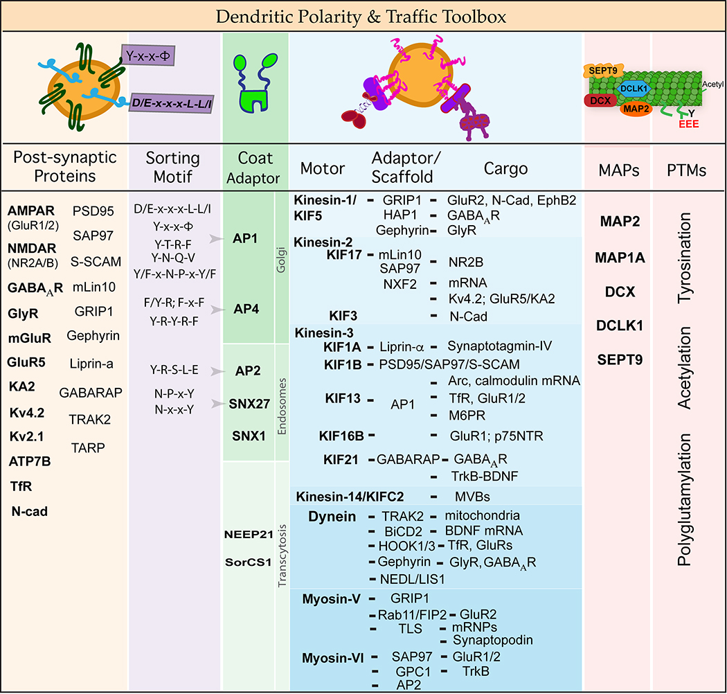
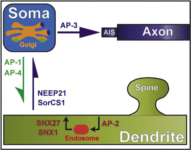
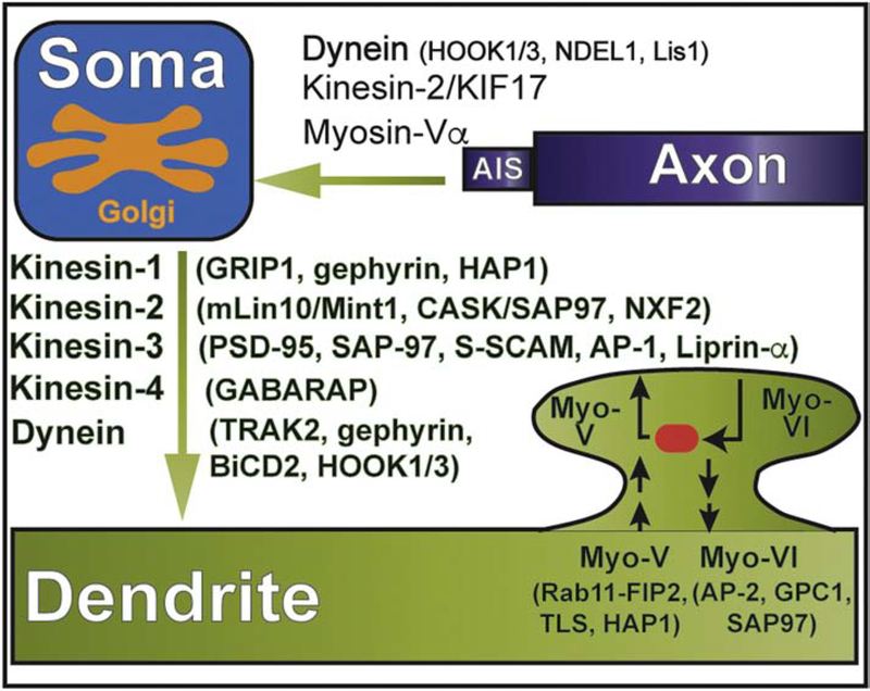
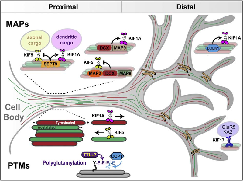
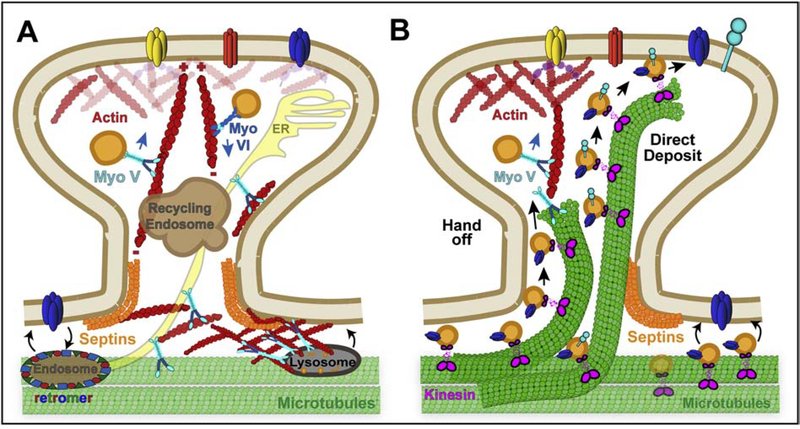
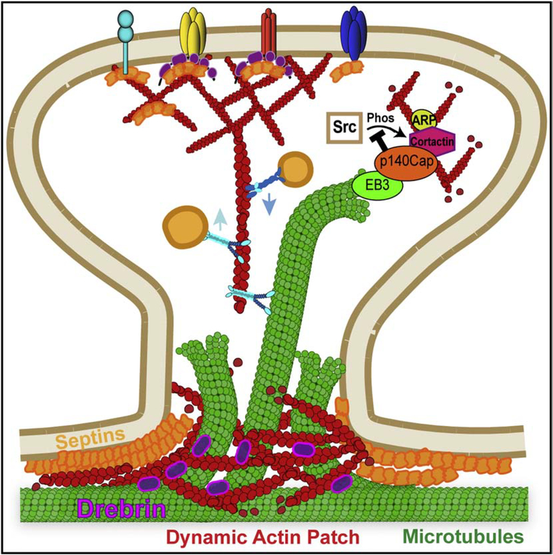
Neuronal dendrites are highly branched and specialized compartments with distinct structures and secretory organelles (e.g., spines, Golgi outposts), and a unique cytoskeletal organization that includes microtubules of mixed polarity. Dendritic membranes are enriched with proteins, which specialize in the formation and function of the post-synaptic membrane of the neuronal synapse. How these proteins partition preferentially in dendrites, and how they traffic in a manner that is spatiotemporally accurate and regulated by synaptic activity are long-standing questions of neuronal cell biology. Recent studies have shed new insights into the spatial control of dendritic membrane traffic, revealing new classes of proteins (e.g., septins) and cytoskeleton-based mechanisms with dendrite-specific functions. Here, we review these advances by revisiting the fundamental mechanisms that control membrane traffic at the levels of protein sorting and motor-driven transport on microtubules and actin filaments. Overall, dendrites possess unique mechanisms for the spatial control of membrane traffic, which might have specialized and co-evolved with their highly arborized morphology.

Keywords: Actin; Adaptor proteins; Axons; Dendrites; Kinesins; Membrane traffic; Microtubules; Myosins; Scaffolds; Septins; Sorting.


Figures

Figure 1.
Figure 1.
Dendritic polarity and traffic toolbox. Dendrites are enriched with post-synaptic membrane proteins including the AMPA (GluR1/2, GluR2/3) and NMDA (NR1, NR2A/B) receptors of the excitatory synapse, the GABAA and glycine receptors (GlyR) of the inhibitory synapse, kainate receptors such as GluR5 and KA2, potassium (Kv4.2, Kv2.1) and copper (ATP7B) ion channels, and the transferrin receptor (TfR). Dendritic localization and targeting of these receptors and channels involves interaction with adaptor and scaffold proteins (e.g., PSD95, SAP97, GRIP1, mLin10, Liprin-α, GABARAP), which interface with membrane- and cytoskeleton-based mechanisms of intracellular traffic. Specificity in the trafficking of dendritic membrane proteins is achieved during sorting at the trans-Golgi and endosomes, transport by kinesin, dynein and myosin motors, and additional guidance from microtubule-associated proteins (MAPs) and post-translational modifications (PTMs) of the cytoskeleton which provide spatial cues. This figure summarizes and tabulates the sorting motifs that interact with membrane coat adaptors, the cytoskeletal motors and their interactions with adaptor/scaffold and cargo proteins, and the MAPs and PTMs that function in dendritic membrane polarity and traffic.
Figure 2.
Figure 2.
Sorting of somatodendritic and axonal proteins by membrane adaptors. Schematic shows the major compartments of a neuronal cell and the clathrin adaptors (AP-1, AP-3, AP-4) involved in the sorting of somatodendritic and axonal proteins in the trans-Golgi of the cell body. Adaptors proteins (NEEP21, SorCS1) that mediate the sorting of axonal proteins for retrieval from dendrites (transcytosis) and the endocytic recycling of dendritic membrane proteins (AP-2, SNX27, SNX1) are also outlined.
Figure 3.
Figure 3.
Cytoskeletal motors and adaptors in the polarized and local traffic of dendritic proteins. Schematic shows the microtubule (kinesin, dynein) and actin motors (myosins, myo) involved in the retrieval of somatodendritic proteins from the axon initial segment and transport from the cell body to dendrites. In addition, the myosin motors that drive membrane traffic in dendritic spines are shown. Kinesin motors are summarized under their respective families (e.g. kinesin-1) and the adaptor/scaffold proteins that mediate interaction with dendritic cargo are provided in parentheses.
Figure 4.
Figure 4.
Spatial control of dendritic membrane traffic by MAPs and microtubule PTMs. Schematic depicts dendritic MAPs and microtubule PTMs with roles in the regulation of kinesin-driven membrane traffic. SEPT9 and DCLK1 localize in proximal and distal dendrites, respectively. SEPT9 impedes traffic of axonal cargo of kinesin-1 into dendrites, while it promotes the anterograde transport of dendritic cargo of kinesin-3. DCLK1, DCX and MAP9 also promote the microtubule binding and motility of kinesin-3. In vitro motility assays indicate that MAP2, DCX and MAP9 inhibit kinesin-1 motility, but it is unknown whether these MAPs can impede entry of kinesin-1 and its axonal cargo into dendrites. Dendrites contain tyrosinated and acetylated microtubules, which are oriented with their plus-ends away and toward the cell body, respectively. The kinesin-3/KIF1A motor associates preferentially with tyrosinated plus-end out microtubules and kinesin-1/KIF5 interacts with acetylated plus-end in microtubules. Dendritic microtubules are also modified with short glutamate chains, which are maintained by the tubulin glutamylase TTLL7 (tubulin tyrosine like ligase 7) and the deglutamylase CCP1 (cytosolic carboxypeptidase 1). Note that kinesin-2/KIF17 transports the kainate receptors GluR5 and KA2 in distal dendrites, which is indicative of a spatial specificity in dendritic motor-cargo traffic.
Figure 5.
Figure 5.
Membrane traffic in dendritic spines. (A) The actin motors myosin V and VI mediate the transport of post-synaptic cargo toward and away from the dendritic spine head, respectively. At the base of the dendritic spines and along the dendritic shaft, endolysosomes are entrapped and anchored to actin patches by myosin V, which provides a mechanism for fusion with the spine membrane in response to synaptic activity. Dendritic membrane proteins recycle through retromer-positive endosomes, which localize along the dendritic shaft membrane. (B) Dynamic microtubules that enter into dendritic spines deliver vesicles to the spine membrane by either a handoff mechanism, which involves vesicle switching from microtubules to actin filaments, or direct delivery of microtubule plus-end-associated vesicles to the membrane. Microtubule-dependent transport of vesicles also occurs in the dendritic shaft, where vesicles with post-synaptic receptors are delivered by microtubule motors to synapses and membrane sites of the dendritic shaft.
Figure 6.
Figure 6.
Actin-microtubule crosstalk in dendritic spines. Microtubule plus ends are captured at the base of dendritic spines by dynamic patches of actin filaments and possibly other cytoskeletal elements such as septins, which are enriched at the membrane curvature along the base and neck of dendritic spines. Microtubule-actin crosstalk is mediated by proteins such as drebrin, which interacts with both actin and microtubules, and myosin V, which can diffuse on the microtubule lattice and thereby, could transition vesicular cargo from microtubules to actin filaments. Notably, microtubules have the capacity to regulate the actin organization of dendritic spines through proteins that associate with microtubule plus ends. For example, p140Cap/SNIP associates with the microtubule plus-end protein EB3 and interacts with cortactin that promotes Arp2/3-induced assembly of branched actin filaments. Microtubule plus ends with p140Cap/SNIP can promote actin assembly by inhibiting the Src kinase, which is a negative regulator of cortactin.

Similar articles

Cited by

References

    1. Adrian M, Kusters R, Wierenga CJ, Storm C, Hoogenraad CC, Kapitein LC, 2014. Barriers in the brain: resolving dendritic spine morphology and compartmentalization. Front Neuroanat 8, 142. - PMC - PubMed
    1. Aguirre-Chen C, Bulow HE, Kaprielian Z, 2011. C. elegans bicd-1, homolog of the Drosophila dynein accessory factor Bicaudal D, regulates the branching of PVD sensory neuron dendrites. Development 138, 507–518. - PMC - PubMed
    1. Al-Bassam S, Xu M, Wandless TJ, Arnold DB, 2012. Differential trafficking of transport vesicles contributes to the localization of dendritic proteins. Cell Rep 2, 89–100. - PMC - PubMed
    1. Alberi S, Boda B, Steiner P, Nikonenko I, Hirling H, Muller D, 2005. The endosomal protein NEEP21 regulates AMPA receptor-mediated synaptic transmission and plasticity in the hippocampus. Mol Cell Neurosci 29, 313–319. - PubMed
    1. Ali MY, Krementsova EB, Kennedy GG, Mahaffy R, Pollard TD, Trybus KM, Warshaw DM, 2007. Myosin Va maneuvers through actin intersections and diffuses along microtubules. Proc Natl Acad Sci U S A 104, 4332–4336. - PMC - PubMed

Publication types