. 2020 Apr 13;16(4):e1008426.
doi: 10.1371/journal.ppat.1008426. eCollection 2020 Apr.

Killer cell proteases can target viral immediate-early proteins to control human cytomegalovirus infection in a noncytotoxic manner

Affiliations

Killer cell proteases can target viral immediate-early proteins to control human cytomegalovirus infection in a noncytotoxic manner

Liling Shan et al. PLoS Pathog. .

Abstract

Human cytomegalovirus (HCMV) is the most frequent viral cause of congenital defects and can trigger devastating disease in immune-suppressed patients. Cytotoxic lymphocytes (CD8+ T cells and NK cells) control HCMV infection by releasing interferon-γ and five granzymes (GrA, GrB, GrH, GrK, GrM), which are believed to kill infected host cells through cleavage of intracellular death substrates. However, it has recently been demonstrated that the in vivo killing capacity of cytotoxic T cells is limited and multiple T cell hits are required to kill a single virus-infected cell. This raises the question whether cytotoxic lymphocytes can use granzymes to control HCMV infection in a noncytotoxic manner. Here, we demonstrate that (primary) cytotoxic lymphocytes can block HCMV dissemination independent of host cell death, and interferon-α/β/γ. Prior to killing, cytotoxic lymphocytes induce the degradation of viral immediate-early (IE) proteins IE1 and IE2 in HCMV-infected cells. Intriguingly, both IE1 and/or IE2 are directly proteolyzed by all human granzymes, with GrB and GrM being most efficient. GrB and GrM cleave IE1 after Asp398 and Leu414, respectively, likely resulting in IE1 aberrant cellular localization, IE1 instability, and functional impairment of IE1 to interfere with the JAK-STAT signaling pathway. Furthermore, GrB and GrM cleave IE2 after Asp184 and Leu173, respectively, resulting in IE2 aberrant cellular localization and functional abolishment of IE2 to transactivate the HCMV UL112 early promoter. Taken together, our data indicate that cytotoxic lymphocytes can also employ noncytotoxic ways to control HCMV infection, which may be explained by granzyme-mediated targeting of indispensable viral proteins during lytic infection.


Conflict of interest statement

The authors have declared that no competing interests exist.

Figures
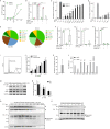
Fig 1
Fig 1. Cytotoxic lymphocytes can inhibit HCMV dissemination in a noncytotoxic manner and can induce IE degradation in infected cells.
(A) CMV+ donor-derived fibroblasts were infected with GFP-HCMV (MOI = 0.05) and harvested at indicated time points for GFP quantification by flow cytometry (data represent the mean ± SD, each performed in triplicate). (B) Fibroblasts were infected with GFP-HCMV (MOI = 0.08) and co-cultured with autologous CD8+ T cells (E:T = 2.5:1) from CMV+ donors (data represent the mean with range of 3 donors, each performed in triplicate), CMV- donors (data represent the mean with range of 2 donors, each performed in triplicate), or non-autologous NK cells (E:T = 2.5:1) for 9–14 days. (C) GFP-HCMV (MOI = 0.08) infected CMV+ donor-derived fibroblasts were co-cultured with autologous CD8+ T cells at indicated E:T ratios for 9 days (data represent the mean ± SD, each performed in triplicate). (D) GFP-HCMV (MOI = 0.08) infected CMV+ donor-derived fibroblasts were co-cultured with non-autologous NK cells at indicated E:T ratios for 9 days (data represent the mean ± SD, n = 3). (E) Absolute numbers (IFN-γ Spot Forming Units SFU/106 cells) of HCMV-specific CD8+ T cells (stimulated with overlapping peptide pools UL144, gB, pp65, IE1, IE2, US3, UL28, pp71) derived from two CMV+ donors as determined by FluoroSpot IFN-γ assay. (F) Fibroblasts were infected with GFP-HCMV (MOI = 0.08) and co-cultured with autologous CD8+ T cells (E:T = 2.5:1) in the absence or presence of MHC-I blocking antibody (data represent the mean ± SD of 3 CMV+ donors, each performed in triplicate). (G) Fibroblasts were infected with GFP-HCMV (MOI = 0.08) and co-cultured with autologous CD8+ T cells (E:T = 2.5:1) in the absence or presence of IFN-γ blocking antibody (data represent the mean with range of 2 CMV+ donors, each performed in triplicate). (H) Fibroblasts were infected with GFP-HCMV (MOI = 0.08) and co-cultured with autologous CD8+ T cells (E:T = 2.5:1) in the absence or presence of IFN-α and IFN-β blocking antibodies (data represent the mean with range of 2 CMV+ donors, each performed in triplicate). (I) FACS histogram visualizing PFN protein levels in WT-NK cells and PFN-knockout (KO) NK cells. (J) GFP-HCMV (MOI = 0.08) infected CMV+ donor-derived fibroblasts were co-cultured with non-autologous WT or PFN-KO NK cells at indicated E:T ratios for 9 days (data represent the mean ± SD, n = 3). (K) Fibroblasts were infected with GFP-HCMV (MOI = 0.08) and co-cultured with autologous CD8+ T cells (E:T = 2.5:1) of one CMV+ donor for 10 days. Then, CD8+ T cells were either left or washed out and fibroblasts were cultured for another 10 days. GFP+ cells were measured (data represent the mean ± SD, n = 3). (L) Fibroblasts were infected with GFP-HCMV (MOI = 0.08) and co-cultured with autologous CD8+ T cells (E:T = 2.5:1) of one CMV+ donor for 10 days. Then, CD8+ T cells were either left or washed out and fibroblasts were cultured for another 10 days. Viral mRNA was measured (data represent the mean ± SD, n = 3). (M) Fibroblasts were infected with Merlin delta US2-11-HCMV (MOI = 0.8) and treated with autologous IE1-specific CD8+ T cells (from one donor) at indicated E:T ratios for 24 h, followed by immunoblotting using antibodies against IE1/2, pp65 and Hsp90β. (N) IE1/2 and Hsp90β band intensities (see M) were semi-quantified. IE/Hsp90β ratio in absence of CD8+ T cells was set to 100% (data represent the mean ± SD, n = 3). (O) HFFs were infected with GFP-HCMV (MOI = 0.4) and treated with LAK cells at indicated E:T ratios for 6 h, followed by immunoblotting using antibodies against IE1/2 and GFP. (P) HFFs were infected with GFP-HCMV (MOI = 0.4) and treated with NK cells at indicated E:T ratios for 6 h, followed by immunoblotting using antibodies against IE1 and GFP. White and black triangles indicate full length IE proteins and IE degradation products, respectively. Statistical analysis was performed using one-way ANOVA with Turkeys multiple comparisons test (B, F, G, H) or Student t test (C, D, J, K, L, N). *P<0.05, **P<0.01, ****P<0.0001.
Fig 2
Fig 2. Immune effector cells target IE1 and IE2.
HeLa cells were transfected with N-terminal HA-tagged IE1, IE2, or empty vector combined with GFP. 20 h post-transfection, (A) LAK cells or (B) NK cells (YT-Indy) were added with increasing E:T ratios for 6 h. (C) Caspase inhibitor ZVAD-fmk was added (100 μM) before challenging by NK cells. Lysates were subjected to immunoblotting using either an anti-HA or anti-GFP antibody. White and black triangles indicate full length IE proteins and IE degradation products, respectively.
Fig 3
Fig 3. All granzymes directly cleave HCMV IE1 and/or IE2.
(A) HFFs were infected with HCMV (AD169) at a MOI of 1.0. 3 days p.i., nuclear and cytosolic fractions were generated and protein concentrations were measured. Nuclear fractions (0.5 μg) were incubated with 300 nM purified human granzymes or their corresponding catalytically inactive control SA mutants, or left untreated for 4 h at 37°C and immunoblotted using an antibody against IE. (B) Cell-free lysates of HA-IE1/2 transfected HeLa cells were incubated with increasing concentrations of purified human granzymes or SA mutants (300 nM) for 3 h at 37°C and subjected to immunoblotting with an antibody against HA. (C) Schematic overview of putative human granzyme cleavage sites in IE1 and IE2 (NLS, nuclear localization signal; AD, activation domain). (D) SDS-PAGE and full protein staining of purified IE1 and IE2 incubated with five human granzymes at concentrations ranging from 0–300 nM for 3 h at 37°C. White and black triangles indicate full length IE proteins and IE cleavage products, respectively. The arrow indicates the granzymes.
Fig 4
Fig 4. GrB and GrM may inhibit IE1 function.
(A, B) HeLa cells were transfected with HA-IE1WT, IE1D398A or IE1L414A and empty vector for 24 h. Cell-free lysates were incubated with increasing concentrations of GrB (A, left panel) or GrM (B, left panel) or SA mutant (150 nM) at 37°C for 3 h and immunoblotted using an anti-HA antibody. Sequence alignment of the amino acid regions surrounding IE1D398 and IE1L414 from different HCMV strains (A and B, right panel). Amino acid in red is the P1 cleavage site and different amino acids among sequences are shown in blue. (C, D) IE1WT, IE1D398A (C) or IE1L414A (D) were labeled with fluorescent green lysines during in vitro transcription/translation and incubated at 37°C for 3 h with increasing concentrations of GrB (C) or GrM (D) or SA mutants (200 nM). Samples were separated by SDS-PAGE. (E, F) Left panel: Schematic overview of IE1, IE11-398, and IE1399-491 (E), IE1, IE11-414, and IE1415-491 (F) constructs. NLS, nuclear localization signal; AD, acidic domain; DB, DNA-binding domain. Right panel: IE1 variants were expressed in HeLa cells and lysates were subjected to immunoblotting, using an anti-HA antibody. (G, H) Immunofluorescence images of HeLa cells transfected with HA and GFP-tagged IE1, IE11-398 and IE1399-491 (G), HA and GFP-tagged IE1, IE11-414 and IE1415-491 (H) constructs. GFP-tagged proteins were visualized in green and nuclei were stained by co-transfection with H2B-mCherry in red. (I, J) HEK293T cells were co-transfected with pGL4.74 hRL-TK (20 ng), pGL4.24 10×IRE (100 ng), H2B-mCherry (30 ng) and increasing amounts of expression plasmids of IE1, IE11-398, or/and IE1399-491 (I), IE1, IE11-414, or/and IE1415-491 (J). All conditions were performed in triplicates. 24 h post-transfection, cells were incubated with 5 ng/ml IFN-β for 6 h, after which dual luciferase reporter assay was used to assess luciferase activity. Relative luciferase activity, i.e. Firefly/Renilla are depicted and the values in the absence of IE (fragments) were set to 100% of IRE activation. Bars represent the mean ± SD of three independent experiments. (K, L) lysates used in the luciferase reporter assay were subjected to immunoblotting using an anti-HA antibody. (M, N) HeLa cells were transfected with either both N- and C-terminal HA-tagged IE1 (M) or only C-terminal HA-tagged IE1 (N), complemented with H2B-GFP. After 24 h transfection, NK (YT-Indy) cells were added at indicated E:T ratios for 6 h incubation in the presence of ZVAD-fmk. Lysates were subjected to immunoblotting, using antibodies against HA and GFP. (O) HeLa cells were transfected with N- and C-terminal HA-tagged IE1 and H2B-GFP. 24 h post-transfection, cells were treated with or without MG132 (250 nM) for 30 min before challenging with NK (YT-Indy) cells at 8:1 ratio in the presence of ZVAD-fmk. Lysates were subjected to immunoblotting, using antibodies against HA, GFP, and ubiquitin. White and black triangles indicate full length IE1 proteins and IE1 cleavage products, respectively. All immunoblots are representative of at least three separate experiments.
Fig 5
Fig 5. GrB and GrM abolish IE2 function.
(A, B) HeLa cells were transfected with HA-IE2WT, IE2D184A, or IE2L173A, or empty vector for 24 h. Cell-free lysates were incubated with increasing concentrations of GrB (A, left panel) or GrM (B, left panel) or SA (150 nM) at 37°C for 3 h. Lysates were subjected to immunoblotting, using an anti-HA antibody. Sequence alignment of the amino acid regions surrounding IE2D184 and IE2L173 from different HCMV strains (A and B, right panel). Amino acids in red represent P1 cleavage sites. (C, D) IE2WT, IE2D184A (C) or IE2L173A (D) were labeled with fluorescent green lysines during in vitro transcription/translation and incubated at 37°C for 3 h with increasing concentrations of GrB (C) or GrM (D) or SA (200 nM). Samples were separated by SDS-PAGE. (E, F) Left panel: Schematic overview of the IE2, IE21-184, and IE2185-579 (E), IE2, IE21-173, and IE2174-579 (F) constructs. NLS, nuclear localization signal; DB, DNA-binding domain. Right panel: IE2 variants were expressed in HeLa cells and lysates were subjected to immunoblotting, using an anti-HA antibody. (G, H) Immunofluorescence images of HeLa cells transfected with HA-tagged IE2, IE21-184, and IE2185-579 (G), HA-tagged IE2, IE21-173, and IE2174-579 (H) constructs. HA-tagged proteins were visualized in green and nuclei were stained by co-transfection with H2B-mCherry in red. (I, J) HeLa cells were co-transfected with pGL4.74 TK-hRL (20 ng) (Renilla control), pGL4.11-EP Luc2 (100 ng) (UL112 Early Promoter, EP), H2B-mCherry (30 ng) and increasing amounts of IE2, IE21-184, and IE2185-579 (I, left panel), or IE2, IE21-173, and IE2174-579 (J, left panel). All conditions were performed in triplicate. Dual luciferase reporter assay was used to measure UL112 early promoter activation after 24 h transfection. pcDNA3.1-IE2 (50 ng), H2B-mCherry (30 ng), pGL4.74 hRL-TK (20 ng), pGL4.11-EP (100 ng), increasing amounts of pcDNA3.1-IE21-184 or/and pcDNA3.1-IE2185-579 (I, right panel), or pcDNA3.1-IE21-173 or/and pcDNA3.1-IE2174-579 (J, right panel), complemented with empty vector up to 400 ng total DNA were transfected in HeLa cells. All conditions were performed in triplicate. After 24 h transfection, dual luciferase reporter assay was used to measure early promoter activation. Relative luciferase activity, i.e. Firefly/Renilla, is depicted and the values from conditions in absence of IE were set to 100% early promoter activation. Bars represent the mean ± SD of three independent experiments. (K, L) Lysates used in the luciferase reporter assay were subjected to immunoblotting, using an anti-HA antibody. White and black triangles indicate full length IE2 proteins and IE2 cleavage products, respectively. All immunoblots are representative of at least three separate experiments. (M) HEK293T cells were co-transfected with H2B-mCherry (30 ng), pGL4.74 hRL-TK (20 ng), pGL4.11-EP (UL112 Early Promoter) (100 ng), with or without pcDNA3.1-HA-IE2 (10 ng), complemented with empty vector to a total of 400 ng DNA per well. After 24 h transfection, cells were cultured with or without NK cells (YT-Indy) for 23 h. Dual luciferase reporter assay was used to measure UL112 early promoter activation. Corrected relative Firefly/Renilla ratios in the absence of NK cells were set to 100%. Data represent the mean ± SD, n = 3. Student t test was used for statistical analysis, **P<0.01.

Similar articles

Cited by

References

    1. Crough T, Khanna R. Immunobiology of human cytomegalovirus: from bench to bedside. Clin Microbiol Rev. 2009;22(1): 76–98, Table of Contents. 10.1128/CMR.00034-08 - DOI - PMC - PubMed
    1. dos Santos CJ, Stangherlin LM, Figueiredo EG, Correa C, Teixeira MJ, da Silva MC. High prevalence of HCMV and viral load in tumor tissues and peripheral blood of glioblastoma multiforme patients. J Med Virol. 2014;86(11): 1953–1961. 10.1002/jmv.23820 - DOI - PubMed
    1. Sinclair J. Human cytomegalovirus: Latency and reactivation in the myeloid lineage. J Clin Virol. 2008;41(3): 180–185. 10.1016/j.jcv.2007.11.014 - DOI - PubMed
    1. McVoy MA. Cytomegalovirus vaccines. Clin Infect Dis. 2013;57 Suppl 4: S196–199. - PMC - PubMed
    1. Steininger C. Clinical relevance of cytomegalovirus infection in patients with disorders of the immune system. Clin Microbiol Infect. 2007;13(10): 953–963. 10.1111/j.1469-0691.2007.01781.x - DOI - PubMed

Publication types

MeSH terms