. 2020 Apr 7;11(2):e00524-20.
doi: 10.1128/mBio.00524-20.

Visualizing Association of the Retroviral Gag Protein with Unspliced Viral RNA in the Nucleus

Affiliations

Visualizing Association of the Retroviral Gag Protein with Unspliced Viral RNA in the Nucleus

Rebecca J Kaddis Maldonado et al. mBio. .

Abstract

Packaging of genomic RNA (gRNA) by retroviruses is essential for infectivity, yet the subcellular site of the initial interaction between the Gag polyprotein and gRNA remains poorly defined. Because retroviral particles are released from the plasma membrane, it was previously thought that Gag proteins initially bound to gRNA in the cytoplasm or at the plasma membrane. However, the Gag protein of the avian retrovirus Rous sarcoma virus (RSV) undergoes active nuclear trafficking, which is required for efficient gRNA encapsidation (L. Z. Scheifele, R. A. Garbitt, J. D. Rhoads, and L. J. Parent, Proc Natl Acad Sci U S A 99:3944-3949, 2002, https://doi.org/10.1073/pnas.062652199; R. Garbitt-Hirst, S. P. Kenney, and L. J. Parent, J Virol 83:6790-6797, 2009, https://doi.org/10.1128/JVI.00101-09). These results raise the intriguing possibility that the primary contact between Gag and gRNA might occur in the nucleus. To examine this possibility, we created a RSV proviral construct that includes 24 tandem repeats of MS2 RNA stem-loops, making it possible to track RSV viral RNA (vRNA) in live cells in which a fluorophore-conjugated MS2 coat protein is coexpressed. Using confocal microscopy, we observed that both wild-type Gag and a nuclear export mutant (Gag.L219A) colocalized with vRNA in the nucleus. In live-cell time-lapse images, the wild-type Gag protein trafficked together with vRNA as a single ribonucleoprotein (RNP) complex in the nucleoplasm near the nuclear periphery, appearing to traverse the nuclear envelope into the cytoplasm. Furthermore, biophysical imaging methods suggest that Gag and the unspliced vRNA physically interact in the nucleus. Taken together, these data suggest that RSV Gag binds unspliced vRNA to export it from the nucleus, possibly for packaging into virions as the viral genome.IMPORTANCE Retroviruses cause severe diseases in animals and humans, including cancer and acquired immunodeficiency syndromes. To propagate infection, retroviruses assemble new virus particles that contain viral proteins and unspliced vRNA to use as gRNA. Despite the critical requirement for gRNA packaging, the molecular mechanisms governing the identification and selection of gRNA by the Gag protein remain poorly understood. In this report, we demonstrate that the Rous sarcoma virus (RSV) Gag protein colocalizes with unspliced vRNA in the nucleus in the interchromatin space. Using live-cell confocal imaging, RSV Gag and unspliced vRNA were observed to move together from inside the nucleus across the nuclear envelope, suggesting that the Gag-gRNA complex initially forms in the nucleus and undergoes nuclear export into the cytoplasm as a viral ribonucleoprotein (vRNP) complex.

Keywords: Gag proteins; RNA trafficking; Rous sarcoma virus; genomic RNA packaging; live cell imaging; nucleocytoplasmic trafficking; retrovirus assembly.


Figures

FIG 1
FIG 1
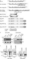
Schematic diagram and characterization of MS2-containing proviral constructs. (A) RNAs and proteins used in this paper. The pRC.V8, pRC.V8-24xMS2, and pRC.V8 Gag-CFP expression plasmids are derived from pRCAN, which contains the hygromycin gene (hygro) in place of the src coding sequence. Twenty-four tandem repeats of the 19 nucleotide MS2 phage RNA stem-loops (36) were inserted into pRC.V8 upstream of the hygromycin coding region to create pRC.V8-24xMS2, causing the loops to be present in both spliced and unspliced vRNA. To create pRC.V8 Gag-CFP, CFP was fused to the C terminus NC, and CFP contains stop codons to prevent the translation of a Gag-CFP-pr-pol protein. pRC.V8 Gag-CFP-24xMS2 was created by inserting 24 copies of MS2 loops into the BstBI and SpeI sites in pRC.V8 Gag-CFP, and the loops are only present in the unspliced vRNA. The pMS2-YFP-NLS expression plasmid, under the control of the polymerase II (Pol II) promoter, contains an NLS that targets MS2-YFP-NLS coat protein to the nucleus, where it binds RNA cotranscriptionally (34). pNES1-YFP-MS2-NLS encodes the Rev NES and well as an NLS to help reduce background fluorescence in the nucleus (49). Wild-type Gag-CFP, wild-type Gag.ΔPR, and Gag.L219A-CFP were previously described (12, 31). Gag.L219A-CFP contains a point mutation in the coding sequence of the p10 domain nuclear export signal, causing the mutant protein to accumulate in the nucleus and within nuclear foci and nucleoli (12, 14). (B) Whole-cell lysates (lanes 1 to 6) and viral particles (lanes 7 to 12) were collected 48 h posttransfection (pt). Western blotting against RSV (rabbit polyclonal antibody) (top row) and MS2 coat protein (3H4 monoclonal antibody; bottom row) was performed to assess the viral protein production and MS2-YFP-NLS incorporation in viral particles. (C) Whole-cell lysates were collected every 3 days for 12 days. Western blotting for RSV Gag was performed at the end of the collection period. On both days 3 and 12 pt, RC.V8 and RC.V8-24xMS2 (RC.V8-24xMS2) cells produced Gag protein. The contrast and brightness were adjusted across the entire image to remove background from the film.
FIG 2
FIG 2
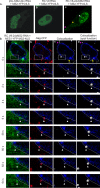
Subcellular localization of MS2-YFP-NLS and wild-type Gag-CFP in QT6 cells. (A) In the presence of the pSL-MS2-24x, MS2-YFP-NLS forms puncta in the nucleus. In the absence of MS2 RNA stem-loops, MS2-YFP-NLS (RC.V8 panel) remains diffuse in the nucleus. When MS2-YFP-NLS is coexpressed with RC.V8-24xMS2, MS2-YFP-NLS foci are present in the nucleus (white arrow), in the cytosol (yellow arrow), and at the plasma membrane (magenta arrow). (B) Various time points from a live-cell time-lapse of QT6 cells were transfected with pRC.V8-24xMS2, pNES1-YFP-MS2-NLS, wild-type pGag-CFP, and pSun1-mCherry. Cells were imaged every 2 s over a period of approximately 10.5 min. vRNA (green) colocalizes (white) with Gag-CFP (red) at the plasma membrane (magenta arrow). (a to c) A focus of NES1-YFP-MS2-NLS-tagged vRNA (arrowhead) (a) and a focus of Gag-CFP (arrowhead) (b) colocalize (c) in the nucleus, which is outlined with Sun1-mCherry (false-colored blue/white outline in all images). (c) Regions of colocalized signal were visualized via the generation of a colocalization channel (white). Colocalized Gag-vRNA foci are indicated by white arrows. (d) The colocalization channel was used to generate spots of Gag-vRNA complexes in the nucleus and the surrounding area that can be tracked over time.
FIG 3
FIG 3
Subcellular localization of wild-type Gag-CFP and unspliced vRNA in QT6 cells. (A) Cross-sections of a z-stack of QT6 cell expressing the proviral pRC.V8 Gag-CFP construct. The Gag-CFP (red) colocalized with unspliced vRNA (green) labeled using RNA smFISH. The crosshairs show a complex of Gag and vRNA that is located in the nucleus. Left, cross-section of an overlay of Gag (red) and vRNA (green). Right, white signal indicates areas of Gag-vRNA colocalization. (B) The z-stack from panel A was generated into a 3D volume surface rendering. Left, x,y orthogonal clipping plane of a cell displaying unspliced RSV RNA (green) labeled via smFISH and Gag-CFP (red) complexes within the DAPI-stained nucleus (blue). Right, colocalization between unspliced vRNA and Gag-CFP is displayed as a white surface rendering an x,y cut of the same cell. (C) Stills from a live-cell time-lapse movie of QT6 cells transfected with pRC.V8 Gag-CFP-24xMS2, untagged wild-type pGag.ΔPR, pNES1-YFP-MS2-NLS, and pSun1-mCherry. Cells were imaged every 2 s. Unspliced vRNA (green) colocalizes (white) with Gag-CFP (red) in the inner nuclear rim (blue/white outline). (a to c) Foci of NES1-YFP-MS2-NLS-tagged vRNA (arrowhead) (a) and foci of Gag-CFP (arrowhead) (b) colocalize (c) in the nucleus, which is outlined with Sun1-mCherry (false-colored blue in all images). (c) Regions of colocalized signal were visualized via the generation of a colocalization channel (white). Colocalized Gag-vRNA foci are indicated by white arrows. (d) The colocalization channel was used to generate spots of Gag-vRNA complexes in the nucleus and the surrounding area that can be tracked over time.
FIG 4
FIG 4
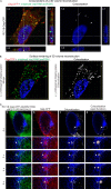
Colocalization between Gag and unspliced vRNA in infected cells. Cross-sections of RC.V8-infected cells transfected with Gag-SNAP-tag JF646 (red) and unspliced vRNA labeled via smFISH (green). (A) An example of a cell containing Gag-unspliced viral RNA complexes in the nucleus, cytoplasm, and plasma membrane. Left, a complex of Gag (red) and unspliced vRNA (green) within the nucleus (white outline) is outlined in the yellow crosshairs. Right, a white colocalization channel was generated between Gag and unspliced vRNA. Foci of colocalization are also present in the cytoplasm and at the plasma membrane. (B) Left, a second example of a cell in which Gag (red) colocalizes with a large unspliced vRNA focus (green) in the nucleus (white outline). Right, a colocalization channel in white shows where the complex resides in the nucleus. Fifteen cells were imaged from three biological replicates.
FIG 5
FIG 5
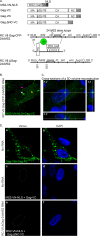
BiFC between Gag and unspliced vRNA. (A) RC.V8 Gag-CFP-24xMS2 contains 24 copies of the MS2 stem-loops between gag-cfp and pr, allowing for the labeling of only the unspliced vRNA. Because the stem-loops are closer to Ψ, this allows MS2-VN-NLS to bind to Gag-VC if Gag binds to the vRNA, causing a positive BiFC signal. (B) A single optical slice of a QT6 cell expressing RC.V8 Gag-CFP-24xMS2, MS2-VN, and Gag-VC. BiFC foci representing Gag-unspliced vRNA complexes are present in the nucleus (white arrow), in the cytoplasm (yellow arrow), and at the plasma membrane (magenta arrow). (C) A cross-section of a z-stack of the same cell. The crosshairs show a complex of Gag and unspliced vRNA in the nucleus. (D) (a and b) Gag-VN and Gag-VC form BiFC foci (green) in the nucleus, in the cytoplasm, and at the plasma membrane. (c and d) In the absence of RC.V8, Gag-CFP-24xMS2, MS2-VN-NLS, and Gag-VC do not fluoresce. (e and f) BiFC fluorescence was negative when a functional Gag was absent. Combinations expected to have a positive BiFC signal are labeled in green text, and those with negative signal are have white labels.
FIG 6
FIG 6
FRET efficiency. All images presented demonstrated the highest FRET efficiency measured during the experiment to better visualize the change in Gag-CFP signal from prebleach to postbleach. (A to C) Examples of FRET between a Gag-CFP (donor) focus and unspliced vRNA labeled with MS2-YFP (acceptor) focus (white arrows) in the nucleus (A), in the cytoplasm (B), and at the plasma membrane (C). (D) The FRET efficiencies of Gag and unspliced vRNA in the nucleus (11% ± 3%; P = 0.0031), in the cytoplasm (9% ± 2%, P = 0.0180), and at the plasma membrane (11% ± 2%, P = 0.0008) were statistically significant over that of the CFP/YFP control (4% ± 0.5%).
FIG 7
FIG 7
Subcellular localization of vRNA and Gag.L219A in QT6 cells. (A) (a) In the presence of Gag.L219A-CFP, MS2-YFP-NLS remains diffuse in the nucleus in the absence of MS2 stem-loop-containing RNA. (b) RC.V8-24xMS2 RNA foci (9 ± 0.9 foci per nucleus) colocalize with Gag.L219A foci (44 ± 4 foci per nucleus) at a higher level in the nucleus. (c) Unspliced (US) RC.V8-24xMS2 RNAs (11 ± 2 foci per nucleus) colocalized with Gag.L219A-CFP (80 ± 7 foci per nucleus) in the nucleus higher than both spliced and US vRNA (RC.V8-24xMS2). RC.V8-24xMS2 US RNA (FISH) colocalization with Gag.L219A is statistically higher (P = 0.0219) than RC.V8-24xMS2 colocalization with Gag.L219A. Statistical analyses were performed in Prism (GraphPad) using an unpaired two-tailed t test. (B) A cross-section of a z-stack of a cell expressing RC.V8-24xMS2, MS2-YFP-NLS, and Gag.L219A-CFP and stained with DAPI was generated. The focus of interest (located in between the yellow lines) is outlined in x,y (left), y,z (right), and x,z (bottom) positions. (C) A z-stack of a cell expressing RC.V8-24xMS2, MS2-YFP-NLS, and Gag.L219A-CFP was imaged and used to create a 3D volume surface rendering. Left, x,y cut of a cell displaying RC.V8-24xMS2 (green) and Gag.L219A-CFP (red) complexes within the DAPI-stained nucleus (blue). Right, colocalization between the vRNA and Gag.L219A is displayed as a white surface rendering in an x,y cut of the same cell. (D) A z-stack the nucleus of a cell expressing RC.V8-24xMS2, MS2-YFP-NLS, Gag.L219A-CFP, and Sun1-mCherry that was imaged using structured illumination microscopy (SIM) was used to create a 3D volume rendering. The 3D rendering is rotated at 90°. (E) A colocalization channel (white) was generated to visualize Gag.L219A-vRNA complexes, and a surface rendering was created from the z-stack of the same cell in panel D.

Similar articles

Cited by

References

    1. Scheifele LZ, Garbitt RA, Rhoads JD, Parent LJ. 2002. Nuclear entry and CRM1-dependent nuclear export of the Rous sarcoma virus Gag polyprotein. Proc Natl Acad Sci U S A 99:3944–3949. doi:10.1073/pnas.062652199. - DOI - PMC - PubMed
    1. Garbitt-Hirst R, Kenney SP, Parent LJ. 2009. Genetic evidence for a connection between Rous sarcoma virus Gag nuclear trafficking and genomic RNA packaging. J Virol 83:6790–6797. doi:10.1128/JVI.00101-09. - DOI - PMC - PubMed
    1. Pincetic A, Leis J. 2009. The mechanism of budding of retroviruses from cell membranes. Adv Virol 2009:6239691–6239699. doi:10.1155/2009/623969. - DOI - PMC - PubMed
    1. Mattei S, Schur FK, Briggs JA. 2016. Retrovirus maturation–an extraordinary structural transformation. Curr Opin Virol 18:27–35. doi:10.1016/j.coviro.2016.02.008. - DOI - PubMed
    1. Dubois N, Marquet R, Paillart JC, Bernacchi S. 2018. Retroviral RNA dimerization: from structure to functions. Front Microbiol 9:527. doi:10.3389/fmicb.2018.00527. - DOI - PMC - PubMed

Publication types

LinkOut - more resources