. 2020 May 15;295(20):6958-6971.
doi: 10.1074/jbc.RA120.013189. Epub 2020 Apr 7.

Activation of JNK signaling promotes all- trans- retinal-induced photoreceptor apoptosis in mice

Affiliations

Activation of JNK signaling promotes all- trans- retinal-induced photoreceptor apoptosis in mice

Chunyan Liao et al. J Biol Chem. .

Abstract

Disrupted clearance of all-trans-retinal (atRAL), a component of the visual (retinoid) cycle in the retina, may cause photoreceptor atrophy in autosomal recessive Stargardt disease (STGD1) and dry age-related macular degeneration (AMD). However, the mechanisms underlying atRAL-induced photoreceptor loss remain elusive. Here, we report that atRAL activates c-Jun N-terminal kinase (JNK) signaling at least partially through reactive oxygen species production, which promoted mitochondria-mediated caspase- and DNA damage-dependent apoptosis in photoreceptor cells. Damage to mitochondria in atRAL-exposed photoreceptor cells resulted from JNK activation, leading to decreased expression of Bcl2 apoptosis regulator (Bcl2), increased Bcl2 antagonist/killer (Bak) levels, and cytochrome c (Cyt c) release into the cytosol. Cytosolic Cyt c specifically provoked caspase-9 and caspase-3 activation and thereby initiated apoptosis. Phosphorylation of JNK in atRAL-loaded photoreceptor cells induced the appearance of γH2AX, a sensitive marker for DNA damage, and was also associated with apoptosis onset. Suppression of JNK signaling protected photoreceptor cells against atRAL-induced apoptosis. Moreover, photoreceptor cells lacking Jnk1 and Jnk2 genes were more resistant to atRAL-associated cytotoxicity. The Abca4-/-Rdh8-/- mouse model displays defects in atRAL clearance that are characteristic of STGD1 and dry AMD. We found that JNK signaling was activated in the neural retina of light-exposed Abca4-/-Rdh8-/- mice. Of note, intraperitoneal administration of JNK-IN-8, which inhibits JNK signaling, effectively ameliorated photoreceptor degeneration and apoptosis in light-exposed Abca4-/-Rdh8-/- mice. We propose that pharmacological inhibition of JNK signaling may represent a therapeutic strategy for preventing photoreceptor loss in retinopathies arising from atRAL overload.

Keywords: DNA damage; apoptosis; photoreceptor; retina; retinal metabolism.


Conflict of interest statement

The authors declare that they have no conflicts of interest with the contents of this article

Figures

Figure 1.
Figure 1.
Evidence that atRAL induces apoptosis of 661W photoreceptor cells. A, cell viability, 6 h after exposure of 661W photoreceptor cells to serial concentrations of atRAL (2.5, 5, 10 and 20 μm), was measured using MTS assay. Control cells were treated with vehicle (DMSO) alone. Bar heights indicate MTS absorbance at 490 nm and reflect cell viability. Data are presented as mean ± S.D. (error bars) of three independent experiments, and statistical analyses were performed by using one-way ANOVA with Tukey's post-test. ***, p < 0.001. B, TUNEL staining of 661W photoreceptor cells treated with 5 μm atRAL or vehicle (DMSO) alone for 6 h. Stained cells were visualized by using an Olympus FV1000 confocal fluorescence microscope. Red arrows indicate TUNEL-positive cells. Scale bars, 20 μm. Ctrl, control. C, percentage of TUNEL-positive cells relative to the total number of cells. 661W photoreceptor cells were incubated with 5 μm atRAL for 6 h. Cells treated for 6 h with an equal volume of DMSO served as a vehicle control. Data are presented as mean ± S.D. (error bars) of three independent experiments, and statistical analyses were performed by using Student's t test. ***, p < 0.001. D, fluorescence measurement of ΔΨm in 661W photoreceptor cells treated with 5 μm atRAL or vehicle (DMSO) alone for 6 h. The cells were stained by rhodamine-123 for 20 min and observed by using the confocal fluorescence microscope. Scale bars, 10 μm. E, Western blot analysis of Bcl2 in lysates of 661W photoreceptor cells exposed to 5 μm atRAL or vehicle (DMSO) alone for 6 h. GAPDH was used as an internal control. Molecular mass markers (kDa) are indicated to the left of the blots. F, expression levels of Bcl2 protein were normalized to those of GAPDH and presented as fold changes relative to vehicle (DMSO) controls. Data are presented as mean ± S.D. (error bars) of three independent experiments, and statistical analyses were performed by using Student's t test. **, p < 0.01. G, Western blotting was used to analyze protein levels of mitochondrial Bak in 661W photoreceptor cells incubated with 5 μm atRAL for 6 h. Control cells were treated with vehicle (DMSO) alone. COX IV was used as a loading control. H, protein levels of Cyt c in the cytosol of 661W photoreceptor cells exposed to 5 μm atRAL for 6 h were detected by Western blotting. Control cells were incubated with vehicle (DMSO) alone. GAPDH was used as a loading control. I, Western blot analysis of pro-caspase-9, cleaved caspase-9, and cleaved caspase-3 in lysates of 661W photoreceptor cells exposed to 5 μm atRAL or vehicle (DMSO) alone for 6 h. GAPDH was used as an internal control. J, immunofluorescence staining of cleaved caspase-3 in 661W photoreceptor cells incubated for 6 h with 5 μm atRAL or vehicle (DMSO) alone. Nuclei were stained blue with DAPI. Scale bars, 20 μm. K, immunoblot analysis of Bcl2, Flag (Bcl2), and cleaved caspase-3 in lysates of Bcl2 overexpressing 661W photoreceptor cells incubated with 5 μm atRAL or vehicle (DMSO) alone for 6 h, respectively. Cells transfected with vector pcDNA3.1 served as a control. GAPDH was used as a loading control. L, cell viability, 6 h after exposure of Bcl2 overexpressing 661W photoreceptor cells to 5 μm atRAL or vehicle (DMSO) alone, was determined by MTS assay. Cells transfected with vector pcDNA3.1 were used as a control. M, immunoblot analysis of cleaved caspase-3 in 661W photoreceptor cells exposed to 5 μm atRAL for 6 h in the absence or presence of 20 μm caspase inhibitor Z-VAD-FMK. Note that cells were pretreated with 20 μm Z-VAD-FMK for 1 h. Cells treated with Z-VAD-FMK or vehicle (DMSO) served as controls. GAPDH was utilized as an internal control. N, cell viability was probed by MTS assay. 661W photoreceptor cells were pretreated with caspase-3 inhibitor Z-VAD-FMK (20 μm) for 1 h, followed by incubation with or without 5 μm atRAL for 6 h. Control cells were cultured with atRAL or vehicle (DMSO) alone. Each value represents mean ± S.D. (error bars) of three independent experiments, and statistical analyses were performed by using one-way ANOVA with Tukey's post-test.
Figure 2.
Figure 2.
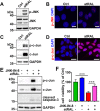
atRAL activates JNK signaling in 661W photoreceptor cells. A, immunoblot analysis of p-JNK and JNK in lysates of 661W photoreceptor cells treated with 5 μm atRAL or vehicle (DMSO) alone for 6 h. GAPDH was used as an internal control. Molecular mass markers (kDa) are indicated to the left of the blots. Ctrl, control. B, immunofluorescence staining for p-JNK in 661W photoreceptor cells incubated with 5 μm atRAL or vehicle (DMSO) alone for 6 h. Nuclei were stained blue with DAPI. Scale bars, 10 μm. C, Western blot analysis of p-c-Jun and c-Jun in lysates of 661W photoreceptor cells exposed to 5 μm atRAL or vehicle (DMSO) alone for 6 h. GAPDH was used as an internal control. D, immunofluorescence staining for p-c-Jun in 661W photoreceptor cells exposed to 5 μm atRAL or vehicle (DMSO) alone for 6 h. Nuclei were stained blue with DAPI. Scale bars, 10 μm. E, immunoblot analysis of p-c-Jun, c-Jun, and cleaved caspase-3 in 661W photoreceptor cells treated with 5 μm atRAL for 6 h in the absence or presence of 1 μm JNK-specific inhibitor JNK–IN-8. Note that cells were pretreated with 1 μm JNK–IN-8 for 1 h. Cells treated with vehicle (DMSO) or JNK–IN-8 served as controls. GAPDH was utilized as an internal control. F, cell viability was detected by MTS assay. 661W photoreceptor cells were pretreated with JNK-specific inhibitor JNK–IN-8 (1 μm) for 1 h, followed by incubation with and without 5 μm atRAL for 6 h. Control cells were treated with atRAL or vehicle (DMSO) alone. Each value represents mean ± S.D. (error bars) of three independent experiments, and statistical analyses were performed by using one-way ANOVA with Tukey's post-test.
Figure 3.
Figure 3.
atRAL induces DNA damage response in 661W photoreceptor cells. A, immunoblot analysis of γH2AX in 661W photoreceptor cells treated with 5 μm atRAL or vehicle (DMSO) alone for 6 h. GAPDH was used as an internal control. Molecular mass markers (kDa) are indicated to the left of the blots. Ctrl, control. B and C, immunofluorescence staining for γH2AX (B) and p-ATM (C) in 661W photoreceptor cells incubated for 6 h with 5 μm atRAL or vehicle (DMSO) alone. Nuclei were stained blue with DAPI. Scale bars, 10 μm. D, Western blot analysis of γH2AX in 661W photoreceptor cells treated with 5 μm atRAL for 6 h in the absence or presence of 1 μm JNK-specific inhibitor JNK–IN-8. Note that cells were pretreated with 1 μm JNK–IN-8 for 1 h. Cells treated with vehicle (DMSO) or JNK–IN-8 served as controls. GAPDH was utilized as an internal control.
Figure 4.
Figure 4.
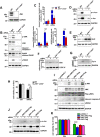
Genetic deletion of Jnk1 and Jnk2 genes rescues 661W photoreceptor cells from apoptosis caused by atRAL via blocking JNK signaling. A, Western blot analysis of JNK in lysates of WT and Jnk1−/−Jnk2−/− 661W photoreceptor cells. GAPDH was utilized as an internal control. Molecular mass markers (kDa) are indicated to the left of the blots. B, immunoblot analysis of p-JNK, JNK, cleaved caspase-3, PARP, cleaved PARP and γH2AX in lysates of WT and Jnk1−/−Jnk2−/− 661W photoreceptor cells cultured with 5 μm atRAL or vehicle (DMSO) for 6 h, respectively. GAPDH was used as an internal control. C, protein levels of cleaved caspase-3, cleaved PARP, and γH2AX were normalized to those of GAPDH and presented as fold changes relative to vehicle (DMSO) controls. Ctrl, control. Data are presented as mean ± S.D. (error bars) of three independent experiments, and statistical analyses were performed by using two-way ANOVA with Sidak's post-test. ***, p < 0.001. D, immunoblot analysis of p-c-Jun and c-Jun in lysates of WT and Jnk1−/−Jnk2−/− 661W photoreceptor cells exposed to 5 μm atRAL or vehicle (DMSO) for 6 h, respectively. GAPDH was used as an internal control. E, Western blot analysis of Bcl2 in lysates of WT and Jnk1−/−Jnk2−/− 661W photoreceptor cells exposed to 5 μm atRAL or vehicle (DMSO) for 6 h, respectively. GAPDH was used as an internal control. F, Western blotting was used to analyze protein expression of mitochondrial Bak in WT and Jnk1−/−Jnk2−/− 661W photoreceptor cells treated with 5 μm atRAL or vehicle (DMSO) for 6 h, respectively. GAPDH was used as an internal control. G, immunoblot analysis of Cyt c in the cytosol of WT and Jnk1−/−Jnk2−/− 661W photoreceptor cells exposed to 5 μm atRAL or vehicle (DMSO) for 6 h, respectively. GAPDH was used as an internal control. H, cell viability, 6 h after incubating WT and Jnk1−/−Jnk2−/− 661W photoreceptor cells with 5 μm atRAL or vehicle (DMSO) alone, respectively, was measured by MTS assay. Each value represents mean ± S.D. (error bars) of three independent experiments, and statistical analyses were performed by using two-way ANOVA with Sidak's post-test. **, p < 0.01. I, Western blot analysis of p-JNK, JNK, Flag (JNK), cleaved caspase-3, and γH2AX in atRAL-loaded Jnk1−/−Jnk2−/− 661W photoreceptor cells re-expressing JNK1, JNK2, or both of them. Jnk1−/−Jnk2−/− 661W photoreceptor cells transfected with vector pcDNA3.1 were used as a control. Cells were treated with 5 μm atRAL or vehicle (DMSO) alone for 6 h. GAPDH served as a loading control. J, immunoblot analysis of p-c-Jun and c-Jun in atRAL-loaded Jnk1−/−Jnk2−/− 661W photoreceptor cells re-expressing JNK1, JNK2, or both of them. Jnk1−/−Jnk2−/− 661W photoreceptor cells transfected with vector pcDNA3.1 were used as a control. Cells were incubated with 5 μm atRAL or vehicle (DMSO) alone for 6 h. GAPDH was utilized as a loading control. K, cell viability was examined by MTS assay. Jnk1−/−Jnk2−/− 661W photoreceptor cells re-expressing JNK1, JNK2, or both of them were treated with 5 μm atRAL or vehicle (DMSO) alone for 6 h. Jnk1−/−Jnk2−/− 661W photoreceptor cells transfected with vector pcDNA3.1 were used as a control. Each value represents mean ± S.D. (error bars) of three independent experiments, and statistical analyses were performed by using two-way ANOVA with Sidak's post-test. ***, p < 0.001.
Figure 5.
Figure 5.
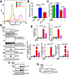
atRAL provokes ROS (including mitochondrial ROS) generation in 661W photoreceptor cells. A, intracellular ROS production, 6 h after incubating 661W photoreceptor cells with 5 μm atRAL or vehicle (DMSO) alone, was measured by H2DCFDA staining coupled with flow cytometry. B, quantification of H2DCFDA-positive cells by flow cytometry. Each value is shown as a percentage of the total cell population and presented as mean ± S.D. (error bars) of three independent experiments. Statistical analyses were performed by using Student's t test. **, p < 0.01. C, intracellular ROS generation, 6 h after exposure of 661W photoreceptor cells to 5 μm atRAL, was visualized using the fluorescent probe H2DCFDA by confocal laser-scanning fluorescence microscopy. H2DCFDA emits green fluorescence upon reaction with ROS. Cells treated with DMSO alone served as a vehicle control (Ctrl). Nuclei were stained with Hoechst 33342 (blue). Scale bars, 10 μm. D, mitochondrial ROS production in 661W photoreceptor cells exposed to 5 μm atRAL for 6 h was determined by MitoSOX Red staining. Control cells were incubated with DMSO alone. Nuclei were labeled by Hoechst 33342 (blue). Scale bars, 10 μm.
Figure 6.
Figure 6.
Antioxidant NAC ameliorates atRAL-induced apoptosis of 661W photoreceptor cells by suppressing JNK signaling. A, intracellular ROS levels were measured by flow cytometry using the fluorescence probe H2DCFDA. 661W photoreceptor cells were cultured for 6 h with 5 μm atRAL or vehicle (DMSO) alone. Alternatively, 661W photoreceptor cells were pretreated with 2 mm NAC for 2 h and incubated with 5 μm atRAL for 6 h. Subsequently, the cells were stained by 10 μm H2DCFDA for 10 min at 37 °C, and the levels of intracellular ROS were determined by flow cytometry. B, quantity of H2DCFDA-positive cells was assayed by flow cytometry and expressed as a percentage of the total cell population. Each value represents mean ± S.D. (error bars) of three independent experiments, and statistical analyses were performed by using one-way ANOVA with Tukey's post-test. **, p < 0.01; ***, p < 0.001. C, cell viability was examined by MTS assay. 661W photoreceptor cells were pretreated with antioxidant NAC (2 mm) for 2 h, followed by incubation with or without 5 μm atRAL for 6 h. Control cells were treated with atRAL or vehicle (DMSO) alone. Each value represents mean ± S.D. (error bars) of three independent experiments, and statistical analyses were performed by using one-way ANOVA with Tukey's post-test. D, immunoblot analysis of p-JNK, JNK, pro-caspase-9, cleaved caspase-9, cleaved caspase-3, PARP, cleaved PARP, and γH2AX in lysates of 661W photoreceptor cells treated for 6 h with 5 μm atRAL in the absence or presence of 2 mm NAC. Note that cells were pretreated with 2 mm NAC for 2 h. Cells treated with NAC or vehicle (DMSO) alone served as controls. GAPDH was utilized as an internal control. Molecular mass markers (kDa) are indicated to the left of the blots. E, ratios of p-JNK/JNK protein level and protein levels of cleaved caspase-9, cleaved caspase-3, cleaved PARP, and γH2AX were shown as fold changes relative to vehicle (DMSO) controls. Levels of each protein were normalized to those of GAPDH. Note that the normalization for band intensity of p-JNK or JNK against that of GAPDH was individually made before calculating the p-JNK/JNK ratio. Each value represents mean ± S.D. (error bars) of three independent experiments, and statistical analyses were performed by using one-way ANOVA with Tukey's post-test. **, p < 0.01; ***, p < 0.001. F, immunoblot analysis of p-c-Jun and c-Jun in lysates of 661W photoreceptor cells incubated with 5 μm atRAL for 6 h in the presence or absence of 2 mm NAC. Note that cells were pretreated with 2 mm NAC for 2 h. Control cells were treated with NAC or vehicle (DMSO) alone. GAPDH served as an internal control. G, Western blot analysis of cytosolic Cyt c in 661W photoreceptor cells exposed to 5 μm atRAL for 6 h with or without 2 mm NAC. Note that cells were pretreated with 2 mm NAC for 2 h. Cells treated with NAC or vehicle (DMSO) alone served as controls. GAPDH was used as an internal control.
Figure 7.
Figure 7.
Light-induced photoreceptor degeneration in Abca4−/−Rdh8−/− mice involves activation of JNK signaling. Four-week-old C57BL/6J WT and Abca4−/−Rdh8−/− mice were exposed to LED light with an intensity of 10,000 lx for 2 h after their pupils were dilated with 1% tropicamide. Eyeballs, 1, 3, and 5 days after light exposure, were harvested for subsequent studies. C57BL/6J WT and Abca4−/−Rdh8−/− mice raised normally in the dark for 7 days in the absence of 10,000 lx LED light served as controls. A, histology of mouse retina was examined by H&E staining. Scale bars, 20 μm. B, apoptotic cells in mouse retina were detected by TUNEL staining. Scale bars, 20 μm. C, immunoblot analysis of p-JNK, JNK, γH2AX and cleaved caspase-3 in extracts from mouse neural retina. GAPDH was used as an internal control. Molecular mass markers (kDa) are indicated to the left of the blots. D, genotyping of Abca4−/−Rdh8−/− mice on a C57BL/6J genetic background was carried out by PCR amplification of tail DNAs with specific primers. Abca4 deletion in Abca4−/−Rdh8−/− mice was identified by producing a 455-bp amplicon with the A0 and N1 primers, yet C57BL/6J WT mice generated a 619-bp amplicon with the ABCR1 and ABCR2 primers. Rdh8 deletion in Abca4−/−Rdh8−/− mice was corroborated by giving rise to a 450-bp amplicon with the Neo1 and DMR11 primers, whereas C57BL/6J WT mice produced an 800-bp amplicon with the DMR5 and DMR6 primers. To further confirm the absence of Rd8 mutation of the Crb1 gene in C57BL/6J WT and Abca4−/−Rdh8−/− mice used in the study, DNA samples extracted from mouse tail biopsies were amplified for Wt allele and mutant Rd8 allele using primers mCrb1 mF1, mCrb1 mF2, and mCrb1 mR. C57BL/6N mice carrying Rd8 mutation in Crb1 gene served as a control. Amplicon sizes were 220 bp for Wt allele and 244 bp for Rd8 allele. A 100-bp DNA ladder was used as a size standard. OS, outer segment; IS, inner segment; ONL, outer nuclear layer; OPL, outer plexiform layer; INL, inner nuclear layer; IPL, inner plexiform layer; GCL, ganglion cell layer.
Figure 8.
Figure 8.
JNK-specific inhibitor JNK–IN-8 alleviates light-induced photoreceptor degeneration and apoptosis in Abca4−/−Rdh8−/− mice. 48-h dark-adapted Abca4−/−Rdh8−/− mice at 4 weeks of age were intraperitoneally injected with JNK–IN-8 or DMSO (vehicle) (4 mg/kg body weight). One hour later, pupils of mice were dilated with 1% tropicamide, and the mice were exposed to LED light with the intensity of 10,000 lx for 2 h, followed by once-daily treatment with JNK–IN-8 or DMSO (vehicle) (4 mg/kg body weight) for 4 days. Eyeballs, 5 days after light illumination, were collected for subsequent studies. Control Abca4−/−Rdh8−/− mice were administered intraperitoneally with JNK–IN-8 or DMSO (vehicle) (4 mg/kg body weight) in the absence of light exposure. A, morphology of mouse retina was analyzed by H&E staining. Scale bars, 20 μm. B, apoptotic cells in mouse retina were examined by TUNEL staining. Scale bars, 20 μm. C and D, immunoblot analysis of p-c-Jun, c-Jun, cleaved caspase-3, and γH2AX in extracts from mouse neural retina. GAPDH served as a loading control. Molecular mass markers (kDa) are indicated to the left of the blots.
Figure 9.
Figure 9.
Proposed mechanisms showing atRAL-induced apoptosis in photoreceptor cells via activating JNK signaling. In photoreceptor cells, atRAL evokes collapsed ΔΨm and JNK activation. The latter is at least partially mediated by the ROS (including mitochondrial ROS) production. Phosphorylation of JNK results in decreased protein expression of Bcl2 and mitochondrial damage through increased Bak protein that may cause Bak activation. Cyt c is released from ruptured mitochondria into the cytosol where it stimulates the formation of apoptosomes consisting of Cyt c, caspase-9, and Apaf-1 (51, 52). Caspase-9 activation cleaves and activates caspase-3 to induce apoptosis. However, p-JNK, the active form of JNK, partially translocates into the nucleus where it phosphorylates the histone H2AX to form γH2AX, a sensitive marker for DNA double-strand breaks. Generation of γH2AX, which localizes to sites of DNA strand breaks, accounts for the onset of irreversible DNA damage correlated with apoptotic cell death (53). It should be mentioned that the ROS production caused by atRAL is one of the initiators to activate JNK signaling, and JNK activation is also not the only way for atRAL-stimulated DNA damage.

Similar articles

Cited by

References

    1. Liu X., Chen J., Liu Z., Li J., Yao K., and Wu Y. (2016) Potential therapeutic agents against retinal diseases caused by aberrant metabolism of retinoids. Invest. Ophthalmol. Vis. Sci. 57, 1017–1030 10.1167/iovs.15-18429 - DOI - PubMed
    1. Maeda A., Maeda T., Golczak M., and Palczewski K. (2008) Retinopathy in mice induced by disrupted all-trans-retinal clearance. J. Biol. Chem. 283, 26684–26693 10.1074/jbc.M804505200 - DOI - PMC - PubMed
    1. Maeda T., Golczak M., and Maeda A. (2012) Retinal photodamage mediated by all-trans-retinal. Photochem. Photobiol. 88, 1309–1319 10.1111/j.1751-1097.2012.01143.x - DOI - PMC - PubMed
    1. Liao Y., Zhang H., He D., Wang Y., Cai B., Chen J., Ma J., Liu Z., and Wu Y. (2019) Retinal pigment epithelium cell death is associated with NLRP3 inflammasome activation by all-trans retinal. Invest. Ophthalmol. Vis. Sci. 60, 3034–3045 10.1167/iovs.18-26360 - DOI - PubMed
    1. Maeda A., Maeda T., Golczak M., Chou S., Desai A., Hoppel C. L., Matsuyama S., and Palczewski K. (2009) Involvement of all-trans-retinal in acute light-induced retinopathy of mice. J. Biol. Chem. 284, 15173–15183 10.1074/jbc.M900322200 - DOI - PMC - PubMed

Publication types

MeSH terms

Substances