. 2020 Jun;11(6):433-445.
doi: 10.1007/s13238-020-00695-w. Epub 2020 Apr 5.

Primary cilia mediate Klf2-dependant Notch activation in regenerating heart

Affiliations

Primary cilia mediate Klf2-dependant Notch activation in regenerating heart

Xueyu Li et al. Protein Cell. 2020 Jun.

Abstract

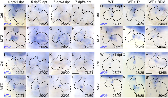
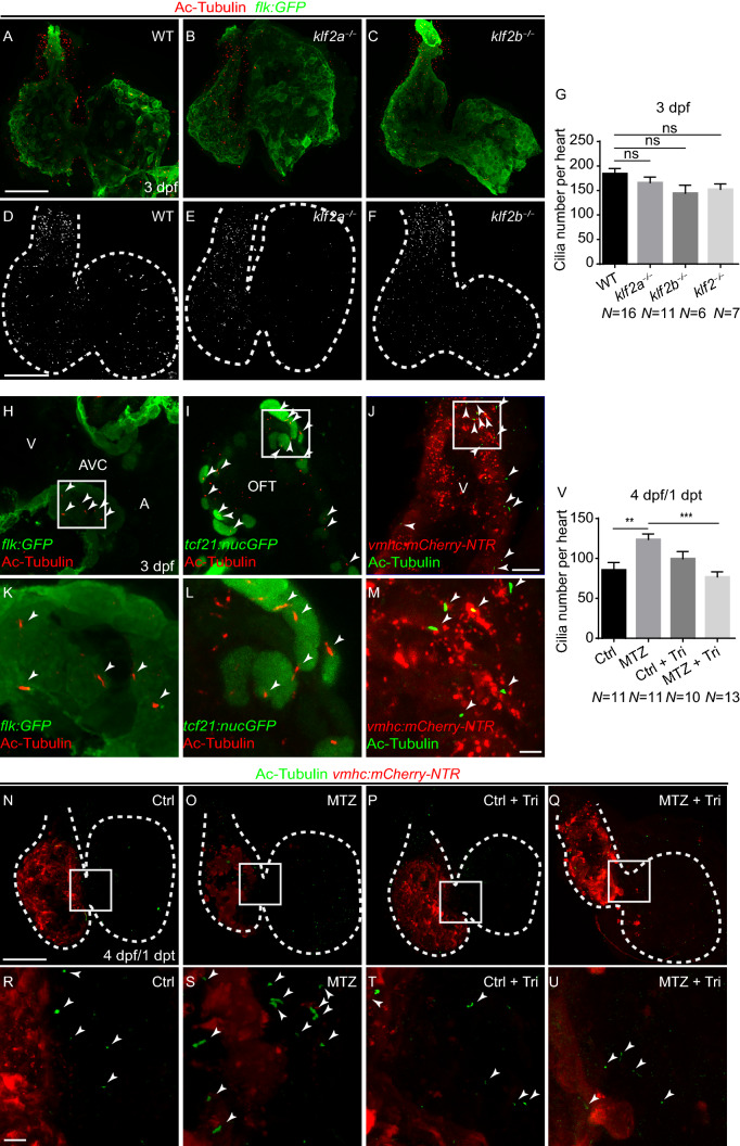
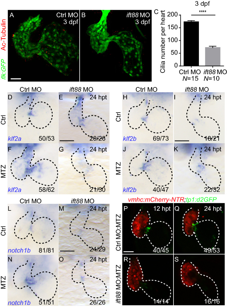
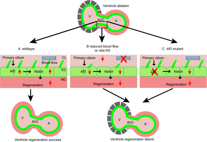
Unlike adult mammalian heart, zebrafish heart has a remarkable capacity to regenerate after injury. Previous study has shown Notch signaling activation in the endocardium is essential for regeneration of the myocardium and this activation is mediated by hemodynamic alteration after injury, however, the molecular mechanism has not been fully explored. In this study we demonstrated that blood flow change could be perceived and transmitted in a primary cilia dependent manner to control the hemodynamic responsive klf2 gene expression and subsequent activation of Notch signaling in the endocardium. First we showed that both homologues of human gene KLF2 in zebrafish, klf2a and klf2b, could respond to hemodynamic alteration and both were required for Notch signaling activation and heart regeneration. Further experiments indicated that the upregulation of klf2 gene expression was mediated by endocardial primary cilia. Overall, our findings reveal a novel aspect of mechanical shear stress signal in activating Notch pathway and regulating cardiac regeneration.

Keywords: Notch signaling; heart regeneration; hemodynamics; klf2; primary cilia.


Figures

Figure 1
Figure 1
Reduced blood flow changesklf2aandklf2bexpression during ventricle regeneration. (A–H) Whole-mount in situ hybridizations showing klf2a upregulation in ablated hearts (E–H) compared to control hearts (A–D) at 4–7 dpf/1–4 dpt. (I–P) Whole-mount in situ hybridizations showing klf2b upregulation in ablated hearts (M-P) compared to control hearts (I–L) at 4–7 dpf/1–4 dpt. (Q–V) Whole-mount in situ hybridizations indicated klf2a upregulation after ablation (Q, T) was blocked in Tricaine treated hearts (R, U) and BDM treated hearts (S, V) at 1 dpt. (W–B’) Whole-mount in situ hybridizations indicated klf2b upregulation after ablation (W, Z) was slightly enhanced in Tricaine treated hearts (X, A’) and BDM treated hearts (Y, B’) at 1 dpt. Scale bars, 50 µm. dpt, days post treatment. Dashed lines outline the hearts. Numbers indicate the ratio of representative staining observed
Figure 2
Figure 2
klf2mutants result in compensation of homologue expression, blunted Notch signaling activation and reduced ventricle regeneration. (A and B) Schematic diagrams of the zebrafish klf2a and klf2b loci with the sgRNA target site sequence (red arrowheads). (C, D) Functional domain diagrams of wildtype Klf2a or Klf2b and predicted truncated protein in corresponding mutants. (E–L) Whole-mount in situ hybridizations showing klf2a expression pattern in the control and ablated hearts of wildtype, klf2a−/− mutants, klf2b−/− mutants and klf2−/− double mutants at 24 hpt. (M) Quantification of the fold change of klf2a expression in wildtype and klf2 mutant larvae at 4 dpf by real time PCR. 4 independent experiments. Mean + s.e.m. ANOVA analysis, *P < 0.05, **P < 0.01, ***P < 0.001. (N–U) Whole-mount in situ hybridizations showing klf2b expression pattern in the control and ablated hearts of wildtype, klf2a−/− mutants, klf2b−/− mutants and klf2−/− double mutants at 24 hpt. (V) Quantification of the fold change of klf2b expression in wildtype and klf2 mutant larvae at 4 dpf by real time PCR. 4 independent experiments. Mean + s.e.m. ANOVA analysis, **P < 0.01. (W) Quantification of the heart recovery rate (black bars) in ablated wildtype and klf2 mutants at 4 dpt. The number of larvae analyzed for each condition is indicated. Binomial test (versus WT), ****P < 0.0001. (X–M’) Confocal stack projections of ablated Tg(vmhc:mCherry-NTR; tp1:d2GFP) hearts showing Notch signaling pattern in wildtype (X–A’), klf2a−/− mutants (B’–E’), klf2b−/− mutants (F’–I’) and klf2−/− double mutants (J’–M’) at 12, 24, 36, 48 hpt. (N’–U’) Whole-mount in situ hybridizations indicated notch1b upregulation as in ablated wildtype hearts (N’, O’) was blocked in klf2a−/− mutant hearts (P’, Q’), klf2b−/− mutant hearts (R’, S’) and klf2−/− double mutant hearts (T’, U’) at 24 hpt. Scale bars, 50 µm. hpt, hours post treatment. Dashed lines outline the hearts. Numbers indicate the ratio of representative staining observed
Figure 3
Figure 3
Endocardial primary cilia exist in the hearts at later stages. (A–F) Confocal stack projections of immunofluorescence showing cardiac cilia in wildtype and klf2 mutants at 3 dpf. Green, anti-GFP immunostaining; red, acetylated tubulin immunostaining. (D–F) greyscale of red channel only. (G) Quantification of cardiac cilia number in wildtype and klf2 mutants at 3 dpf (N = 16, 11, 6, 7 respectively). Mean + s.e.m. ANOVA analysis, ns, not significant. (H–M) Acetylated tubulin immunostaining showing primary cilia in endocardial cells (H, K), epicardial cells (I, L), and cardiomyocytes (J, M) at 3 dpf. (K–M) magnified white boxes in (H–J). (N–U) Confocal stack projections of immunofluorescence showing cardiac cilia in controlTricaine or BDM treatment or ablated Tg(vmhc:mCherry-NTR) hearts with or without Tricaine treatment at 1 dpt. (R–U) magnified white boxes in (N–Q). Red, mCherry fluorescence; green, acetylated tubulin immunostaining. (V) Quantification of cardiac cilia number in control or ablated hearts with or without Tricaine treatment at 1 dpt/4 dpf (N = 11, 11, 10, 13 respectively). Mean + s.e.m. ANOVA analysis, **P < 0.01, ***P < 0.001. Scale bars, (A–F, N–Q) 50 µm, (H–J) 10 µm, (K–M, R–U) 5 µm. dpf, days post fertilization, dpt, days post treatment. Dashed lines outline the hearts. Arrowheads point to cilia. A, atrium; AVC, atrioventricular canal; OFT, outflow tract; V, ventricle
Figure 4
Figure 4
Cilia knockdown inhibits Notch signaling andklf2activation during ventricle regeneration. (A, B) Acetylated tubulin immunostaining in Tg(flk:GFP) heart showing cardiac cilia in control morphants (A) and ift88 morphants (B) at 3 dpf. Green, anti-GFP immunostaining; red, acetylated tubulin immunostaining. dpf, days post fertilization. (C) Quantification of cardiac cilia number in control morphants and ift88 morphants at 3 dpf (N = 15, 10 respectively). Mean + s.e.m. Student’s t-test, ****P < 0.0001. (D–G) Whole-mount in situ hybridizations indicated klf2a upregulation in control morphant ablated hearts (F) compared to control hearts (D) at 24 hpt, whereas this activation was blocked in ablated ift88 morphant hearts (G). (H–K) Whole-mount in situ hybridizations indicated klf2b upregulation in control morphant ablated hearts (J) compared to control hearts (H) at 24 hpt, whereas this activation was blocked in ablated ift88 morphant hearts (K). (L–O) Whole-mount in situ hybridizations indicated notch1b upregulation in control morphant ablated hearts (N) compared to control hearts (L) at 24 hpt, whereas this activation was blocked in ablated ift88 morphant hearts (O). (P–S) Confocal stack projections of ablated Tg(vmhc:mCherry-NTR; tp1:d2GFP) hearts indicated Notch signaling activation was inhibited in ift88 morphants at 12–24 hpt. Scale bars, 50 µm. hpt, hours post treatment. Dashed lines outline the hearts. Numbers indicate the ratio of representative staining observed
Figure 5
Figure 5
Diagrams of hemodynamic-responsive Klf2-dependant Notch activation in ventricle regeneration. (A) During normal ventricle regeneration process, primary cilia on the endocardial cells (green) sense the oscillatory blood flow (blue arrows), which leads to upregulation of the klf2 and subsequent activation of Notch signaling in the endocardium. This Notch activation is essential for myocardium (red) regeneration. (B) When blocking blood flow or impairing cilia development, endocardial klf2 expression and Notch signaling activation are inhibited, which lead to failure of ventricle regeneration. (C) In klf2 mutants, the lack of endocardial klf2 gene expression affects the activation of Notch signaling, and the damaged ventricle cannot regenerate. A, atrium; AVC, atrioventricular canal; CL, cardiac lumen; EC, endocardium; KD, knockdown; MC, myocardium; V, ventricle

Similar articles

Cited by

References

    1. Atkins GB, Jain MK. Role of Kruppel-like transcription factors in endothelial biology. Circ Res. 2007;100(12):1686–1695. doi: 10.1161/01.RES.0000267856.00713.0a. - DOI - PubMed
    1. Austin-Tse C, Halbritter J, Zariwala MA, Gilberti RM, Gee HY, Hellman N, Pathak N, Liu Y, Panizzi JR, Patel-King RS, et al. Zebrafish ciliopathy screen plus human mutational analysis identifies C21orf59 and CCDC65 defects as causing primary ciliary dyskinesia. Am J Hum Genet. 2013;93(4):672–686. doi: 10.1016/j.ajhg.2013.08.015. - DOI - PMC - PubMed
    1. Baratchi S, Khoshmanesh K, Woodman OL, Potocnik S, Peter K, McIntyre P. Molecular sensors of blood flow in endothelial cells. Trends Mol Med. 2017;23(9):850–868. doi: 10.1016/j.molmed.2017.07.007. - DOI - PubMed
    1. Chang N, Sun C, Gao L, Zhu D, Xu X, Zhu X, Xiong JW, Xi JJ. Genome editing with RNA-guided Cas9 nuclease in zebrafish embryos. Cell Res. 2013;23(4):465–472. doi: 10.1038/cr.2013.45. - DOI - PMC - PubMed
    1. Clark BS, Cui S, Miesfeld JB, Klezovitch O, Vasioukhin V, Link BA. Loss of Llgl1 in retinal neuroepithelia reveals links between apical domain size, Notch activity neurogenesis. Development. 2012;139(9):1599–1610. doi: 10.1242/dev.078097. - DOI - PMC - PubMed

Publication types

MeSH terms

LinkOut - more resources