Rbfox-1 contributes to CaMKIIα expression and intracerebral hemorrhage-induced secondary brain injury via blocking micro-RNA-124
- PMID: 32248729
- PMCID: PMC7922744
- DOI: 10.1177/0271678X20916860
Rbfox-1 contributes to CaMKIIα expression and intracerebral hemorrhage-induced secondary brain injury via blocking micro-RNA-124
Abstract
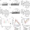
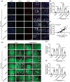
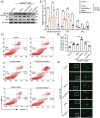
RNA-binding protein fox-1 homolog 1 (Rbfox-1), an RNA-binding protein in neurons, is thought to be associated with many neurological diseases. To date, the mechanism on which Rbfox-1 worsens secondary cell death in ICH remains poorly understood. In this study, we aimed to explore the role of Rbfox-1 in intracerebral hemorrhage (ICH)-induced secondary brain injury (SBI) and to identify its underlying mechanisms. We found that the expression of Rbfox-1 in neurons was significantly increased after ICH, which was accompanied by increases in the binding of Rbfox-1 to Ca2+/calmodulin-dependent protein kinase II (CaMKIIα) mRNA and the protein level of CaMKIIα. In addition, when exposed to exogenous upregulation or downregulation of Rbfox-1, the protein level of CaMKIIα showed a concomitant trend in brain tissue, which further suggested that CaMKIIα is a downstream-target protein of Rbfox-1. The upregulation of both proteins caused intracellular-Ca2+ overload and neuronal degeneration, which exacerbated brain damage. Furthermore, we found that Rbfox-1 promoted the expression of CaMKIIα via blocking the binding of micro-RNA-124 to CaMKIIα mRNA. Thus, Rbfox-1 is expected to be a promising therapeutic target for SBI after ICH.
Keywords: CaMKIIα; Rbfox-1; intracerebral hemorrhage; micro-RNA-124; secondary brain injury.
Conflict of interest statement
Figures


Similar articles
-
Role of miR-148a in Mitigating Hepatic Ischemia-Reperfusion Injury by Repressing the TLR4 Signaling Pathway via Targeting CaMKIIα in Vivo and in Vitro.Cell Physiol Biochem. 2018;49(5):2060-2072. doi: 10.1159/000493716. Epub 2018 Sep 21. Cell Physiol Biochem. 2018. Retraction in: Cell Physiol Biochem. 2022 Oct 31;56(5):610. doi: 10.33594/000000582. PMID: 30244246 Retracted.
-
Exploration of MST1-Mediated Secondary Brain Injury Induced by Intracerebral Hemorrhage in Rats via Hippo Signaling Pathway.Transl Stroke Res. 2019 Dec;10(6):729-743. doi: 10.1007/s12975-019-00702-1. Epub 2019 Apr 2. Transl Stroke Res. 2019. PMID: 30941717
-
Miro1 Regulates Neuronal Mitochondrial Transport and Distribution to Alleviate Neuronal Damage in Secondary Brain Injury After Intracerebral Hemorrhage in Rats.Cell Mol Neurobiol. 2021 May;41(4):795-812. doi: 10.1007/s10571-020-00887-2. Epub 2020 Jun 4. Cell Mol Neurobiol. 2021. PMID: 32500352 Free PMC article.
-
To RNA-binding and beyond: Emerging facets of the role of Rbfox proteins in development and disease.Wiley Interdiscip Rev RNA. 2023 Sep 4:e1813. doi: 10.1002/wrna.1813. Online ahead of print. Wiley Interdiscip Rev RNA. 2023. PMID: 37661850 Review.
-
Intracranial hemorrhage: mechanisms of secondary brain injury.Acta Neurochir Suppl. 2011;111:63-9. doi: 10.1007/978-3-7091-0693-8_11. Acta Neurochir Suppl. 2011. PMID: 21725733 Free PMC article. Review.
Cited by
-
FGF21 alleviates endothelial mitochondrial damage and prevents BBB from disruption after intracranial hemorrhage through a mechanism involving SIRT6.Mol Med. 2023 Dec 4;29(1):165. doi: 10.1186/s10020-023-00755-x. Mol Med. 2023. PMID: 38049769 Free PMC article.
-
Tanshinone IIA Alleviates Traumatic Brain Injury by Reducing Ischemia‒Reperfusion via the miR-124-5p/FoxO1 Axis.Mediators Inflamm. 2024 Mar 21;2024:7459054. doi: 10.1155/2024/7459054. eCollection 2024. Mediators Inflamm. 2024. PMID: 38549714 Free PMC article.
-
TMEM16F may be a new therapeutic target for Alzheimer's disease.Neural Regen Res. 2023 Mar;18(3):643-651. doi: 10.4103/1673-5374.350211. Neural Regen Res. 2023. PMID: 36018189 Free PMC article.
-
BMAL1 attenuates intracerebral hemorrhage-induced secondary brain injury in rats by regulating the Nrf2 signaling pathway.Ann Transl Med. 2021 Nov;9(21):1617. doi: 10.21037/atm-21-1863. Ann Transl Med. 2021. PMID: 34926661 Free PMC article.
-
Pan-cancer analysis of mRNA stability for decoding tumour post-transcriptional programs.Commun Biol. 2022 Aug 20;5(1):851. doi: 10.1038/s42003-022-03796-w. Commun Biol. 2022. PMID: 35987939 Free PMC article.
References
-
- Lee L, Lo YT, See AAQ, et al.. Long-term recovery profile of patients with severe disability or in vegetative states following severe primary intracerebral hemorrhage. J Crit Care 2018; 48: 269–275. - PubMed
-
- Zhu MX, Lu C, Xia CM, et al.. Simvastatin pretreatment protects cerebrum from neuronal injury by decreasing the expressions of phosphor-CaMK II and AQP4 in ischemic stroke rats. J Mol Neurosci 2014; 54: 591–601. - PubMed
Publication types
MeSH terms
Substances
LinkOut - more resources
Full Text Sources
Molecular Biology Databases
Miscellaneous

