Phenotypic and functional characterization of natural killer cells in rheumatoid arthritis-regulation with interleukin-15
- PMID: 32246007
- PMCID: PMC7125139
- DOI: 10.1038/s41598-020-62654-z
Phenotypic and functional characterization of natural killer cells in rheumatoid arthritis-regulation with interleukin-15
Abstract
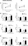
Rheumatoid arthritis (RA) is an autoimmune disease characterized by synovial inflammation and joint destruction. Previous studies have shown that natural killer (NK) cells may play an important role in the pathogenesis of RA. Interleukin (IL)-15, a pro-inflammatory cytokine which induces proliferation and differentiation of NK cells, is overexpressed in RA. In this present study, we examine various NKRs and adhesion molecule expression on NK cells from RA patients and their response to IL-15 stimulation. We also sought to study cytokine-induced memory-like (CIML) NK cells in RA patients. We established that 1. RA patients had higher NK cell percentages in peripheral blood and their serum IL-15 levels were higher compared to healthy volunteers; 2. NK cells from RA patients showed lower NKp46 expression and an impaired CD69 response to IL-15; 3. NK cells from RA patients showed higher CD158b and CD158e expression but lower CD62L expression; 4. exogenous IL-15 up-regulated CD69, CD158b, CD158e but down-regulated NKp46 and CD62L expression in RA; 5. As to CIML NK cells, restimulation - induced NK cytotoxicity and IFN-γ production was impaired in RA patients, 6. Reduced NKp46, perforin, and granzyme B expression on NK cells was found in RA patients with bone deformity and erosion, 7. RA disease activity (DAS28) showed inverse correlation with the percentages of CD56+CD3- NK cells, and NKp46 and perforin expression on NK cells, respectively. Taken together, our study demonstrated differential expression of various NK receptors in RA patients. NKp46, CD158e, and perforin expression on NK cells may serve as markers of RA severity.
Conflict of interest statement
The authors declare no competing interests.
Figures


Similar articles
-
Differential expression of NK receptors CD94 and NKG2A by T cells in rheumatoid arthritis patients in remission compared to active disease.PLoS One. 2011;6(11):e27182. doi: 10.1371/journal.pone.0027182. Epub 2011 Nov 15. PLoS One. 2011. PMID: 22102879 Free PMC article.
-
Decreased expression of NKG2D, NKp46, DNAM-1 receptors, and intracellular perforin and STAT-1 effector molecules in NK cells and their dim and bright subsets in metastatic melanoma patients.Melanoma Res. 2014 Aug;24(4):295-304. doi: 10.1097/CMR.0000000000000072. Melanoma Res. 2014. PMID: 24769842
-
Altered Natural Killer Cell Subsets in Seropositive Arthralgia and Early Rheumatoid Arthritis Are Associated with Autoantibody Status.J Rheumatol. 2016 Jun;43(6):1008-16. doi: 10.3899/jrheum.150644. Epub 2016 Apr 1. J Rheumatol. 2016. PMID: 27036380
-
The role of NK cells in rheumatoid arthritis.Inflamm Res. 2021 Dec;70(10-12):1063-1073. doi: 10.1007/s00011-021-01504-8. Epub 2021 Sep 27. Inflamm Res. 2021. PMID: 34580740 Review.
-
Natural killer cells and their role in rheumatoid arthritis: friend or foe?ScientificWorldJournal. 2012;2012:491974. doi: 10.1100/2012/491974. Epub 2012 Apr 1. ScientificWorldJournal. 2012. PMID: 22547986 Free PMC article. Review.
Cited by
-
Leveraging whole blood based functional flow cytometry assays to open new perspectives for rheumatoid arthritis translational research.Sci Rep. 2022 Jul 16;12(1):12166. doi: 10.1038/s41598-022-16622-4. Sci Rep. 2022. PMID: 35842449 Free PMC article.
-
Angiogenic Properties of NK Cells in Cancer and Other Angiogenesis-Dependent Diseases.Cells. 2021 Jun 29;10(7):1621. doi: 10.3390/cells10071621. Cells. 2021. PMID: 34209508 Free PMC article. Review.
-
Reevaluation of NOD/SCID Mice as NK Cell-Deficient Models.Biomed Res Int. 2021 Nov 10;2021:8851986. doi: 10.1155/2021/8851986. eCollection 2021. Biomed Res Int. 2021. PMID: 34805408 Free PMC article.
-
Identifying the causal relationship between immune factors and osteonecrosis: a two-sample Mendelian randomization study.Sci Rep. 2024 Apr 23;14(1):9371. doi: 10.1038/s41598-024-59810-0. Sci Rep. 2024. PMID: 38654114 Free PMC article.
-
Natural killer cells in inflammatory autoimmune diseases.Clin Transl Immunology. 2021 Feb 1;10(2):e1250. doi: 10.1002/cti2.1250. eCollection 2021. Clin Transl Immunology. 2021. PMID: 33552511 Free PMC article. Review.
References
-
- Goldring SR. Pathogenesis of bone and cartilage destruction in rheumatoid arthritis. Rheumatology (Oxford) 2003;42(Suppl 2):ii11–116. - PubMed
Publication types
MeSH terms
Substances
LinkOut - more resources
Full Text Sources
Medical
Research Materials

