

Comprehensive chromosomal aberrations in a case of a patient with TCF3-HLF-positive BCP-ALL
- PMID: 32245383
- PMCID: PMC7118981
- DOI: 10.1186/s12920-020-0709-y
Comprehensive chromosomal aberrations in a case of a patient with TCF3-HLF-positive BCP-ALL
Abstract
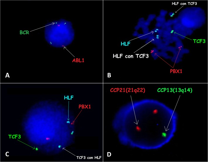
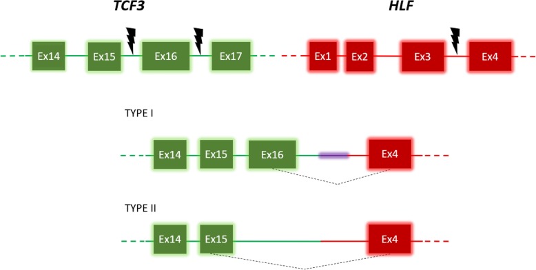
Background: The use of high-throughput analytical techniques has enabled the description of acute lymphoblastic leukaemia (ALL) subtypes. The TCF3-HLF translocation is a very rare rearrangement in ALL that is associated with an extremely poor prognosis. The TCF3-HLF fusion gene in the described case resulted in the fusion of the homeobox-related gene of TCF3 to the leucine zipper domain of HLF. The TCF3-HLF fusion gene product acts as a transcriptional factor leading to the dedifferentiation of mature B lymphocytes into an immature state (lymphoid stem cells). This process initiates the formation of pre-leukaemic cells. Due to the rarity of this chromosomal aberration, only a few cases have been described in the literature. The advantage of this work is the presentation of an interesting case of clonal evolution of cancer cells and the cumulative implications (diagnostic and prognostic) of the patient's genetic alterations.
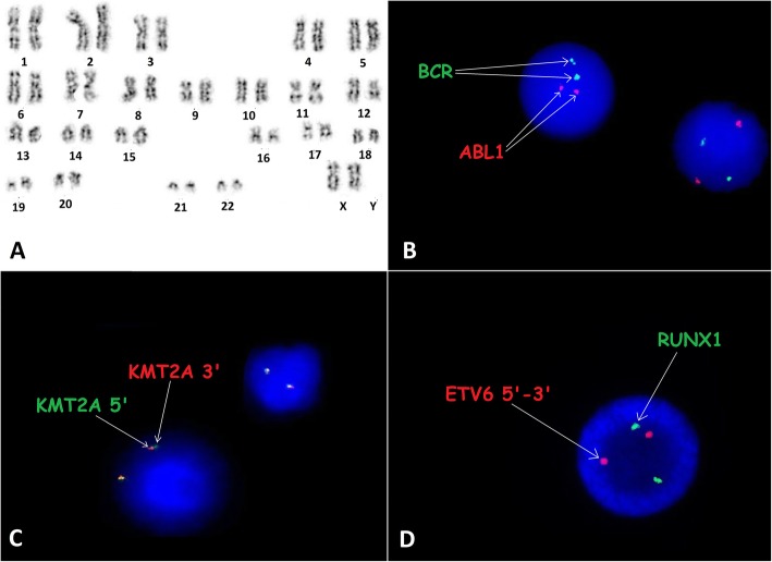
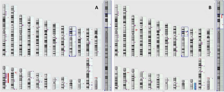
Case presentation: This work presents a patient with diagnosed with TCF3-HLF-positive ALL. Moreover, the additional genetic alterations, which play a key role in the pathogenesis of ALL, were detected in this patient: deletion of a fragment from the long arm of chromosome 13 (13q12.2-q21.1) containing the RB1 gene, intragenic deletions within the PAX5 gene and NOTCH1 intragenic duplication.
Conclusions: A patient with coexistence of chromosomal alterations and the TCF3-HLF fusion has not yet been described. Identifying all these chromosomal aberrations at the time of diagnosis could be sufficient to determine the cumulative effects of the described deletions on the activity of other oncogenes or tumour suppressors, as well as on the clinical course of the disease. On the other hand, complex changes in the patient's karyotype and clonal evolution of cancer cells call into question the effectiveness of experimental therapy.
Keywords: Acute lymphoblastic leukaemia; Case report; Gene fusion; Molecular abnormalities; RB1; TCF3-HLF.
Conflict of interest statement
The authors declare that they have no competing interests.
Figures
Similar articles
-
A novel TCF3-HLF fusion transcript in acute lymphoblastic leukemia with a t(17;19)(q22;p13).Cancer Genet. 2012 Dec;205(12):669-72. doi: 10.1016/j.cancergen.2012.10.004. Epub 2012 Nov 20. Cancer Genet. 2012. PMID: 23181981
-
The proto-oncogene HLF and the related basic leucine zipper protein TEF display highly similar DNA-binding and transcriptional regulatory properties.Blood. 1996 Jun 1;87(11):4607-17. Blood. 1996. PMID: 8639829
-
TCF3 gene rearrangements in pediatric B-cell acute lymphoblastic leukemia-A single center experience.Int J Lab Hematol. 2023 Aug;45(4):533-540. doi: 10.1111/ijlh.14072. Epub 2023 Apr 14. Int J Lab Hematol. 2023. PMID: 37058324
-
E2A-HLF chimeric transcription factors in pro-B cell acute lymphoblastic leukemia.Curr Top Microbiol Immunol. 1997;220:45-53. doi: 10.1007/978-3-642-60479-9_3. Curr Top Microbiol Immunol. 1997. PMID: 9103674 Review. No abstract available.
-
Mixed Phenotype Acute Leukemia with t(12;17)(p13;q21)/TAF15-ZNF384 and Other Chromosome Abnormalities.Cytogenet Genome Res. 2016;149(3):165-170. doi: 10.1159/000448447. Epub 2016 Sep 9. Cytogenet Genome Res. 2016. PMID: 27607436 Review.
References
-
- Harvey RC, Mullighan CG, Wang X, Dobbin KK, Davidson GS, Bedrick EJ, et al. Identification of novel cluster groups in pediatric high-risk B-precursor acute lymphoblastic leukemia with gene expression profiling: correlation with genome-wide DNA copy number alterations, clinical characteristics, and outcome. Blood. 2010;116(23):4874–4884. doi: 10.1182/blood-2009-08-239681. - DOI - PMC - PubMed
Publication types
MeSH terms
Substances
Grants and funding
LinkOut - more resources
Full Text Sources
Molecular Biology Databases
Research Materials
Miscellaneous
