. 2020 Apr 21;117(16):8859-8869.
doi: 10.1073/pnas.2001165117. Epub 2020 Apr 2.

CtIP promotes the motor activity of DNA2 to accelerate long-range DNA end resection

Affiliations

CtIP promotes the motor activity of DNA2 to accelerate long-range DNA end resection

Ilaria Ceppi et al. Proc Natl Acad Sci U S A. .

Abstract

To repair a DNA double-strand break by homologous recombination, 5'-terminated DNA strands must first be resected to reveal 3'-overhangs. This process is initiated by a short-range resection catalyzed by MRE11-RAD50-NBS1 (MRN) stimulated by CtIP, which is followed by a long-range step involving EXO1 or DNA2 nuclease. DNA2 is a bifunctional enzyme that contains both single-stranded DNA (ssDNA)-specific nuclease and motor activities. Upon DNA unwinding by Bloom (BLM) or Werner (WRN) helicase, RPA directs the DNA2 nuclease to degrade the 5'-strand. RPA bound to ssDNA also represents a barrier, explaining the need for the motor activity of DNA2 to displace RPA prior to resection. Using ensemble and single-molecule biochemistry, we show that CtIP also dramatically stimulates the adenosine 5'-triphosphate (ATP) hydrolysis-driven motor activity of DNA2 involved in the long-range resection step. This activation in turn strongly promotes the degradation of RPA-coated ssDNA by DNA2. Accordingly, the stimulatory effect of CtIP is only observed with wild-type DNA2, but not the helicase-deficient variant. Similarly to the function of CtIP to promote MRN, also the DNA2 stimulatory effect is facilitated by CtIP phosphorylation. The domain of CtIP required to promote DNA2 is located in the central region lacking in lower eukaryotes and is fully separable from domains involved in the stimulation of MRN. These results establish how CtIP couples both MRE11-dependent short-range and DNA2-dependent long-range resection and define the involvement of the motor activity of DNA2 in this process. Our data might help explain the less severe resection defects of MRE11 nuclease-deficient cells compared to those lacking CtIP.

Keywords: DNA; DNA end resection; helicase; homologous recombination; nuclease.


Conflict of interest statement

The authors declare no competing interest.

Figures

Fig. 1.
Fig. 1.
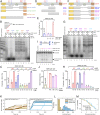
pCtIP promotes DNA2-dependent long-range DNA end resection pathway. (A) DNA end resection by WRN, DNA2, and human RPA (176 nM) using 2.2-kilobase pair (kbp)-long randomly labeled dsDNA substrate in the presence or absence of pCtIP. The reaction buffer contained 50 mM NaCl. Reaction products were separated by 1% agarose gel electrophoresis. Panel shows a representative experiment. Red asterisks indicate random labeling. (B) Representative 15% denaturing polyacrylamide gel showing the degradation kinetics of a Y-structured (45 nt/48 bp) DNA by DNA2 with or without pCtIP, in the presence of human RPA (15 nM) and 100 mM NaCl. The red asterisk indicates the position of the labeling. (C) Quantitation of overall substrate utilization from experiments such as shown in B. n = 3; error bars, SEM. (D) A representative experiment showing DNA unwinding by nuclease-deficient DNA2 D277A with or without pCtIP on oligonucleotide-based Y-structured (45 nt/48 bp) DNA. Reactions were supplemented with human RPA (7.5 nM) and 50 mM NaCl and analyzed on 10% native acrylamide gel electrophoresis. A red asterisk indicates the position of the labeling. (E) Quantitation of overall substrate unwinding from experiments such as shown in D. n = 3; error bars, SEM.
Fig. 2.
Fig. 2.
pCtIP dramatically promotes degradation of long stretches of ssDNA by DNA2. (A) Representative 1% agarose gel showing degradation kinetics of 3′ 32P-labeled ssDNA fragments (derived from λDNA) of various lengths by DNA2 without or with pCtIP in the presence of 864 nM human RPA. The sizes of the corresponding dsDNA fragments are indicated on the left. A red asterisk indicates the position of the labeling. (B) Quantitation of products smaller than 300 nt from experiments such as shown in A. n = 3; error bars, SEM. (C) Representative experiment as in A with various concentrations of pCtIP incubated for 8 min. (D) Quantitation of data such as shown in C; n = 3; error bars, SEM. The degradation activity of pCtIP alone is the same as in Fig. 3A (lane 7). (E) Experiment such as in A with various concentrations of DNA2 incubated for 8 min in the presence or absence of pCtIP. (F) Quantitation of data such as shown in E; n = 3; error bars, SEM. (G) Degradation of 2.2-kilonucleotide (knt)-long randomly labeled ssDNA substrate by DNA2 in the presence or absence of pCtIP. The reaction buffer contained 50 mM NaCl and, where indicated, 352 nM RPA. Reaction products were separated by 1% agarose gel electrophoresis. The panel shows a representative experiment. Red asterisks indicate random labeling of substrate DNA. (H) Quantitation of data such as shown in G; n = 3; error bars, SEM.
Fig. 3.
Fig. 3.
The motor activity of DNA2 mediates the accelerated ssDNA degradation with pCtIP. (A) Degradation of 3′ end-labeled ssDNA fragments (derived from λDNA) of various lengths by DNA2 in the presence or absence of pCtIP without or with ATP, as indicated. All reactions contained human RPA (864 nM). The experiment was incubated at 37 °C for 8 min. ATP is required for the stimulatory effect of pCtIP on DNA2. A red asterisk indicates the position of the labeling. (B) Quantitation of data such as shown in A. n = 3; error bars, SEM. The grey circles represent data points from indepedent experiments. (C) Degradation of ssDNA fragments by WT, helicase-deficient K654R, or nuclease-deficient D277A DNA2 variants without or with pCtIP. All reactions contained human RPA (864 nM). The experiment was incubated at 37 °C for 8 min. HelDead, helicase-dead; NucDead, nuclease-dead. (D) Quantitation of small degradation products from experiments such as shown in C. n = 3; error bars, SEM. (E) ATP hydrolysis by WT, helicase-deficient K654R, or nuclease-deficient D277A DNA2 alone (10 nM) or with pCtIP (40 nM). Reactions contained 10.3-kbp-long substrate denatured at 95 °C for 5 min, 395.5 nM human RPA where indicated and 20 mM NaCl. (F) Degradation of 3′ end-labeled ssDNA fragments derived from λDNA by human or yeast DNA2/Dna2 without or with human pCtIP or yeast pSae2. Reactions with human DNA2 were carried out at 37 °C for 8 min with human RPA (864 nM). Reactions with yeast Dna2 were performed at 25 °C for 1 min with yeast RPA (1.09 µM). (G) Analysis of DNA2 interaction with pCtIP. DNA2-FLAG was immobilized on M2 anti-FLAG affinity resin and incubated with purified recombinant pCtIP (phosphorylation sites depicted as P in green circles). The Western blot was performed with anti-FLAG and anti-CtIP antibodies.
Fig. 4.
Fig. 4.
Single-molecule experiments demonstrate accelerated DNA2 motor activity in the presence of pCtIP. (A) Sketch of the employed magnetic tweezers assay and the DNA construct carrying a 40-nt 5′ flap to allow loading of either helicase. (B) Representative DNA unwinding events of the DNA2 nuclease-dead mutant (D277A, 25 nM) in the absence (orange) and presence (green) of pCtIP (25 nM). Both reactions were also supplemented with 25 nM human RPA. (C) Histograms of the observed unwinding velocities for nuclease-dead DNA2 (n = 40 traces for each case). (D) Representative DNA unwinding events of BLM (25 nM) in the absence (magenta) and presence (purple) of pCtIP (25 nM). (E) Histogram of the observed unwinding velocities for BLM (n = 852 events). The force in all experiments was F = 19 ± 3 pN (SD).
Fig. 5.
Fig. 5.
Separate domains of pCtIP promote the MRN and DNA2 nucleases. (A) A schematic representation of the primary structure of WT pCtIP and internal deletion variants (pCtIP Δ1 to Δ4) purified from Sf9 cells in the presence of phosphatase inhibitors. Main ATM phosphorylation sites are indicated in red, and CDK phosphorylation sites are indicated in blue. (B) Degradation ssDNA fragments of various lengths by DNA2 without or with pCtIP variants, as indicated. All reactions contained human RPA (864 nM). A red asterisk indicates the position of the radioactive label. (C) Quantitation of data such as shown in B. n = 3–4; error bars, SEM. (D) A schematic representation of purified recombinant CtIP fragments (F1 to F3) expressed in E. coli. Full-length pCtIP is again shown as a reference. (E) Quantitation of ssDNA fragment degradation into products smaller than ∼300 nt by DNA2 without or with F1 to F3 CtIP fragments. n = 3; error bars, SEM. (F) A schematic representation of internal deletion variants (pCtIP Δ1A to Δ1D) purified from Sf9 cells in the presence of phosphatase inhibitors. Full-length pCtIP is shown as a reference. (G) Degradation of ssDNA fragments of various lengths by DNA2 without or with various concentrations of WT pCtIP or Δ1A to Δ1D mutants, in the presence of human RPA (864 nM). (H) Quantitation of data such as shown in G. n = 3; error bars, SEM. (I) Endonuclease assay with MRN (25 nM) and WT full-length or pCtIP Δ1B and Δ1C variants. A red asterisk indicates the position of the radioactive label. (J) Quantitation of data such as shown in I. n = 3; error bars, SEM. (K) Representative DNA unwinding events using the DNA2 nuclease-dead mutant (D277A, 25 nM) in the presence of pCtIP Δ1C (25 nM) (yellow) and pCtIP Δ1B (25 nM) (cyan). Both reactions were also supplemented with 25 nM RPA. (L) Histograms of the observed unwinding velocities for nuclease-dead DNA2 (n > 25 traces for each case) in the presence of pCtIP Δ1C (yellow) and pCtIP Δ1B (cyan) with mean values of 16 ± 1 bp/s (SEM) and 85 ± 3 bp/s (SEM), respectively. (M) Cumulative probability distributions (shown as survival probability) of the processivity of the individual DNA unwinding events by nuclease-dead DNA2 D277A in the presence of pCtIP Δ1C (Left) and pCtIP Δ1B (Right) with mean values of 3.6 ± 0.3 kbp (SEM) and 3.8 ± 0.3 kbp (SEM), respectively.
Fig. 6.
Fig. 6.
pCtIP phosphorylation facilitates its capacity to promote DNA2. (A) Degradation of ssDNA fragments of various lengths with DNA2 alone (20 nM) or with pCtIP WT, Δ1, Δ2, Δ3, or Δ4, mock-treated or λ-treated, in the presence of human RPA (864 nM). A red asterisk indicates the position of the labeling. (B) Quantitation of small degradation products from experiments such as shown in A. n = 4; error bars, SEM. (C) Representative DNA unwinding events of the DNA2 nuclease-dead mutant (D277A, 25 nM) in the presence of pCtIP (25 nM) treated with λ phosphatase (Left) and mock buffer (Right). Both reactions were also supplemented with 25 nM RPA. (D) Histograms of the observed unwinding velocities for nuclease-dead DNA2 (n > 25 traces for each case) in the presence of pCtIP treated with λ phosphatase (pink) and mock buffer (violet) with mean values of 16 ± 2 bp/s (SEM) and 74 ± 3 bp/s (SEM), respectively. (E) Cumulative (Cum.) probability distributions (such as survival probability) of the processivity of the individual unwinding events of nuclease-dead DNA2 D277A in the presence of pCtIP treated with λ phosphatase (pink) and mock buffer (violet) with mean values of 3.8 ± 0.3 kbp (SEM) and 3.9 ± 0.3 kbp (SEM), respectively. (F) A schematic representation of purified recombinant pCtIP Δ5 lacking the first 160 amino acids. pCtIP WT is again shown as a reference. (G) Degradation of ssDNA fragments of various lengths with DNA2 alone (20 nM) or with pCtIP WT or Δ5, mock-treated or λ-treated, in the presence of human RPA (864 nM). (H) Quantitation of data such as shown in G. n = 3–4; error bars, SEM.
Fig. 7.
Fig. 7.
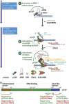
Model for pCtIP functions in DNA end resection. (A) A schematic depicting the role of pCtIP in short-range resection by MRE11 within the MRN complex and in long-range resection by DNA2-BLM. In long-range resection, pCtIP stimulates both the helicase activity of BLM to unwind dsDNA and the translocase activity of DNA2 downstream to facilitate degradation of unwound, RPA-coated ssDNA. Unwinding of dsDNA and ssDNA degradation in long-range resection is likely coordinated and was depicted as separate for easy visualization. (B) A schematic representation of the primary structure of pCtIP and the domains required for the stimulation of MRN or DNA2 nucleases.

Similar articles

Cited by

References

    1. Ranjha L., Howard S. M., Cejka P., Main steps in DNA double-strand break repair: An introduction to homologous recombination and related processes. Chromosoma 127, 187–214 (2018). - PubMed
    1. Cejka P., DNA end resection: Nucleases team up with the right partners to initiate homologous recombination. J. Biol. Chem. 290, 22931–22938 (2015). - PMC - PubMed
    1. Bonetti D., Colombo C. V., Clerici M., Longhese M. P., Processing of DNA ends in the maintenance of genome stability. Front. Genet. 9, 390 (2018). - PMC - PubMed
    1. Cannavo E., et al. , Regulatory control of DNA end resection by Sae2 phosphorylation. Nat. Commun. 9, 4016 (2018). - PMC - PubMed
    1. Huertas P., Jackson S. P., Human CtIP mediates cell cycle control of DNA end resection and double strand break repair. J. Biol. Chem. 284, 9558–9565 (2009). - PMC - PubMed

Publication types